0.5pt
Two-Dimensional Transition Metal Silicate Formed on Ru (0001) by Hydrogenation
Abstract
Two-dimensional (2D) transition metal silicates are interesting materials because of their potential ferromagnetism together with their inherent piezoelectric response to their structural symmetry. Substrate-assisted bottom-up synthesis of these materials offers flexibility in their chemical composition. However, synthesizing free-standing layers has been challenging due to strong overlayer-substrate interactions which hinders exfoliation of the overlayer. Here, using density functional theory calculations, we systematically investigate the hydrogenation of such overlayers as a way to decrease the substrate-overlayer interactions. Using the Fe2Si2OO/Ru(0001) structure as our starting point, we study hydrogenation levels up to Fe2Si2O9H4/Ru(0001). Structural and thermodynamic properties are studied at different hydrogenation levels to describe the conditions under which exfoliation of the Fe-silicate is feasible. Simulated surface core-level binding energy shifts of core electrons show that Fe is primarily in the 3+ state throughout the hydrogenation process. Simulated reflection adsorption infrared spectroscopy (RAIRS) yields distinctive shifts in vibrational peaks with increasing hydrogenation which can guide future experiments.
I Introduction
Two-dimensional (2D) materials research has grown immensely in recent years: a key fact fueling these developments is that these materials can be isolated from their bulk counterparts. These 2D materials show distinct physical and chemical properties from their bulk counterparts due to effects such as quantum confinement and significantly increased exposed surface area. Top-down approaches, such as mechanical exfoliation, are often used to isolate 2D layers from the bulk. Bottom-up approaches, however, can enable control over the structure and chemical composition of the synthesized layers and hence provide more flexibility. By selecting an appropriate substrate, surface-assisted bottom-up synthesis can create structures that are not available in bulk phases, e.g., 2D bilayer silicates Büchner and Heyde (2017). For the large-scale application of these bottom-up fabricated materials, they must be high quality and are air-stable.
Recently, 2D transition metal silicates such as Fe, Ni, and Ti-silicates have been synthesized on metal substrates Włodarczyk et al. (2013); Zhou et al. (2019); Fischer et al. (2015); Li et al. (2017); Doudin et al. (2021). These materials have the chemical formula of M2Si2O8, where a honeycomb Si2O5 layer with corner-sharing tetrahedra is stacked on top of a transition metal oxide layer which has both edge and corner-sharing 5-fold coordinated, square pyramidal polyhedra. These transition metal silicates on metal substrates resemble crystal structures of naturally existing sheet silicates (phyllosilicates), particularly that of dehydroxylated nontronite Włodarczyk et al. (2013); Tissot et al. (2016). These structures are also very similar despite the nominal preferences of Ni, Fe, and Ti for different oxidation states. The synthesis procedure for these transition metal silicates starts with depositing Si or SiO and the transition metal at modest temperatures on the substrate or by depositing the Si on an alloy that contains the target transition metal and then following annealing near 950 K Zhou et al. (2019).
To our knowledge, there has been no concerted effort to yield a VDW layer from these materials after their syntheses: The 2D transition metal silicate forms strong chemical bonds with the substrate, which prevents the isolation of the 2D layer Zhou et al. (2019). An efficient strategy can be to hydrogenate these materials with a hydrogen source such as hydrogen gas, plasma, or water. The dehydroxylation of kaolinite to yield metakaolin (M2Si2O9) is done in large-scale industrial processes White et al. (2010), hence these hydrogenation reactions can be chemically accessible.
In this work, using density functional theory (DFT), we study the hydrogenation mechanism of 2D Fe-silicates on Ru (0001). 2D Fe-silicate on Ru (0001) Włodarczyk et al. (2013) and Pd (111)Saritas et al. (2021a) were synthesized previously. We show that Fe-silicate/substrate bond can be substantially weakened by the hydrogenation, such that the film may be isolated to yield a 2D Van der Waals (VDW) layer. Most importantly, we show that the interface oxygens, which are the oxygens chemically bound on the substrate surface, behave in a way that is similar to the hydroxide groups during the water adsorption and dissociation on Ru (0001) Feibelman (2002); Michaelides et al. (2003); Tatarkhanov et al. (2009). With increasing hydrogenation, oxygen atoms originally near Ru (0001) hcp and bridge sites migrate closer to atop sites in our materials, and this is similar to hydroxide groups forming on the atop Ru (0001) sites in the case of the water adsorption. Fully hydrogenated Fe-silicate forms a compound with the chemical formula of Fe2Si2O9H4, thus incorporating the adatom oxygen as well which was initially located at the hcp site of Ru (0001). Fe2Si2O9H4 on Ru (0001) has a binding energy of 0.101 meV/Å2, which classifies the compound as potentially exfoliable Mounet et al. (2018). Our analyses show that the Fe atom has a charge state of nearly for all Fe2Si2O9Hn () on Ru(0001) complexes we studied. We perform density of states, workfunction, core level binding energy shifts, and simulated reflection adsorption infrared spectroscopy (RAIRS) calculations plus structural analyses to corroborate our findings with ongoing experiments.
II Methods
All DFT calculations were performed using the plane-wave Vienna ab initio Simulation Package (VASP) Kresse and Furthmüller (1996a, b), using the Perdew-Burke-Ernzerhof exchange-correlation functional with D3 semi-empirical vdw correction (PBE+D3) Perdew et al. (1996); Grimme et al. (2010). We used a kinetic energy cutoff of 520 eV, periodic boundary conditions, projector-augmented wave (PAW) pseudopotentials Kresse and Joubert (1999) and dipole correction Neugebauer and Scheffler (1992). We used Gaussian smearing of 0.2 eV to better capture the metallic nature of the film on the substrate together with a dense reciprocal grid density of 661. Geometry relaxations were performed until the forces on all atoms were smaller than 10-2 eV/Å with an electronic convergence of 10-5 eV. Each simulation cell consists of a 22 supercell of the 5-layer thick Ru (0001) surface primitive cell and a vacuum layer larger than 15 Å. All the calculations were made with spin-polarization due to the presence of the Fe atom. All Fe atoms were initialized in the high-spin ferromagnetic configuration.
Harmonic vibrational frequencies at were calculated using the frozen phonon method using central differences with 0.015 Å displacements along with Cartesian directions. Zone-center phonon modes were obtained from the eigenvalues of the dynamical matrix. Experimentally, the RAIRS method is only sensitive to changes in the dipole moment vertical to the substrate surface. Therefore, simulated RAIRS intensities were obtained from the changes in the square of the dipole moments along the z-axis at each phonon mode Karhánek et al. (2010). The vibrational frequencies were scaled with a factor of 1.0341 derived from a comparison between the theoretical and experimental vibrational frequencies of -quartz Löffler et al. (2010).
To calculate simulated surface core-level binding energy shifts (SCLS), , we used the initial-state approximation via DFTKöhler and Kresse (2004). SCLS can explain the relative positions of the core levels of an adsorbate, hence can be compared to XPS experiments. In this approach, the core-level binding energy of an electron is first calculated as ) where is the Kohn-Sham eigenstate of the core level, and are Fermi level and the workfunction of the system. Therefore, are calculated as the difference in the core-level binding energies on substrate, , and under vacuum, : Lizzit et al. (2001). SCLS can occur due to multiple factors such as transitioning to a different oxidation state in the adsorbate due to charge transfer or modification of the interface dipole Egelhoff (1987). When comparing Fe-silicates with different amounts of hydrogens on Ru (0001), or under similar varying conditions, it is expected that the interface dipole is modified at every hydrogenation level. We aim to understand how the charge states of Fe and O atoms change with increasing hydrogenation. Therefore, the effect of interface dipole on SCLS should be subtracted to the best approximation. These considerations have also been observed in XPS studies where core levels of all species at the overlayer can shift depending on the strength of the interfacial dipole, which can introduce inconsistencies between measurements of different samples Jhang et al. (2020). For physisorbed silica bilayer on Ru(0001), XPS study shows that upon removal of the chemisorbed oxygen, O(22)/Ru(0001), silica bilayer associated O-1 and Si-2 core levels are shifted uniformly by 0.75 eV, indicating that the effect of the interface dipole on the core level binding energies of adsorbates is uniform Wang et al. (2017). Therefore, SCLS of a species with a well-defined oxidation state, such as Si4+, can be taken as a reference across different samples each having different interface dipoles. Si4+ is a good reference, because Si4+-p valence levels of Fe-silicates on Ru(0001) are energetically stable by nearly 20 eV below the Fermi level Sup and we find that Si- valence states have a charge transfer of only 0.001 e from the Ru(0001) substrate. Therefore, the change in the Si-2 core level shifts can be approximated to be primarily due to modification of the interfacial dipole, because the charge transfer is very limited. Therefore, we use a modified definition of the SCLS for our oxidation state analysis where is the change in the SCLS of Si-2 electrons compared to a reference system, which we choose as Fe2Si2OO/Ru(0001). The correction due to anchors at the reference energy across samples with varying amounts of hydrogens, hence the effect of interfacial dipole on the other core-levels is subtracted. To simulate the spectra we use Gaussians with a full half-width maximum of 0.8 eV Jain et al. (2018); Baer et al. (2020).
The chemical potential of the hydrogen gas at standard conditions is required to calculate the Gibbs formation enthalpies. Using DFT we calculate the H2(g) dimer at 0 K and find that it has a dissociation energy of 4.54 eV compared to the experimental value of 4.48 eV Darwent and (U.S.). Then we write the reference chemical potential of H at standard conditions as, . Experimental entropy of H2 gas under standard conditions, is taken from NIST Webbook Cox, J.D.; Wagman, D.D.; Medvedev (1984) as 130.680 J/mol*K. Using this and the DFT energy of H2 gas at 0 K, of -6.774 eV, we find under standard conditions as -3.571 eV. To define the hydrogen chemical potential under varying pressure at 298 K, we define , where is the hydrogen gas partial pressure under standard conditions. Surface binding energies are calculated from
| (1) |
where , , and are the total energies of isolated adsorbate, substrate, and the chemisorption system, respectively. In this definition, a positive binding energy means that the process is exothermic.
III Results and Discussion
We use Fe2Si2OO/Ru(0001) slab as the starting point of our theoretical investigation of its hydrogenation as shown in Fig. 1(a). This dehydroxylated system was previously studied by Włodarczyk et al. (2013) and here, we first reproduce their findings. As shown in Fig. 1, Fe2Si2OO/Ru(0001) is made of Fe-O layer at the interface with the substrate and has a nontronite-like as previously explained. Here, O/Ru(0001) stands for the pristine Ru(0001) surface plus an adatom O located at every other hcp site in (22) pattern. This O/Ru(0001) phase is also known as the low coverage phase of adsorbed O on Ru (0001) Lizzit et al. (2001). Therefore, Fe2Si2O8 film is best understood as being bout to the O/Ru(0001) surface, hence combined making up the Fe2Si2OO/Ru(0001) complex. Oxygens in the Fe2Si2OO/Ru(0001) complex can be separated into five groups depending on their positions and vertical distances to the Ru substrate: The adatom O, Oad, is closest to the Ru substrate and Oad has a vertical distance, z(Oad-Ru), of 1.12 Å to the top Ru layer. However, the the shortest Ru-Oad bond, min(d(Oad-Ru)), is 2.03 Å, . This 2.03 Å d(Oad-Ru) bond length compares well to the experimental value of 1.99 Å in RuO2 Bolzan et al. (1997). Oxygens above the atop and hcp Ru sites, (Oatop and Ohcp) as shown in Fig. 1, are further away from the substrate compared to Oad and have an average z(O-Ru) of 1.8 Å. The corner-sharing oxygens (Oc) are, however, further distant to the Ru substrate by 3.57 Å, and positioned at the shared corners of the FeO5 polyhedra. The remaining oxygen atoms that are furthest away from the Ru substrate, by at least 4.2 Å, make up the Si-O tetrahedra. In the structure reported by Włodarczyk et al. (2013), the Oad is positioned below the Si-O-Si bridging oxygens along the -axis. However, the Fe2Si2O8 can slide over this substrate and can use a different registry. We construct our simulation using these constraints and slide the Fe2Si2O8 along the substrate surface and find that the O adatom is positioned below the Si-O-Si is indeed the most stable configuration. We find that the binding energy of Fe2Si2O8 on O/Ru(0001) substrate is 0.149 eV/Å2 (1.88 eV per Fe) which agrees nicely with the 0.160 eV/Å2 (-15.48 kJmol-1/Å2) based on the DFT+D functional Grimme (2006) Włodarczyk et al. (2013).
III.1 Binding of a single H on Fe2Si2OO/Ru(0001)
To study the effect of hydrogenation on the Fe2Si2OO/Ru(0001) system, we first introduce a single hydrogen atom and investigate its binding energy to different oxygen sites. In the serpentine-silicate analogs such as nontronite, kaolinite, lizardite, and talc, the hydrogens are always found to be bonded to metal oxide groups rather than the Si-O tetrahedral groups. Here, as well in our prior work Saritas et al. (2021b), we find this to be true as shown in Fig. 1(b). Therefore, we do not consider the oxygens that are part of Si-O network as potential hydrogen binding sites. Hence, out of a total 9 starting oxygen sites, we will study hydrogen binding on the remaining 4 oxygen sites. These sites are, in the order of their distance to the Ru substrate, adsorbed oxygen Oad, oxygens above atop and hcp Ru (0001) sites, Oatop and Ohcp, and finally the corner sharing Fe-O-Fe oxygen Oc. We study the binding energy of single H at a time separately to these four oxygen sites in this section. We find that the hydrogen binding energy in the Fe2Si2OO/Ru(0001) + 1/2 H2 Fe2Si2OOH/Ru(0001) reaction is -1.22, -0.65, -0.25 and -0.07 eV for Oc, Oatop, Ohcp and Oad respectively. It is interesting to note that the H-binding energy at Oc site is significantly larger than the binding at other sites considered. The distinctly lower hydrogen binding energy at the Oc site was also observed at the nontronites under vacuum Saritas et al. (2021b). This will be further discussed in Sec. III.2.
The difference in the hydrogen binding energies at Oatop and Ohcp sites can be understood via the mechanism of water dissociation at Ru (0001) surfaces Michaelides et al. (2003). Water on Ru (0001) is known to partially dissociate into H and OH at 100-170 K Feibelman (2002). Its structure has been characterized experimentally Feibelman (2002), and theoretical research confirmed this structure with a dissociation barrier of 0.5 eV which agrees with the experimental dissociation temperature Michaelides et al. (2003); Tatarkhanov et al. (2009). Water dissociation on Ru (0001) is energetically quite favorable with an enthalpy of -0.85 eV per dissociated H2O Michaelides et al. (2003). Theoretical calculations find those intact H2O molecules, before their eventual dissociation on Ru (0001), form a bilayer with two types of molecules: one nearly parallel to the surface and the second with an O-H bond nearly perpendicular to the surface. However, it is thermodynamically favorable to dissociate every one of two water molecules on the substrate. Upon the dissociation, the OH, H, and H2O all bind parallel to the surface, hence forming a monolayer, and are adsorbed on atop Ru (0001) sites Michaelides et al. (2003). OH and H2O form a honeycomb lattice by forming hydrogen bonds parallel to the substrate, whereas the H atoms are positioned in the center of each honeycomb site Feibelman (2002). Based on this, in the case of Fe2Si2OOH/Ru(0001), H binding on Oatop should be more favorable because of the collective preference of H2O and OH groups to be located at the atop Ru sites. For OH and H2O on Ru (0001), it was noted that the surface adsorption mainly occurs thanks to an H-/O- hybridized orbital and Ru- orbitals when H2O is located at favorable atop-site Michaelides et al. (2003). When the OH or H2O is not located on an atop Ru site, this overlap may not be as strong as in the case of atop case, hence leading to the difference in H-binding energies at Oatop and Ohcp sites.
The reason for low hydrogen binding energy at Oad can also be explained by similar reasoning of the difference in hydrogen binding energies at Oatop and Ohcp. After binding hydrogen to Oad, we find that the geometry relaxation yields a structure where the Oad migrates from its original hcp position towards a bridge site between two top layers of Ru atoms. We tried various orientations of the Oad-H bond to the substrate surface and found that the vertical orientation of Oad-H group towards the substrate is not energetically favorable. Relaxed geometry of Oad-H group is angled such that its H atom forms a hydrogen bond with the Oatop with a d(Oatop-H) distance of 1.96 Å. This is similar to the partially dissociated water on Ru (0001), however, the migration of the Oad to an atop site can be hindered by the presence of the interface oxygen atoms of the Fe-silicate layer. This indicates that Fe2Si2OO/Ru(0001) has a similar complexity for hydrogen bonding networks which facilitates a particular orientation of the OH groups.
III.2 Complete hydrogenation of Fe2Si2OO/Ru(0001) to Fe2Si2O9H4/Ru(0001)
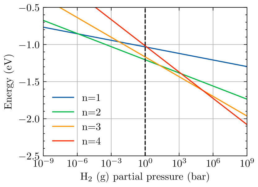
Having studied how a single H atom bonds to Fe2Si2OO/Ru(0001), we now consider further hydrogenation. We considered up to four H bound to the four favorable sites of Fe2Si2OO/Ru(0001) as explained in the previous section. At each hydrogenation level, we considered all possible combinations of different sites. We find that the qualitative energetic ordering of the binding energies for single hydrogen adsorption applies while achieving complete hydrogenation. In Fig. 2, we plot the Gibbs free energies of hydrogenation for the
reaction and show that the formation of Fe2Si2O9H2 is thermodynamically favorable under ambient conditions. According to Fig. 2, higher H pressures in the hundreds of kbar range are needed to completely hydrogenate Fe2Si2O9H4. Alternatively, one can consider H atoms generated using a plasma source or an H atom source Tschersich et al. (2008).
Introducing more than single hydrogen to Fe2Si2OO/Ru(0001) causes some changes in the geometry of the complex which facilitates gradual migration of Oad towards the Fe-silicate film from the Ru substrate. With the addition of second hydrogen () at Oatop site (the second most favorable hydrogen binding oxygen site), shown in Fig. 1(c), one of the Fe atoms move towards the Oad. Initially at the dehydroxylated case, , the closest d(Fe-Oad) distances are 2.67 and 2.81 Å. However, at one d(Fe-Oad) is 2.07 Å while the other becomes 2.91 Å. Average Ru-Oad bond lengths also increase from 2.01 to 2.10 Å as increases from 0 to 2. Therefore, these collective changes can indicate that Ru-Oad bonds are weakened or Fe-Oad bonds got stronger with hydrogenation. To understand whether Oad is more strongly bound to the substrate or the Fe-silicate at , we explore two different scenarios: We find that the Fe2Si2O8HO/Ru(0001) Fe2Si2O8H2 + O/Ru(0001) reaction yields a surface binding energy of 1.76 eV/Fe (0.14 eV/Å2), whereas the Fe2Si2O9HO/Ru(0001) Fe2Si2O9H2 + Ru(0001) reaction has a surface binding energy of 2.62 eV/Fe (0.21 eV/Å2). Therefore, at , the Ru-Oad bonds are harder to break than the Fe-Oad bonds, hence Oad still prefers being on the substrate rather than leaving the substrate with the film upon exfoliation. Therefore, we conclude that the addition of only two hydrogens per formula unit Fe-silicate to this slab still yields very large binding energy between the overlayer and substrate.
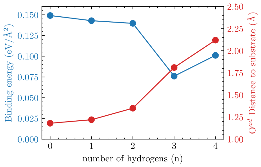
The third hydrogen can be bonded to the Ohcp, as shown in Fig. 1(d). Therefore, the Ohcp-H group migrates towards an atop Ru site by 0.8 Å, while the Si-tetrahedra slide about 1.1 Å on the film in the same direction as the Ohcp-H group. This causes a significant reorganization for Oad atom which moves from its original hcp site to a bridge site between two top Ru atoms while (Oad-Ru) increases from 1.35 Å to 1.80 Å. To explain these significant changes at the step, we plot the surface binding energies and z(Oad-Ru) as a function of hydrogenation level in Fig. 3. As shown in Fig. 3, the vertical distance of the Oad to the Ru substrate, z(Oad-Ru), increases as 1.18, 1.22, 1.35, 1.80, 1.98 Åwith increasing the number of hydrogens, , from 0 to 4. Therefore, there is a trend of Oad moving away from the substrate even before this significant reorganization at happens. Up to , we see no change in the lateral position of the Oad, meaning that its Ru(0001)-hcp site is still strongly favorable for Oad and Oad is most strongly interacting with the Ru substrate. Similar to the case, to quantify whether the Oad would like to bond to the film or the substrate at , we again compare two different scenarios: We find that the Fe2Si2O9H3 on Ru(0001) has a surface binding energy of 0.149 eV/Å2, whereas Fe2Si2O8H3 on O/Ru(0001) has a surface binding energy of 0.076 eV/Å2. Therefore, at hydrogenation level, the Oad adsorption on Ru significantly weakens. However, according to Fig. 2, hydrogen gas partial pressures of near 1 kbar or with hydrogen richer conditions/sources are needed to achieve Fe2Si2O9H3/Ru(0001).
With the addition of the fourth hydrogen to the Oad site, as shown in Fig. 1(e), the vertical distance of this Oad atom to the Ru substrate increases to 1.98 Å. This is a substantial increase over the 1.18 Å z(Oad-Ru) at . Fe2Si2O9H4 film on the Ru substrate has a binding energy of 1.27 eV/Si (0.10 eV/Å2). Interestingly, this indicates an increase over the binding energy at case. At this point, it should be noted that hydrogen gas partial pressures of 105 bar are needed to thermodynamically stabilize Fe2Si2O9H4/Ru(0001), which indicates about four orders of magnitude richer H gas richer conditions compared to the onset of Fe2Si2O9H3/Ru(0001) formation according to Fig. 2. The 0.10 eV/Å2 binding energy suggests that the interaction is not fully a van-der Waals type but includes a degree of chemical adsorption. Nevertheless, materials with up to 0.15 eV/Å2 binding are often regarded as potentially exfoliable, therefore it may be possible to extract a 2D Fe-silicate layer using special techniques Gibertini et al. (2019).
To explain the differences in the hydrogen binding energies at different oxygen sites, it can be useful to study the changes in densities of states of Fe-silicates upon hydrogenation. Therefore, in Fig. 5, we show the detailed partial densities of states of hydrogen binding oxygens at Fe2Si2OO/Ru(0001) structure. The sharp atomic-like O peak is close to Fermi energy at about -1 eV and the large O peak is at -7 eV are the contrasting features of these two oxygens. We find that upon complete hydrogenation at Sup , the sharp atomic-like O peak reduces in energy by about 6-7 eV. This is in contrast to other O- peaks which show much smaller or zero reductions. In Fig. 5, O states are centered at the lowest energy level compared to other hydrogen-binding oxygens, which would mean that the Oad is passivated more strongly. Upon complete hydrogenation at , however, partial densities of states of Oatop and Ohcp become exactly equal.
| property | 0 | 1 | 2 | 3 | 4 |
|---|---|---|---|---|---|
| E (meV/Å2) | 0.149 | 0.142 | 0.139 | 0.076 | 0.101 |
| (eV) | 1.02 | 1.02 | 0.46 | 0.95 | 0.72 |
| z(Oad-Ru) (Å) | 1.18 | 1.22 | 1.35 | 1.81 | 1.98 |
| (e) | 0.75 | 0.71 | 0.46 | 0.95 | 0.76 |
III.3 Changes in the work function with increasing hydrogenation
Hydrogenation of the Fe-silicate can affect the formation of the interface dipole, and the amount of charge transfer between the substrate and the film. Therefore, the workfunction of the substrate-film complex can be modified. First, we calculate that the bare Ru (0001) surface has a work function of =5 eV compared to an experimental value of 5.4 V Böttcher and Niehus (1999); Himpsel et al. (1982). The Fe2Si2OO/Ru(0001) complex has a work function of 6.02 eV, which indicates a change in the workfunction, , by about 1.02 eV which is close to 1.4 eV from Włodarczyk et al. (2013). Our results of the changes in the work functions from bulk, , with increased hydrogenation of the Fe-silicate are given in Table 1. initially decreases as a function of hydrogenation from 1.02, 1.02 to 0.46 eV for =0 to 2, indicating that the charge transfer is significantly decreasing as a function of hydrogenation. However, at =3 the film reorganizes and incorporates the Oad, hence the again increases to 0.95 eV. However, for complete hydrogenation at , the again reduces to 0.72 eV meaning the charge transfer in Fe2Si2O8H2 and Fe2Si2O9H4 are likely similar. As shown in Table 1, changes in correlate with the amount of charge transfer. This is expected considering Fe has a formal charge of 3+ in both compounds if they were under vacuum, but a positive change in the workfunction would still indicate that there is charge transfer from the substrate to the film. In the SI Sup , we show that the work function of bare Fe2Si2O9H4 in a vacuum is about 1.5 eV larger than the work function of Ru substrate, hence a charge transfer from the Ru substrate to the overlayer would be expected even when the overlayer is completely hydrogenated.
III.4 Ionic states of Fe and O with increasing hydrogenation
To further guide experimental synthesis and characterization, we compute additional observables. First, we try to determine the charge state of the Fe ion as a function of the hydrogenation level. Therefore, using LOBSTER code Maintz et al. (2016); Dronskowski and Bloechl (1993) we project the plane-wave wavefunction onto atomic orbitals and study integrated densities of states. We find that the improved basis sets LOBSTER can yield numerically more accurate results for quantities such as integrated densities of states, compared to the internal routines in VASP Maintz et al. (2013).
To understand how the oxidation states of Fe-silicates evolve on Ru, we first explore Fe2Si2O9H4 in a vacuum. In a vacuum, Fe2Si2O9H4 would have no way to exchange charge with its surroundings, hence the simple assignment of formal charges to block elements yields a 3+ charge for Fe. In Fig. 4a, we plot the partial densities of states (DOS) for Fe2Si2O9H4 in a vacuum and show that simply enforcing the octet rule for -block elements yields the correct oxidation state for Fe atoms. We enforce high-spin starting conditions for Fe in all the calculations. As shown in Fig. 4a, the high-spin Fe orbitals are completely occupied. We integrate Fe- curves up to Fermi level and find 4.98/0.86 (spin-up/down) electrons per Fe for Fe2Si2O9H4 in vacuum. However, using the formal charge argument, meaning Fe would be 3+ in Fe2Si2O9H4, would indicate that Fe3+ is , hence would have no spin-down electrons. The fact that the down spin O- electrons are also polarized above the Fermi level and the Fe- up spin orbitals are full indicates that the O- and Fe- are mainly hybridized in the minority spin channel and Fe- down spin electrons in Fig. 4a formally belong to oxygen atoms. We support our findings using crystal orbital Hamilton population analysis (COHP)Dronskowski and Bloechl (1993) in the SI Sup which shows that Fe-O bonding is over the minority spins.
We analyze the partial DOS of Fe2Si2O9H4 on Ru and find that the Fe and O densities are very similar to the vacuum case, hence the integrals up to the Fermi level are 4.96/0.97 (spin-up/down). In a vacuum, down spin Fe- orbitals are centered around -4 eV, while on Ru, this density is concentrated around the Fermi level, which could indicate that Fe atoms interact with the substrate Ru atoms, through O, as the work function of Ru is smaller than Fe2Si2O9H4. The DOS in Fig. 4a, indicates that Fe2Si2O9H4 is a semi-metal. Therefore, a discussion on the choice of the DFT functional can be appropriate. With additional exchange splitting through Hubbard-, an electronic gap would open in Fig. 4a, which would make Fe2Si2O9H4 in a vacuum insulating with a finite gap. Nevertheless, PBE still yields a sizeable exchange splitting of nearly 5 eV and as the spin-up energy levels of Fe- electrons are completely occupied for under vacuum and on Ru (0001), results from PBE and PBE+ would be qualitatively consistent. PBE functional was used to simulate Ti-silicate Fischer et al. (2015) and Fe-silicate Włodarczyk et al. (2013) on Ru (0001) previously and yield accurate structural and vibrational properties.
For the lesser hydrogenated Fe-silicates, however, the situation can be more complicated. For the completely dehydroxylated case, Fe2Si2OO/Ru(0001), assuming the substrate is defined as O/Ru(0001), the overlayer would be Fe2Si2O8. For Fe2Si2O8 in a vacuum, the formal charge assignment method would yield an oxidation state of 4+ on Fe. However, we find that as goes from 0 to 4 for Fe2Si2O8,9HO1,0/Ru(0001), the partial densities of states on Fe changes very weakly such that the total number of electrons on each Fe atom remain between 5.93 and 6.04 calculated using their partial DOS Sup This highlights the difficulties in assigning an integer charge state to transition metal atoms due to charge self-regulation Raebiger et al. (2008).
III.5 Surface core-level binding energy shifts
While it would be useful to assign integer oxidation states to the Fe atoms at different hydrogenation levels of , it is more important to relate the structural and physical changes to the observables from the experiments. For this reason, we calculate surface core-level binding energy shifts (SCLS) using DFT which can directly be compared to experimental x-ray photoelectron spectroscopy (XPS) results. SCLS can arise from two main effects: electrostatic potential, hence the chemical environment of a species, and its oxidation state Fongkaew et al. (2017). Both of these factors can modify the screening of the core electrons, hence leading to varying binding energies. Therefore, chemical changes in a compound, such as an adsorbate on a substrate, can be understood through the SCLS of its species. In this section, we investigate the SCLS’ of Fe and O at various hydrogenation levels of the iron silicate films on Ru(0001) to understand the changes in the oxidation state of Fe ions.
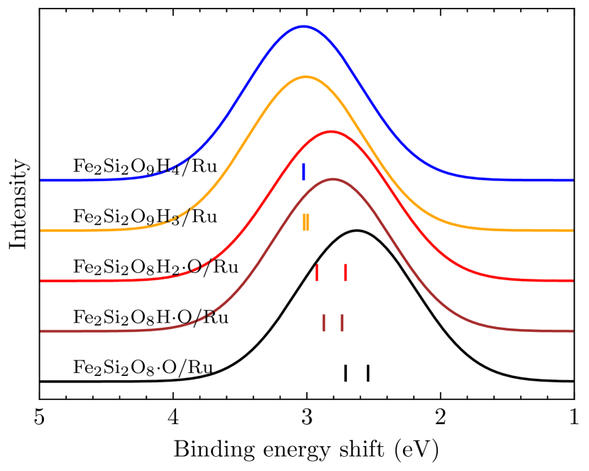
Fig. 6 shows that the SCLS of Fe atoms increases with the hydrogenation level, . Additionally, it can be seen in Fig. 6 that up to , Fe SCLS’ are split by 0.25 eV and for the energies become completely degenerate. The energy split could be due to the difference in their registries to substrate. We find that the distances of Fe atoms to the Oad are 2.81 and 2.64 Å. These differences in surface registries can cause different screening properties. Once the Oad is lifted off from the substrate with increased hydrogenation at and minimum z(O-Ru) distance is about 1.81 Å, this splitting also largely disappears as the effect of surface registry diminishes for Fe atoms. SCLS’ of Fe can explain how the oxidation state of Fe changes with increased hydrogenation. Given that the oxidation states of Fe in Fe2Si2O9H4 under vacuum and Fe2Si2O9H4/Ru should be very close and can be considered close to 3+, the decreased SCLS’ observed in Fig. 6 means that the Fe in Fe2Si2OO/Ru should be oxidizing towards 4+ states Pasquarello et al. (1995). The difference in the Fe SCLS’ in and cases is nearly about 0.4 eV. We can compare our results to bulk La1-xSrxFeO3, where a transition between 4+/3+ can be observed depending on the value of Wang et al. (2019). For case in La1-xSrxFeO3, Fe is formally 3+. At Fe-2p binding energies shift to higher energies by about 1 eV Wang et al. (2019). The 1 eV shift here is larger compared to our finding of nearly 0.4 eV shift between and cases of Fe silicate on Ru. However, an exact quantitative agreement would require more detailed calculations which involve final state and dynamic screening effects, hence our calculations are useful to provide qualitative results.
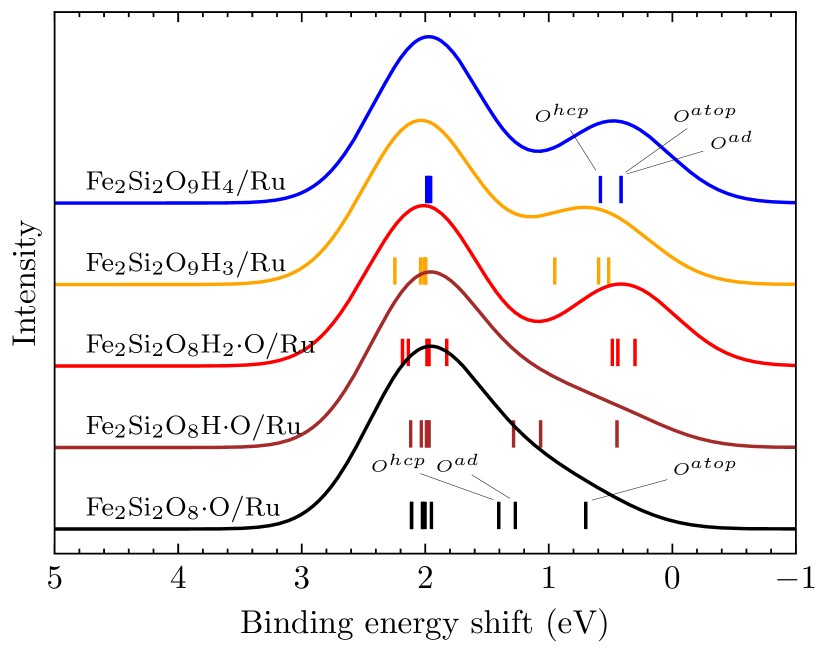
In Fig. 7 we show the core level shifts in oxygen atoms with increased hydrogenation. Our results in Fig. 7 for Fe2Si2OO/Ru(0001) can be compared to previous experiments Włodarczyk et al. (2013); Löffler et al. (2010) and theory Yang et al. (2012) on Fe silicates on Ru and bilayer silicates on Ru. Our results here agree with these previous findings such that there are distinct SCLS’ for Ohcp, Oatop and Oad which result in a shoulder formation in the O-1 XPS with a nearly 2 eV shift compared to the large O-1 peak that is associated with Si-O oxygens. Overall, the SCLS’ around 2 eV in Fig. 7 are rather stationary with increased hydrogenation, meaning that oxygens associated with these peaks have constant charge transfer with increased hydrogenation. However, the SCLS’ of chemisorbed oxygens (Ohcp, Oatop and Oad) typically decrease with increased hydrogenation. For these oxygens, the trend is opposite that of Fe-2 SCLS’ in Fig. 6. These O atoms have decreasing charge transfer from the Ru substrate with increased hydrogenation. This is also correlated to the increasing z(O-Ru) with increased hydrogenation. While the Oad are Oatop 1 SCLS’ are distinct in Fe2Si2OO/Ru(0001), they become isoenergetic for Fe2Si2O9H4/Ru(0001). This can also be related to the fact that with increased hydrogenation, the distance between the film and the substrate increases, and the effect of the surface registry decreases. However, the fact that Ohcp core level shift is different than Oatop and Oad shows that there is still some effect of the surface registry. While the SCLS’ of Fe2Si2OO/Ru(0001) can be seen as a peak and a small shoulder forming at lower binding energies, at the limit of complete hydrogenation, Fe2Si2O9H4/Ru(0001), it can be seen as a combination of two peaks with more distinct energies. Additional hydrogens increase the z(Ru-O) distances and decrease the ionic character of interface oxygens, which can lead to reduced SCLS’. SCLS’ of oxygens around Si-O tetrahedra and Oc remain largely fixed as these oxygens are already well separated from the substrate and interact with the substrate only indirectly.
III.6 Simulated RAIRS
As increased hydrogenation is accompanied by structural changes in the Fe-silicate film, a change of the vibrational spectrum should be observed as the Fe-O layer goes from a 5-fold edge/corner-sharing network to an octahedral edge-sharing network with increased hydrogenation. RAIRS experiments have shown that the Fe2Si2OO/Ru(0001) has a large peak at 1005 cm-1, and through DFT calculations, this peak is associated with the vertical vibrations of bridging Fe-O-Si oxygens Włodarczyk et al. (2013). In Fig. 8(a), we show that our Fe2Si2O8HO/Ru(0001) also has a peak at 1002 cm-1 which is due to the vertical motion of the Fe-O-Si bridging oxygens. This is expected as hydrogenation at 3 does not cause significant distortion of the adsorbed film, hence the vibrational motion of Fe-O-Si bridging oxygen would not be affected. However, at the Oad gets displaced significantly and attaches itself to the Fe-silicate film. This causes a slight shift in the major peak by 13 cm-1 to 1015 cm-1 due to the strengthening of this bond.
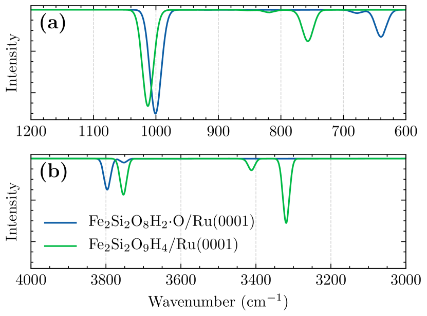
Complete hydrogenation causes a large shift in the smaller peak that is associated with the bending motion of Si-O-Si bridging oxygens. The peak for this mode is around 652 cm-1 for Fe2Si2O8HO/Ru(0001). Włodarczyk et al. (2013) had observed a Si-O-Si bending mode at 652 cm-1, that is the same as ours. However, the Si-O-Si bending mode is observed at 758 cm-1 for Fe2Si2O8HO/Ru(0001), which indicate a shift of the peak nearly 100 cm-1. This indicates a change of character in Si-O tetrahedra which can be due to the differences in the lattice parameters of Fe-silicates Saritas et al. (2021b). The hydrogenation of Fe2Si2OO/Ru(0001) also yields additional O-H stretching modes above 3000 cm-1. IR stretching modes between 3700-3200 cm-1 were observed in various bulk layered silicates Huang (1999). Since the peaks 3000 cm-1 are related to hydrogen stretching, they would be completely absent in the hydrogenation. The Fe2Si2O8HO/Ru(0001) peak at 3800 cm-1 is due to the stretching mode of the H atom bonded to Oc atom, whereas the smaller peak at 3750 cm-1 is due to the H bonded to Oe,atop. With further hydrogenation, both these peaks shift to lower frequencies. The Oe,atop peak shifts rather significantly from 3750 cm-1 to 3425 cm-1 as shown in Fig. 8(b). The H-Oe,hcp and H-Oad peaks are similar as their convolution creates a larger peak around 3300 cm-1.
IV Conclusion
In conclusion, we calculated the structural, energetic, and electronic properties of Fe-silicates on Ru(0001) substrate using density functional theory. It is found that under ambient conditions, the Fe-silicate does hydrogenate with two hydrogens per formula unit, however, further hydrogenation using H2 gas is challenging as it would require very high partial pressures. When complete hydrogenation is achieved, the overlayer structure is potentially exfoliable. We expect that using substrates with weaker oxygen binding energies, the exfoliation energies can be reduced even further. Our subsequent work on Pd substrates will follow. Using partial densities of states and SCLS’ energies, we find that the oxidation state of Fe is mostly 3+ throughout different stages of hydrogenation. Simulated RAIRS analysis shows that complete hydrogenation is noticeable in the vibrational spectrum and helps guide the experimental efforts.
Acknowledgement
We acknowledge the Army Research Office grant W911NF-19-1-0371 for the funding of this work and also the computational resources provided by the institutional clusters at Yale University.
References
- Büchner and Heyde (2017) C. Büchner and M. Heyde, “Two-dimensional silica opens new perspectives,” (2017).
- Włodarczyk et al. (2013) R. Włodarczyk, J. Sauer, X. Yu, J. A. Boscoboinik, B. Yang, S. Shaikhutdinov, and H. J. Freund, J. Am. Chem. Soc. 135, 19222 (2013).
- Zhou et al. (2019) C. Zhou, X. Liang, G. S. Hutchings, Z. S. Fishman, J.-H. Jhang, M. Li, U. D. Schwarz, S. Ismail-Beigi, and E. I. Altman, Chem. Mater. 31, 851 (2019).
- Fischer et al. (2015) F. D. Fischer, J. Sauer, X. Yu, J. A. Boscoboinik, S. Shaikhutdinov, and H. J. Freund, J. Phys. Chem. C 119, 15443 (2015).
- Li et al. (2017) L. Li, H. Tissot, S. Shaikhutdinov, and H. J. Freund, Chem. Mater. 29, 931 (2017).
- Doudin et al. (2021) N. Doudin, K. Saritas, S. Ismail-Beigi, and E. I. Altman, J. Vac. Sci. Technol. A 39, 062201 (2021).
- Tissot et al. (2016) H. Tissot, L. Li, S. Shaikhutdinov, and H. J. Freund, Phys. Chem. Chem. Phys. 18, 25027 (2016).
- White et al. (2010) C. E. White, J. L. Provis, T. Proffen, D. P. Riley, and J. S. Van Deventer, J. Phys. Chem. A 114, 4988 (2010).
- Saritas et al. (2021a) K. Saritas, N. Doudin, E. I. Altman, and S. Ismail-Beigi, Phys. Rev. Mater. 5, 104002 (2021a), arXiv:2011.12938 .
- Feibelman (2002) P. J. Feibelman, Science (80-. ). 295, 99 (2002).
- Michaelides et al. (2003) A. Michaelides, A. Alavi, and D. A. King, J. Am. Chem. Soc. 125, 2746 (2003).
- Tatarkhanov et al. (2009) M. Tatarkhanov, D. F. Ogletree, F. Rose, T. Mitsui, E. Fomin, S. Maier, M. Rose, J. I. Cerdá, and M. Salmeron, J. Am. Chem. Soc. 131, 18425 (2009).
- Mounet et al. (2018) N. Mounet, M. Gibertini, P. Schwaller, D. Campi, A. Merkys, A. Marrazzo, T. Sohier, I. E. Castelli, A. Cepellotti, G. Pizzi, and N. Marzari, Nat. Nanotechnol. 13, 246 (2018), arXiv:1611.05234 .
- Kresse and Furthmüller (1996a) G. Kresse and J. Furthmüller, Comput. Mater. Sci. 6, 15 (1996a).
- Kresse and Furthmüller (1996b) G. Kresse and J. Furthmüller, Phys. Rev. B. Condens. Matter 54, 11169 (1996b).
- Perdew et al. (1996) J. P. Perdew, K. Burke, and M. Ernzerhof, Phys. Rev. Lett. 77, 3865 (1996).
- Grimme et al. (2010) S. Grimme, J. Antony, S. Ehrlich, and H. Krieg, J. Chem. Phys. 132, 154104 (2010).
- Kresse and Joubert (1999) G. Kresse and D. Joubert, Phys. Rev. B 59, 11 (1999).
- Neugebauer and Scheffler (1992) J. Neugebauer and M. Scheffler, Phys. Rev. B 46, 16067 (1992).
- Karhánek et al. (2010) D. Karhánek, T. Bučko, and J. Hafner, J. Phys. Condens. Matter 22, 265006 (2010).
- Löffler et al. (2010) D. Löffler, J. J. Uhlrich, M. Baron, B. Yang, X. Yu, L. Lichtenstein, L. Heinke, C. Büchner, M. Heyde, S. Shaikhutdinov, H. J. Freund, R. Włodarczyk, M. Sierka, and J. Sauer, Phys. Rev. Lett. 105 (2010), 10.1103/PhysRevLett.105.146104.
- Köhler and Kresse (2004) L. Köhler and G. Kresse, Phys. Rev. B - Condens. Matter Mater. Phys. 70, 1 (2004).
- Lizzit et al. (2001) S. Lizzit, A. Baraldi, A. Groso, K. Reuter, M. V. Ganduglia-Pirovano, C. Stampfl, M. Scheffler, M. Stichler, C. Keller, W. Wurth, and D. Menzel, Phys. Rev. B - Condens. Matter Mater. Phys. 63, 34012 (2001), arXiv:0102350 [cond-mat] .
- Egelhoff (1987) W. F. Egelhoff, “Core-level binding-energy shifts at surfaces and in solids,” (1987).
- Jhang et al. (2020) J. H. Jhang, J. A. Boscoboinik, and E. I. Altman, J. Chem. Phys. 152, 084705 (2020).
- Wang et al. (2017) M. Wang, J. Q. Zhong, J. Kestell, I. Waluyo, D. J. Stacchiola, J. A. Boscoboinik, and D. Lu, Top. Catal. 60, 481 (2017).
- (27) See Supplementary Information.
- Jain et al. (2018) V. Jain, M. C. Biesinger, and M. R. Linford, Appl. Surf. Sci. 447, 548 (2018).
- Baer et al. (2020) D. R. Baer, K. Artyushkova, H. Cohen, C. D. Easton, M. Engelhard, T. R. Gengenbach, G. Greczynski, P. Mack, D. J. Morgan, and A. Roberts, J. Vac. Sci. Technol. A Vacuum, Surfaces, Film. 38, 031204 (2020).
- Darwent and (U.S.) B. Darwent and N. S. R. D. S. (U.S.), Bond Dissociation Energies in Simple Molecules, NSRDS-NBS (U.S. Department of Commerce, National Bureau of Standards, 1970).
- Cox, J.D.; Wagman, D.D.; Medvedev (1984) V. Cox, J.D.; Wagman, D.D.; Medvedev, Hemisph. Publ. Corp., New York (1984) p. 1.
- Bolzan et al. (1997) A. A. Bolzan, C. Fong, K. B. J, and C. J. Howard, Acta Crystallogr. B53, 373 (1997).
- Grimme (2006) S. Grimme, J. Comput. Chem. 27, 1787 (2006).
- Saritas et al. (2021b) K. Saritas, N. Doudin, E. I. Altman, and S. Ismail-Beigi, “Stability, Electronic, Magnetic and Piezoelectric Properties of Two-dimensional Silicates,” (2021b).
- Tschersich et al. (2008) K. G. Tschersich, J. P. Fleischhauer, and H. Schuler, J. Appl. Phys. 104, 034908 (2008).
- Gibertini et al. (2019) M. Gibertini, M. Koperski, A. F. Morpurgo, and K. S. Novoselov, Nat. Nanotechnol. 14, 408 (2019).
- Böttcher and Niehus (1999) A. Böttcher and H. Niehus, Phys. Rev. B - Condens. Matter Mater. Phys. 60, 14396 (1999).
- Himpsel et al. (1982) F. Himpsel, K. Christmann, P. Heimann, D. Eastman, and P. J. Feibelman, Surf. Sci. Lett. 115, L159 (1982).
- Maintz et al. (2016) S. Maintz, V. L. Deringer, A. L. Tchougréeff, and R. Dronskowski, J. Comput. Chem. 37, 1030 (2016).
- Dronskowski and Bloechl (1993) R. Dronskowski and P. E. Bloechl, J. Phys. Chem. 97, 8617 (1993).
- Maintz et al. (2013) S. Maintz, V. L. Deringer, A. L. Tchougréeff, and R. Dronskowski, J. Comput. Chem. 34, 2557 (2013).
- Raebiger et al. (2008) H. Raebiger, S. Lany, and A. Zunger, Nature 453, 763 (2008).
- Fongkaew et al. (2017) I. Fongkaew, R. Akrobetu, A. Sehirlioglu, A. Voevodin, S. Limpijumnong, and W. R. Lambrecht, J. Electron Spectros. Relat. Phenomena 218, 21 (2017).
- Pasquarello et al. (1995) A. Pasquarello, M. S. Hybertsen, and R. Car, Phys. Rev. Lett. 74, 1024 (1995).
- Wang et al. (2019) L. Wang, Y. Du, P. V. Sushko, M. E. Bowden, K. A. Stoerzinger, S. M. Heald, M. D. Scafetta, T. C. Kaspar, and S. A. Chambers, Phys. Rev. Materials 3, 025401 (2019).
- Yang et al. (2012) B. Yang, W. E. Kaden, X. Yu, J. A. Boscoboinik, Y. Martynova, L. Lichtenstein, M. Heyde, M. Sterrer, R. Włodarczyk, M. Sierka, J. Sauer, S. Shaikhutdinov, and H. J. Freund, Phys. Chem. Chem. Phys. 14, 11344 (2012).
- Huang (1999) Y. Huang, Chem. Mater. 11, 1210 (1999).