Shear-driven solidification and nonlinear elasticity in epithelial tissues
Abstract
Biological processes, from morphogenesis to tumor invasion, spontaneously generate shear stresses inside living tissue. The mechanisms that govern the transmission of mechanical forces in epithelia and the collective response of the tissue to bulk shear deformations remain, however, poorly understood. Using a minimal cell-based computational model, we investigate the constitutive relation of confluent tissues under simple shear deformation. We show that an initially undeformed fluid-like tissue acquires finite rigidity above a critical applied strain. This is akin to the shear-driven rigidity observed in other soft matter systems. Interestingly, shear-driven rigidity can be understood by a critical scaling analysis in the vicinity of the second order critical point that governs the liquid-solid transition of the undeformed system. We further show that a solid-like tissue responds linearly only to small strains and but then switches to a nonlinear response at larger stains, with substantial stiffening. Finally, we propose a mean-field formulation for cells under shear that offers a simple physical explanation of shear-driven rigidity and nonlinear response in a tissue.
Monolayers of tightly connected cells provide essential physical barriers and filters to all organs in vivo. The tight connections between cells allow the tissue to resist external deformation and withstand stress, while maintaining its integrity. At the single cell level, researchers have used a broad repertoire of experimental techniques[1, 2, 3, 4, 5, 6] to reveal a rich mechanical behavior, including power-law rheology[7] and stress stiffening[8]. At the mesoscopic level, traction force microscopy has allowed the mapping of intercellular forces[9, 10, 11], revealing a rough stress landscape, with spatial fluctuations correlated over several cells[12, 13, 14, 15].
There is increasing consensus that mechanical deformations can directly influence collective cell behavior[16, 17, 18, 19, 20] and play a central role in driving developmental processes[21, 22, 23, 24, 25, 26, 27, 28], physiology[29, 30, 31, 14, 32, 33], and tumor progression[34, 35, 36]. Experiments[30, 37, 38, 39] have shown that epithelial monolayers respond nonlinearly to external mechanical stretch, with observed stress-stiffening and even fracturing. Similar behavior has been observed in tissues deformed by internal active motile forces[40] and in curved epithelial sheets enclosing an expanding lumen[41]. Importantly, these experimental studies have typically focused on probing the behavior of solid-like tissue, where cells do not spontaneously exchange neighbors. On the other hand, the last decade has seen a surge of evidence demonstrating that living tissue can spontaneously undergo transitions between a solid-like (jammed) state and a fluid-like (unjammed) state. [42, 43, 44, 45, 46, 47, 48, 49, 50, 51, 52, 53, 54, 55]. Despite its fundamental importance and direct relevance to biology, the response of a cell collective to mechanical deformation at the tissue level remains poorly understood, especially in the vicinity of the tissue solid-fluid transition.
A growing number of theoretical studies has begun to address this gap. Various groups have used vertex-based models[56, 57] to simulate the linear[58] and nonlinear[59, 60, 61] rheology of a tissue under steady shear. The effects of active tension fluctuations[62, 60] and cell division[63] have been explored. An earlier study[64] has showed that the vertex model exhibits a nonlinear mechanical response qualitatively similar to experiments[37]. Despite this growing body of work, to date there is no systematic study of the mechanical response of an amorphous epithelial tissue near the solid-fluid transition.
Here we use a cell-vertex model to investigate the tissue response to externally imposed shear deformations. We show that a tissue which is fluid-like when undeformed acquires rigidity above a threshold value of the applied strain. This is akin to the shear-driven rigidity of fiber networks and shear jamming in granular matter[65]. The onset of shear-driven rigidity in the liquid state is characterized by a discontinuous jump in the tissue shear modulus, and the size of the jump depends on the distance to the second order liquid-solid critical point of the undeformed system. We find that nonlinear elasticity becomes increasingly dominant closer to the critical point, where the mechanical response is completely nonlinear. This intrinsic critical nonlinearity was also demonstrated in recent work on a vertex models of regular polygons, where it was shown to arise from purely geometric constraints[66]. While Ref.[66] focused on the response to infinitesimal perturbations, demonstrating the failure of linear elasticity, here we examine the nonlinear response in the presence of topological rearrangements that mediate plasticity. We additionally extend the mean-field (MF) formulation of [66] to account for the emergence of shear-induced rigidity in the liquid state. The MF predicts exactly the nonlinear response and stress-stiffening exponents observed in the simulations.
Model.
We model a 2D cell layer using the Voronoi-based implementation[67, 68] of the vertex model[57, 69, 70, 71, 51, 72]. Here, the cell centers are the degrees of freedom and their Voronoi tessellation determine the cellular structure[67].
The mechanics of the cell layer is governed by the energy function[73] . The first term, quadratic in the cell areas , originates from the incompressibility of cell volume, giving rise to a 2D area elasticity constant and preferred area [57, 73]. The second term quadratic in the cell perimeters arises from the contractility of the cell cortex, with an elastic constant [57]. Here is the target cell perimeter[74], representing the interfacial tension set by the competition between the cortical tension and the adhesion between adjacent cells[73]. In this work, we focus on the case where all cells have homogeneous single cell parameters , while noting that the results are easily generalized to a tissue containing cell-to-cell heterogeneity[69] and are not qualitatively affected by this assumption. We choose , the mean cell area, which also serves as the length unit. The resulting non-dimensionalized energy is
| (1) |
with the rescaled area elasticity. Here is a crucial model parameter called target cell shape index. To study tissue response beyond the linear regime[71], we impose quasistatic simple shear using Lees-Edwards boundary conditions[75]. Starting from a strain-free state (), the strain is increased in increments of , while cell center positions are subject to an affine displacement Following each strain step, Eq.(1) is relaxed using the FIRE algorithm[76] until all forces are vanishingly small ( ). For all results presented in this work, we used 84 random initial configurations and cells.
The unstrained tissue is known to exhibit a liquid-solid transition as a function of [74, 71, 77]. When is below the critical cell shape index and the unstrained tissue behaves as a rigid solid, with a finite linear-response shear modulus When , the unstrained tissue is fluid and . This solid-fluid transition at is now well-understood in terms of a Maxwell constraint-counting approach[71, 78] and as driven by geometric incompatibility[74, 71, 79, 80, 81].
Nonlinear shear response.
To characterize the mechanical response at finite , we compute the tissue shear stress[82, 83, 84] where is the vector of the junction shared by cells and is the simulation box size. At each junction, the line tension vector is given by .
The stress-strain relation shown in Fig. 1(a) for a range of values of and reveals three regimes. For infinitesimal strain the solid responds linearly with modulus . In the fluid, . At intermediate strain () we observe strong stiffening. In particular, the liquid acquires a finite rigidity for above a critical value . At larger strains (), the tissue undergoes plastic rearrangements via T1 transitions, resulting in intermittent stick-slip behavior. We define the dynamic yield stress by averaging in the plastic regime (). The yield stress is large in a solid tissue and decreases as increases, vanishing at (see Fig.S1). The main focus of this work is the stress response in the intermediate region of strain stiffening and strain-induced rigidity, which is also the regime most relevant to experiments[37]. We show below that in this regime the linear response () cannot predict what happens at finite strain values.
Shear-induced rigidity transition.
When the unstrained tissue is fluid (), an applied shear strain yields a finite stress (Fig. 1(a)). The line where the instantaneous shear modulus vanishes identifies a strain-induced rigidity transition (Fig.1(b)). In the solid (), we observe stiffening for any finite , and . For , a nonzero value of strain is always required for rigidity and grows monotonically with . Beyond the tissue remains fluid-like regardless of the applied shear strain. This is consistent with the vanishing of for . The shear stiffening of the liquid was also reported in recent work on a regular (crystalline) vertex model[58], in spring-networks[80] and in deformable particle models[85]. The mean-field analysis below provides a universal explanation for this behavior.
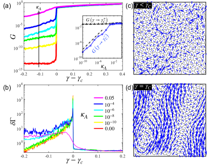
The nature of the strain-induced rigidity transition depends on the value of the area stiffness . This is evident in Fig.2(a), where we plot near the rigidity onset as a function of . At , the onset of rigidity is discontinuous. The jump discontinuity at remains finite well above and becomes vanishingly small and indistinguishable from a continuous increase in at . For the tissue is a marginally rigid solid[79, 80] with (Fig.2(a):inset). This is highlighted by the behavior of the fluctuations near the strain-driven rigidity transition, which are quantified with the non-affinity parameter [86, 87, 88]. Here is the displacement of cell after a strain step and is the affine deformation of the cell located at . As shown in Fig.2(b), at low area elasticity (), grows monotonically with strain and exhibits a sharp peak at , which coincides with the rigidity transition. At higher , there is no pronounced peak in , indicating a smooth cross-over from the marginal solid to a rigid solid, rather than a discontinuous transition.
Relating mechanical response to cell shape.
The strain stiffening behavior above can be understood in terms of shear-induced changes in the structural properties of the cellular network. Past work on vertex models has shown that the observed cell shape index, , is an important metric of the rheological state of the tissue[42, 51]. We have examined the evolution of this order parameter with applied shear. We note, however, that the applied strain does not uniquely define the state of the tissue due to plastic events and non-affine deformations. Instead we use the true strain [89] to quantify the degree of deformation of the tissue. is calculated from the instantaneous deformation tensor of the whole tissue and therefore captures the degree of cumulative strain deformation [90]. The motivation for introducing is similar to that behind the fabric tensor in granular materials[91] or the recoverable strain in rheology[92]. In Fig.3(a,b) we show the stress and the structural order parameter as functions of . It is evident from Fig.3(b) that under shear cell shapes in the fluid stay constant at the energetically preferred value until the fluid strain-stiffens, while in the solid always starts out at the universal value and grows quadratically with . This behavior is well described by
| (2) |
In the next section, we offer a theoretical derivation of this form. A similar functional dependence of the observed cell shape on the cell elongation induced by internally generated active stresses was reported in a recent study of the developing fruit fly[28].
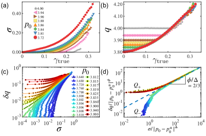
Eq.(2) suggests that the quantity can be used as a morphological order parameter, quantifying the deviation of the measured cell shape from the critical cell shape. Moreover, Figs.3(a,b) suggest that the three state variables () are not independent, and that any two are sufficient to describe the state of the tissue. Therefore, we eliminate and plot as a function (Fig.3(c)) for a large range of . This plot shows typical hallmarks of a critical point, with qualitatively different behavior above and below , suggesting a scaling ansatz
| (3) |
Here are the branches of the universal scaling function for and , respectively, with . This ansatz provides a nearly perfect collapse of the data (Fig.3(d)), with and . For the behavior is controlled by , with for , i.e., , implying . When , the scaling is controlled by . In the limit of (i.e., ), the inverse of tends to a constant, hence . For and , the two universal branches merge and .
A nonlinear constitutive equation for sheared tissue.
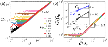
In tissues strained beyond both the stress (Fig.1a) and the shear modulus (Fig.2a) are nonlinear functions of the applied strain . To quantify the nonlinearity and extract a constitutive equation for the tissue, we use , instead of , as a state variable and plot as a function of in Fig.4a for various . At small , is independent of , corresponding to linear elasticity. At higher stress, the elastic response is nonlinear and , with . Using and eliminating , this yields a constitutive relation . The linear and nonlinear regimes are separated by a critical stress threshold . The linear-response modulus also shows power-law scaling in [74, 71]. This behavior can be summarized through a scaling ansatz to describe the behavior of in the vicinity of the critical point
| (4) |
Mean-field model of a sheared tissue.
To gain a theoretical understanding of the strain-driven rigidity and emergence of nonlinear elasticity, we examine a mean-field theory (MFT) formulation of the vertex model[93, 94, 66]. Neglecting cell-cell correlations, we consider the shear deformation of a single -sided polygonal cell. Under affine deformations, the vertex coordinates of a polygon transform according to , where is the deformation tensor given by . We neglect in Eq.(1) the contribution from cell area which is typically small compared to the perimeter term and examine area-preserving affine deformations with . For simple shear and , leaving only and as independent components of .
The perimeter of a deformed polygon can then be expressed in terms of the components of . For example the perimeter of a quadrilateral () is given by
| (5) |
Expressions for any deformed n-gon are given in the SI[90]. For any , the isoperimetric inequality defines the perimeters compatible with a fixed area as , where is the perimeter of a regular polygon with unit area (e.g., for ). The condition , with given by Eq. (5), then defines a manifold in the plane where there exist deformed polygons that statisfy the isoperimetric constraint (Fig.5(a)). The maximum value of along the isoperimetric contour defines the largest simple shear that a cell can sustain by changing its shape, while maintaining its area and perimeter constant. This value is and precisely corresponds to the location of the strain-driven rigidity in the simulations. The exponent is in excellent agreement with the scaling in the vicinity of , shown in Fig.1:inset.
The isoperimetric contours are centered at () and well approximated by an ellipse for small . We introduce polar coordinates with radius and polar angle : and and expand Eq.(5) to to give ( see SI[90])
| (6) |
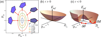
Using Eq.(6), we rewrite the vertex model energy (Eq.(1)) to obtain a Landau-type energy
| (7) |
where is the order parameter, are positive constants, and controls the distance to a continuous phase transition in . For , has a single minimum at (Fig.5b), corresponding to the rigid state. When , the minimum corresponds to the isoperimetrically degenerate liquid state. In the energy landscape these states are connected by a Goldstone mode (Fig.5c).
The MFT explains the origin of the nonlinear elasticity. For , has a single minimum at (corresponding to an undeformed solid state) and deformations away from it can be calculated using Eq.(7)
| (8) |
For small we recover linear elasticity with . At large the response is nonlinear, with . The cross-over stress between the two regimes can be calculated: . These predictions are in excellent agreement with simulations results.
We have used a vertex model to study the nonlinear response of a tissue to shear. Using simulations and MFT, we showed that a tissue that is liquid when unstrained stiffens upon shear. Liquid-solid transitions in VM of biological tissues are driven by geometric frustration and active mechanisms. Recent work by some of us [66] showed that geometric incompatibility controls the response to infinitesimal deformations, providing the underlying unifying mechanism for rigidity in a broad class of underconstrained systems. The present work additionally incorporates active processes that mediate plastic response. Plasticity dominates at higher strains and is likely to underlie the rheology of real tissue. Both works use a MFT to highlight the geometric origin of the degeneracy of the liquid ground state. The same MFT is extended here to investigate the response to deformations. While a Voronoi-based model is used, we have observed the same quantitative behavior using a vertex-based model and the results are independent of the model implementation.
Finally, it was shown in Ref. [66] that at the critical point the VM shares many of the properties of odd elasticity [95] - for instance, spontaneous shear upon uniaxial extension - although this behavior arises from geometry, not from an energy input at the microscale. Exploring the response to deformations other than simple shear and the possible connections with odd elasticity is an important direction for future work.
Acknowledgements.
The authors thank Mark Bowick, Michael Moshe and Arthur Hernandez for illuminating discussions. This work was supported in part by the Northeastern University TIER 1 Grant (J.H. and D.B.), NSF DMR-2046683 (J.H. and D.B.), DMR-2041459 (M.C.M.), PHY-1748958 (D.B. and M.C.M.), the Center for Theoretical Biological Physics NSF PHY-2019745 (J.H. and D.B.) and Mathworks Microgrants. We acknowledge the support of the Northeastern University Discovery Cluster. This project has received funding from the European Research Council (ERC) under the European Union’s Horizon 2020 research and innovation programme (grant agreement No. 885146); and the SOFI CDT, Durham University, EPSRC (EP/L015536/1).References
- Kollmannsberger and Fabry [2011] P. Kollmannsberger and B. Fabry, Linear and nonlinear rheology of living cells, Annual Review of Materials Research 41, 75 (2011).
- Guck et al. [2001] J. Guck, R. Ananthakrishnan, H. Mahmood, T. J. Moon, C. C. Cunningham, and J. Käs, The optical stretcher: A novel laser tool to micromanipulate cells, Biophysical Journal 81, 767 (2001).
- Haase and Pelling [2015] K. Haase and A. E. Pelling, Investigating cell mechanics with atomic force microscopy, Journal of The Royal Society Interface 12, 20140970 (2015), https://royalsocietypublishing.org/doi/pdf/10.1098/rsif.2014.0970 .
- Brill-Karniely [2020] Y. Brill-Karniely, Mechanical measurements of cells using afm: 3d or 2d physics?, Frontiers in Bioengineering and Biotechnology 8, 1265 (2020).
- Fujii et al. [2019] Y. Fujii, Y. Ochi, M. Tuchiya, M. Kajita, Y. Fujita, Y. Ishimoto, and T. Okajima, Spontaneous spatial correlation of elastic modulus in jammed epithelial monolayers observed by afm, Biophysical Journal 116, 1152 (2019).
- Serwane et al. [2017] F. Serwane, A. Mongera, P. Rowghanian, D. A. Kealhofer, A. A. Lucio, Z. M. Hockenbery, and O. Campas, In vivo quantification of spatially varying mechanical properties in developing tissues, Nature methods 14, 181 (2017).
- Hoffman et al. [2006] B. D. Hoffman, G. Massiera, K. M. Van Citters, and J. C. Crocker, The consensus mechanics of cultured mammalian cells, Proceedings of the National Academy of Sciences 103, 10259 (2006), https://www.pnas.org/content/103/27/10259.full.pdf .
- Fernández et al. [2006] P. Fernández, P. A. Pullarkat, and A. Ott, A master relation defines the nonlinear viscoelasticity of single fibroblasts, Biophysical journal 90, 3796 (2006).
- Schwarz and Soiné [2015] U. S. Schwarz and J. R. Soiné, Traction force microscopy on soft elastic substrates: A guide to recent computational advances, Biochimica et Biophysica Acta (BBA) - Molecular Cell Research 1853, 3095 (2015), mechanobiology.
- Tambe et al. [2013] D. T. Tambe, U. Croutelle, X. Trepat, C. Y. Park, J. H. Kim, E. Millet, J. P. Butler, and J. J. Fredberg, Monolayer stress microscopy: Limitations, artifacts, and accuracy of recovered intercellular stresses, PLOS ONE 8, e55172 (2013).
- Butler et al. [2002] J. P. Butler, I. M. Tolic-Nørrelykke, B. Fabry, and J. J. Fredberg, Traction fields, moments, and strain energy that cells exert on their surroundings, American Journal of Physiology-Cell Physiology 282, C595 (2002).
- Mertz et al. [2013] A. F. Mertz, Y. Che, S. Banerjee, J. M. Goldstein, K. A. Rosowski, S. F. Revilla, C. M. Niessen, M. C. Marchetti, E. R. Dufresne, and V. Horsley, Cadherin-based intercellular adhesions organize epithelial cell–matrix traction forces, Proceedings of the National Academy of Sciences 110, 842 (2013).
- Mertz et al. [2012] A. F. Mertz, S. Banerjee, Y. Che, G. K. German, Y. Xu, C. Hyland, M. C. Marchetti, V. Horsley, and E. R. Dufresne, Scaling of traction forces with the size of cohesive cell colonies, Phys. Rev. Lett. 108, 198101 (2012).
- Trepat et al. [2009] X. Trepat, M. R. Wasserman, T. E. Angelini, E. Millet, D. A. Weitz, J. P. Butler, and J. J. Fredberg, Physical forces during collective cell migration, Nature physics 5, 426 (2009).
- Kim et al. [2013] J. H. Kim, X. Serra-Picamal, D. T. Tambe, E. H. Zhou, C. Y. Park, M. Sadati, J.-A. Park, R. Krishnan, B. Gweon, E. Millet, et al., Propulsion and navigation within the advancing monolayer sheet, Nature materials 12, 856 (2013).
- Getsios et al. [2004] S. Getsios, A. C. Huen, and K. J. Green, Working out the strength and flexibility of desmosomes, Nature reviews Molecular cell biology 5, 271 (2004).
- Wang et al. [2009] N. Wang, J. D. Tytell, and D. E. Ingber, Mechanotransduction at a distance: mechanically coupling the extracellular matrix with the nucleus, Nature reviews Molecular cell biology 10, 75 (2009).
- Martino et al. [2018] F. Martino, A. R. Perestrelo, V. Vinarský, S. Pagliari, and G. Forte, Cellular mechanotransduction: From tension to function, Frontiers in Physiology 9, 824 (2018).
- Zhang and Labouesse [2012] H. Zhang and M. Labouesse, Signalling through mechanical inputs – a coordinated process, Journal of Cell Science 125, 3039 (2012).
- Das et al. [2015] T. Das, K. Safferling, S. Rausch, N. Grabe, H. Boehm, and J. P. Spatz, A molecular mechanotransduction pathway regulates collective migration of epithelial cells, Nature cell biology 17, 276 (2015).
- Hayes and Solon [2017] P. Hayes and J. Solon, Drosophila dorsal closure: An orchestra of forces to zip shut the embryo, Mechanisms of Development 144, 2 (2017), roles of physical forces in development.
- Machado et al. [2015] P. F. Machado, J. Duque, J. Étienne, A. Martinez-Arias, G. B. Blanchard, and N. Gorfinkiel, Emergent material properties of developing epithelial tissues, BMC Biology 13, 98 (2015).
- Andrew and Ewald [2010] D. J. Andrew and A. J. Ewald, Morphogenesis of epithelial tubes: Insights into tube formation, elongation, and elaboration, Developmental Biology 341, 34 (2010), special Section: Morphogenesis.
- Etournay et al. [2015] R. Etournay, M. Popović, M. Merkel, A. Nandi, C. Blasse, B. Aigouy, H. Brandl, G. Myers, G. Salbreux, F. Jülicher, and S. Eaton, Interplay of cell dynamics and epithelial tension during morphogenesis of the Drosophila pupal wing, eLife 4, e07090 (2015).
- Guirao et al. [2015] B. Guirao, S. U. Rigaud, F. Bosveld, A. Bailles, J. López-Gay, S. Ishihara, K. Sugimura, F. Graner, and Y. Bellaïche, Unified quantitative characterization of epithelial tissue development, eLife 4, e08519 (2015).
- Lecuit et al. [2011] T. Lecuit, P.-F. Lenne, and E. Munro, Force generation, transmission, and integration during cell and tissue morphogenesis, Annual Review of Cell and Developmental Biology 27, 157 (2011), pMID: 21740231, https://doi.org/10.1146/annurev-cellbio-100109-104027 .
- Tetley et al. [2019] R. J. Tetley, M. F. Staddon, D. Heller, A. Hoppe, S. Banerjee, and Y. Mao, Tissue fluidity promotes epithelial wound healing, Nature Physics 15, 1195 (2019).
- Wang et al. [2020] X. Wang, M. Merkel, L. B. Sutter, G. Erdemci-Tandogan, M. L. Manning, and K. E. Kasza, Anisotropy links cell shapes to tissue flow during convergent extension, Proceedings of the National Academy of Sciences 117, 13541 (2020).
- Fisher et al. [2001] A. B. Fisher, S. Chien, A. I. Barakat, and R. M. Nerem, Endothelial cellular response to altered shear stress, American Journal of Physiology-Lung Cellular and Molecular Physiology 281, L529 (2001).
- Trepat et al. [2007] X. Trepat, L. Deng, S. S. An, D. Navajas, D. J. Tschumperlin, W. T. Gerthoffer, J. P. Butler, and J. J. Fredberg, Universal physical responses to stretch in the living cell, Nature 447, 592 (2007).
- Comelles et al. [2021] J. Comelles, S. SS, L. Lu, E. Le Maout, S. Anvitha, G. Salbreux, F. Jülicher, M. M. Inamdar, and D. Riveline, Epithelial colonies in vitro elongate through collective effects, eLife 10, e57730 (2021).
- Tambe et al. [2011] D. T. Tambe, C. C. Hardin, T. E. Angelini, K. Rajendran, C. Y. Park, X. Serra-Picamal, E. H. Zhou, M. H. Zaman, J. P. Butler, D. A. Weitz, et al., Collective cell guidance by cooperative intercellular forces, Nature materials 10, 469 (2011).
- Vishwakarma et al. [2018] M. Vishwakarma, J. Di Russo, D. Probst, U. S. Schwarz, T. Das, and J. P. Spatz, Mechanical interactions among followers determine the emergence of leaders in migrating epithelial cell collectives, Nature communications 9, 3469 (2018).
- Butcher et al. [2009] D. T. Butcher, T. Alliston, and V. M. Weaver, A tense situation: forcing tumour progression, Nature Reviews Cancer 9, 108 EP (2009).
- Wirtz et al. [2011] D. Wirtz, K. Konstantopoulos, and P. C. Searson, The physics of cancer: the role of physical interactions and mechanical forces in metastasis, Nature Reviews Cancer 11, 512 (2011).
- Jain et al. [2014] R. K. Jain, J. D. Martin, and T. Stylianopoulos, The role of mechanical forces in tumor growth and therapy, Annual Review of Biomedical Engineering 16, 321 (2014), pMID: 25014786, https://doi.org/10.1146/annurev-bioeng-071813-105259 .
- Harris et al. [2012] A. R. Harris, L. Peter, J. Bellis, B. Baum, A. J. Kabla, and G. T. Charras, Characterizing the mechanics of cultured cell monolayers, Proceedings of the National Academy of Sciences 109, 16449 (2012), https://www.pnas.org/content/109/41/16449.full.pdf .
- Khalilgharibi et al. [2019] N. Khalilgharibi, J. Fouchard, N. Asadipour, R. Barrientos, M. Duda, A. Bonfanti, A. Yonis, A. Harris, P. Mosaffa, Y. Fujita, et al., Stress relaxation in epithelial monolayers is controlled by the actomyosin cortex, Nature physics 15, 839 (2019).
- Sadeghipour et al. [2018] E. Sadeghipour, M. A. Garcia, W. J. Nelson, and B. L. Pruitt, Shear-induced damped oscillations in an epithelium depend on actomyosin contraction and e-cadherin cell adhesion, eLife 7, e39640 (2018).
- Prakash et al. [2021] V. N. Prakash, M. S. Bull, and M. Prakash, Motility-induced fracture reveals a ductile-to-brittle crossover in a simple animal’s epithelia, Nature Physics 17, 504 (2021).
- Latorre et al. [2018] E. Latorre, S. Kale, L. Casares, M. Gómez-González, M. Uroz, L. Valon, R. V. Nair, E. Garreta, N. Montserrat, A. Del Campo, et al., Active superelasticity in three-dimensional epithelia of controlled shape, Nature 563, 203 (2018).
- Park et al. [2015] J.-A. Park, J. H. Kim, D. Bi, J. A. Mitchel, N. T. Qazvini, K. Tantisira, C. Y. Park, M. McGill, S.-H. Kim, B. Gweon, J. Notbohm, R. Steward, S. Burger, S. H. Randell, A. T. Kho, D. T. Tambe, C. Hardin, S. A. Shore, E. Israel, D. A. Weitz, D. J. Tschumperlin, E. P. Henske, S. T. Weiss, M. Lisa Manning, J. P. Butler, J. M. Drazen, and J. J. Fredberg, Unjamming and cell shape in the asthmatic airway epithelium, Nat Mater 14, 1040 (2015).
- Garcia et al. [2015] S. Garcia, E. Hannezo, J. Elgeti, J.-F. Joanny, P. Silberzan, and N. S. Gov, Physics of active jamming during collective cellular motion in a monolayer, Proceedings of the National Academy of Sciences 112, 15314 (2015).
- Oswald et al. [2017] L. Oswald, S. Grosser, D. M. Smith, and J. A. Käs, Jamming transitions in cancer, Journal of Physics D: Applied Physics 50, 483001 (2017).
- Paul et al. [2017] C. D. Paul, P. Mistriotis, and K. Konstantopoulos, Cancer cell motility: lessons from migration in confined spaces, Nature Reviews Cancer 17, 131 (2017).
- Malinverno et al. [2017] C. Malinverno, S. Corallino, F. Giavazzi, M. Bergert, Q. Li, M. Leoni, A. Disanza, E. Frittoli, A. Oldani, E. Martini, et al., Endocytic reawakening of motility in jammed epithelia, Nature materials 16, 587 (2017).
- Atia et al. [2018] L. Atia, D. Bi, Y. Sharma, J. A. Mitchel, B. Gweon, S. A. Koehler, S. J. DeCamp, B. Lan, J. H. Kim, R. Hirsch, et al., Geometric constraints during epithelial jamming, Nature physics 14, 613 (2018).
- Mongera et al. [2018] A. Mongera, P. Rowghanian, H. J. Gustafson, E. Shelton, D. A. Kealhofer, E. K. Carn, F. Serwane, A. A. Lucio, J. Giammona, and O. Campàs, A fluid-to-solid jamming transition underlies vertebrate body axis elongation, Nature 561, 401 (2018).
- Ilina et al. [2020] O. Ilina, P. G. Gritsenko, S. Syga, J. Lippoldt, C. A. M. La Porta, O. Chepizhko, S. Grosser, M. Vullings, G.-J. Bakker, J. Starruß, P. Bult, S. Zapperi, J. A. Käs, A. Deutsch, and P. Friedl, Cell–cell adhesion and 3d matrix confinement determine jamming transitions in breast cancer invasion, Nature Cell Biology 10.1038/s41556-020-0552-6 (2020).
- Huebner et al. [2020] R. J. Huebner, A. N. Malmi-Kakkada, S. Sarikaya, S. Weng, D. Thirumalai, and J. B. Wallingford, Cadherin clustering controls heterogeneous, asymmetric junction dynamics during vertebrate axis elongation, bioRxiv 10.1101/2020.02.11.944033 (2020).
- Mitchel et al. [2020] J. A. Mitchel, A. Das, M. J. O’Sullivan, I. T. Stancil, S. J. DeCamp, S. Koehler, O. H. Ocaña, J. P. Butler, J. J. Fredberg, M. A. Nieto, et al., In primary airway epithelial cells, the unjamming transition is distinct from the epithelial-to-mesenchymal transition, Nature communications 11, 1 (2020).
- Petridou et al. [2021] N. I. Petridou, B. Corominas-Murtra, C.-P. Heisenberg, and E. Hannezo, Rigidity percolation uncovers a structural basis for embryonic tissue phase transitions, Cell 184, 1914 (2021).
- Lin et al. [2021] S.-Z. Lin, W.-Y. Zhang, D. Bi, B. Li, and X.-Q. Feng, Energetics of mesoscale cell turbulence in two-dimensional monolayers, Communications Physics 4, 21 (2021).
- Yang et al. [2021] H. Yang, A. F. Pegoraro, Y. Han, W. Tang, R. Abeyaratne, D. Bi, and M. Guo, Configurational fingerprints of multicellular living systems, Proceedings of the National Academy of Sciences 118, 10.1073/pnas.2109168118 (2021).
- Marzio et al. [2021] M. D. Marzio, A. Kılıç, E. Maiorino, J. A. Mitchel, C. Mwase, M. J. O’Sullivan, M. McGill, R. Chase, J. J. Fredberg, J.-A. Park, K. Glass, and S. T. Weiss, Genomic signatures of the unjamming transition in compressed human bronchial epithelial cells, Science Advances 7, eabf1088 (2021).
- Nagai and Honda [2001] T. Nagai and H. Honda, A dynamic cell model for the formation of epithelial tissues, Philosophical Magazine B 81, 699 (2001).
- Farhadifar et al. [2007] R. Farhadifar, J.-C. Röper, B. Aigouy, S. Eaton, and F. Jülicher, The influence of cell mechanics, cell-cell interactions, and proliferation on epithelial packing, Current Biology 17, 2095 (2007).
- Tong et al. [2022] S. Tong, N. K. Singh, R. Sknepnek, and A. Košmrlj, Linear viscoelastic properties of the vertex model for epithelial tissues, PLOS Computational Biology 18, 1 (2022).
- Popović et al. [2021] M. Popović, V. Druelle, N. A. Dye, F. Jülicher, and M. Wyart, Inferring the flow properties of epithelial tissues from their geometry, New Journal of Physics 23, 033004 (2021).
- Duclut et al. [2021] C. Duclut, J. Paijmans, M. M. Inamdar, C. D. Modes, and F. Jülicher, Nonlinear rheology of cellular networks, Cells & Development , 203746 (2021).
- Pasupalak et al. [2021] A. Pasupalak, S. K. Samidurai, Y. Li, Y. Zheng, R. Ni, and M. P. Ciamarra, Unconventional rheological properties in systems of deformable particles, Soft Matter 17, 7708 (2021).
- Krajnc et al. [2021] M. Krajnc, T. Stern, and C. Zankoc, Active instability and nonlinear dynamics of cell-cell junctions, Phys. Rev. Lett. 127, 198103 (2021).
- Xu et al. [2015] G.-K. Xu, Y. Liu, and B. Li, How do changes at the cell level affect the mechanical properties of epithelial monolayers?, Soft Matter 11, 8782 (2015).
- Merzouki et al. [2016] A. Merzouki, O. Malaspinas, and B. Chopard, The mechanical properties of a cell-based numerical model of epithelium, Soft Matter 12, 4745 (2016).
- Bi et al. [2011] D. Bi, J. Zhang, B. Chakraborty, and R. P. Behringer, Jamming by shear, Nature 480, 355 (2011).
- Hernandez et al. [2022] A. Hernandez, M. F. Staddon, M. J. Bowick, M. C. Marchetti, and M. Moshe, Anomalous elasticity of a cellular tissue vertex model, Phys. Rev. E 105, 064611 (2022).
- Bi et al. [2016] D. Bi, X. Yang, M. C. Marchetti, and M. L. Manning, Motility-driven glass and jamming transitions in biological tissues, Phys. Rev. X 6, 021011 (2016).
- Li and Sun [2014] B. Li and S. X. Sun, Coherent motions in confluent cell monolayer sheets, Biophysical Journal 107, 1532 (2014).
- Li et al. [2019] X. Li, A. Das, and D. Bi, Mechanical heterogeneity in tissues promotes rigidity and controls cellular invasion, Phys. Rev. Lett. 123, 058101 (2019).
- Li et al. [2018] X. Li, A. Das, and D. Bi, Biological tissue-inspired tunable photonic fluid, Proceedings of the National Academy of Sciences 115, 6650 (2018), https://www.pnas.org/content/115/26/6650.full.pdf .
- Yan and Bi [2019] L. Yan and D. Bi, Multicellular rosettes drive fluid-solid transition in epithelial tissues, Phys. Rev. X 9, 011029 (2019).
- Das et al. [2021] A. Das, S. Sastry, and D. Bi, Controlled neighbor exchanges drive glassy behavior, intermittency, and cell streaming in epithelial tissues, Phys. Rev. X 11, 041037 (2021).
- Staple et al. [2010] D. B. Staple, R. Farhadifar, J. C. Röper, B. Aigouy, S. Eaton, and F. Jülicher, Mechanics and remodelling of cell packings in epithelia, The European Physical Journal E 33, 117 (2010).
- Bi et al. [2015] D. Bi, J. H. Lopez, J. M. Schwarz, and M. L. Manning, A density-independent rigidity transition in biological tissues, Nature Physics 11, 1074 (2015).
- Allen and Tildesley [1989] M. P. Allen and D. J. Tildesley, Computer Simulation of Liquids (Clarendon Press, New York, NY, USA, 1989).
- Bitzek et al. [2006] E. Bitzek, P. Koskinen, F. Gähler, M. Moseler, and P. Gumbsch, Structural relaxation made simple, Phys. Rev. Lett. 97, 170201 (2006).
- Sussman and Merkel [2018] D. M. Sussman and M. Merkel, No unjamming transition in a voronoi model of biological tissue, Soft Matter 14, 3397 (2018).
- Damavandi et al. [2022] O. K. Damavandi, V. F. Hagh, C. D. Santangelo, and M. L. Manning, Energetic rigidity. i. a unifying theory of mechanical stability, Phys. Rev. E 105, 025003 (2022).
- Moshe et al. [2018] M. Moshe, M. J. Bowick, and M. C. Marchetti, Geometric frustration and solid-solid transitions in model 2d tissue, Physical review letters 120, 268105 (2018).
- Merkel et al. [2019] M. Merkel, K. Baumgarten, B. P. Tighe, and M. L. Manning, A minimal-length approach unifies rigidity in underconstrained materials, Proceedings of the National Academy of Sciences 116, 6560 (2019), https://www.pnas.org/content/116/14/6560.full.pdf .
- Kupferman et al. [2020] R. Kupferman, B. Maman, and M. Moshe, Continuum mechanics of a cellular tissue model, Journal of the Mechanics and Physics of Solids 143, 104085 (2020).
- Ishihara and Sugimura [2012] S. Ishihara and K. Sugimura, Bayesian inference of force dynamics during morphogenesis, Journal of Theoretical Biology 313, 201 (2012).
- Chiou et al. [2012] K. K. Chiou, L. Hufnagel, and B. I. Shraiman, Mechanical stress inference for two dimensional cell arrays, PLOS Computational Biology 8, e1002512 (2012).
- Yang et al. [2017] X. Yang, D. Bi, M. Czajkowski, M. Merkel, M. L. Manning, and M. C. Marchetti, Correlating cell shape and cellular stress in motile confluent tissues, Proceedings of the National Academy of Sciences 114, 12663 (2017).
- VanderWerf et al. [2020] K. VanderWerf, A. Boromand, M. D. Shattuck, and C. S. O’Hern, Pressure dependent shear response of jammed packings of frictionless spherical particles, Phys. Rev. Lett. 124, 038004 (2020).
- Langer and Liu [1997] S. A. Langer and A. J. Liu, Effect of random packing on stress relaxation in foam, The Journal of Physical Chemistry B 101, 8667 (1997).
- DiDonna and Lubensky [2005] B. A. DiDonna and T. C. Lubensky, Nonaffine correlations in random elastic media, Phys. Rev. E 72, 066619 (2005).
- Broedersz and MacKintosh [2014] C. P. Broedersz and F. C. MacKintosh, Modeling semiflexible polymer networks, Rev. Mod. Phys. 86, 995 (2014).
- Gurtin et al. [2010] M. E. Gurtin, E. Fried, and L. Anand, The Mechanics and Thermodynamics of Continua (Cambridge University Press, 2010).
- [90] See Supplemental Material at XXXXXXXXX, which contains additional details on the model, additional results complementing those shown in the main text and includes Refs [93, 96, 97, 98, 99, 100, 101].
- Goddard [1998] J. D. Goddard, Continuum modeling of granular assemblies, in Physics of Dry Granular Media, edited by H. J. Herrmann, J.-P. Hovi, and S. Luding (Springer Netherlands, Dordrecht, 1998) pp. 1–24.
- Singh et al. [2021] P. K. Singh, J. C.-W. Lee, K. A. Patankar, and S. A. Rogers, Revisiting the basis of transient rheological material functions: Insights from recoverable strain measurements, Journal of Rheology 65, 129 (2021), https://doi.org/10.1122/8.0000154 .
- Czajkowski et al. [2018] M. Czajkowski, D. Bi, M. L. Manning, and M. C. Marchetti, Hydrodynamics of shape-driven rigidity transitions in motile tissues, Soft Matter 14, 5628 (2018).
- Hernandez and Marchetti [2021] A. Hernandez and M. C. Marchetti, Poisson-bracket formulation of the dynamics of fluids of deformable particles, Phys. Rev. E 103, 032612 (2021).
- Scheibner et al. [2020] C. Scheibner, A. Souslov, D. Banerjee, P. Surówka, W. T. Irvine, and V. Vitelli, Odd elasticity, Nature Physics 16, 475 (2020).
- Aubouy et al. [2003] M. Aubouy, Y. Jiang, J. A. Glazier, and F. Graner, A texture tensor to quantify deformations, Granular Matter 5, 67 (2003).
- Aigouy et al. [2010] B. Aigouy, R. Farhadifar, D. B. Staple, A. Sagner, J.-C. Röper, F. Jülicher, and S. Eaton, Cell Flow Reorients the Axis of Planar Polarity in the Wing Epithelium of Drosophila, Cell 142, 773 (2010).
- Graner et al. [2008] F. Graner, B. Dollet, C. Raufaste, and P. Marmottant, Discrete rearranging disordered patterns, part I: Robust statistical tools in two or three dimensions, The European Physical Journal E 25, 349 (2008).
- Rauzi et al. [2008] M. Rauzi, P. Verant, T. Lecuit, and P.-F. Lenne, Nature and anisotropy of cortical forces orienting drosophila tissue morphogenesis, Nature Cell Biology 10, 1401 EP (2008).
- Bosveld et al. [2012] F. Bosveld, I. Bonnet, B. Guirao, S. Tlili, Z. Wang, A. Petitalot, R. Marchand, P.-L. Bardet, P. Marcq, F. Graner, and Y. Bellaiche, Mechanical Control of Morphogenesis by Fat/Dachsous/Four-Jointed Planar Cell Polarity Pathway, Science 336, 724 (2012).
- Courtney [2005] T. H. Courtney, Mechanical behavior of materials (Waveland Press, Long Grove, Illinois, 2005).