Optothermal pulling, trapping, and assembly of colloids using nanowire plasmons
Abstract
Optical excitation of colloids can be harnessed to realize soft matter systems that are out of equilibrium. In this paper, we present our experimental studies on the dynamics of silica colloids in the vicinity of a silver nanowire propagating surface plasmon polaritons (SPPs). Due to the optothermal interaction, the colloids are directionally pulled towards the excitation point of the nanowire. Having reached this point, they are spatio-temporally trapped around the excitation location. By increasing the concentration of colloids in the system, we observe multi-particle assembly around the nanowire. This process is thermophoretically driven and assisted by SPPs. Furthermore, we find such an assembly to be sensitive to the excitation polarization at input of the nanowire. Numerically-simulated temperature distribution around an illuminated nanowire corroborates sensitivity to the excitation polarization. Our study will find relevance in exploration of SPPs-assisted optothermal pulling, trapping and assembly of colloids, and can serve as test-beds of plasmon-driven active matter.
1 Introduction
How do colloids1234behave in the vicinity of an optical and/or optothermal potential? How can colloidal dynamics and assembly be controlled by harnessing optical excitation? These are relevant questions in driving soft matter out of equilibrium, where the driving force is provided by an optical excitation. In this study, we present directional pulling of dielectric colloids in the vicinity of an optically-excited metal nanowire. This movement further leads to spatio-temporal trapping of the same colloids. Upon increasing the concentration of the colloids, a two dimensional assembly emerges. This emergent process is found to be sensitive to the excitation polarization of the light illuminating the nanowire.
In this context, understanding the dynamics of colloids under such optical excitation schemes is relevant.567 89Recently, colloidal manipulation using optical pulling forces has also gained significant interest.10111213 Typically, colloids undergo diffusive Brownian motion. 14 Brownian motion is relevant to the understanding of the dynamics of soft matter and biological systems 15 as well as fabricated structures designed to mimic naturally occurring systems.21617 The study of the physics of systems out of equilibrium 1819 is especially of pertinent interest as most of the living matter is far from equilibrium. In this context, colloids have been extensively studied to explore the dynamics of such systems.202122 23 The colloids have the advantage of easy manipulation under external fields. 2425 2627 282930Conventionally, colloids are trapped using a focused laser beam3132. Although powerful, the conventional optical schemes are constrained by the diffraction limit of light.33 As a complement to this, surface plasmon polaritons (SPPs) in metallic nanostructures can facilitate radiative and non-radiative pathways below the diffraction limit of light. 34353637 SPPs are surface electromagnetic waves at metal-dielectric interface.38 They decay into the dielectric medium through radiative and non-radiative channels. Radiative channels can be harnessed for optical trapping and assembly through SPPs momentum, and non-radiative channels of SPPs can be used for optothermal assembly via thermophoretic394041 interactions.
Of relevance to this study is SPPs propagation along the silver nanowire.42 43 When illuminated with a focused laser beam, these single crystalline, chemically-prepared nanowires can propagate SPPs. Given the geometry of the nanowire, quasi-one dimensional confinement of plasmons can lead to interesting optical and optothermal effects at sub-wavelength scales. Although SPPs in the silver nanowire have been studied extensively in the context of nanophotonics4445464748, their interface with soft-matter systems such as colloids is yet to be explored in detail. 49 50 SPPs have been utilized in the past to assemble particles on a large scale. But such studies have mainly relied on extended 2D plasmonic platforms such as metallic films.3537
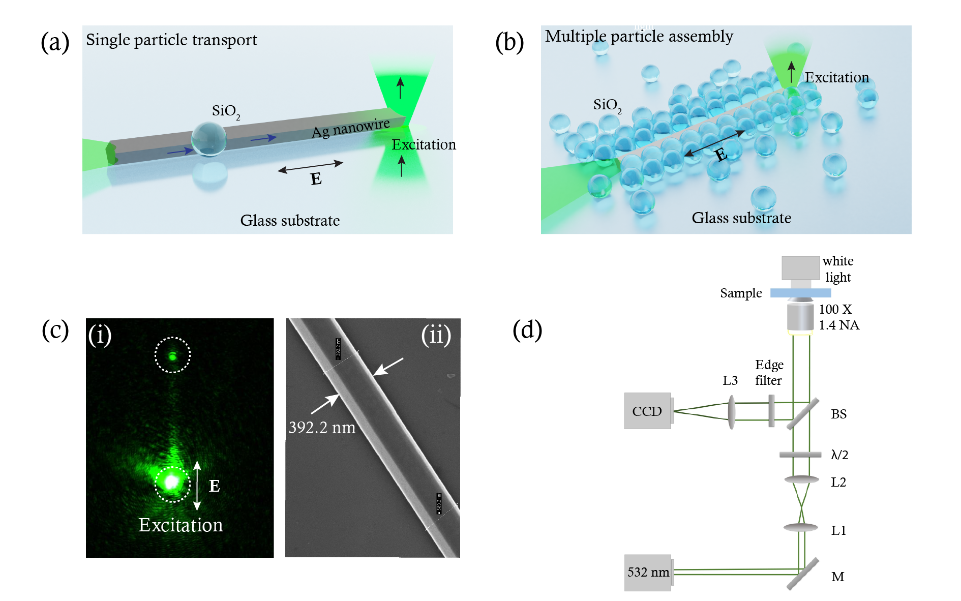
Motivated by this, herein we report the directed pulling and trapping of a single silica colloid as well as assembly formation of colloids by utilizing a single, silver nanowire to create a non-equilibrium environment in the system. A laser beam (532 nm) is focused at the end of a silver nanowire, which leads to the excitation of SPPs. In this study, we explore the transport and trapping of single colloids (figure 1a), and multiparticle assembly (figure 1b) due to the optical excitation of the plasmonic silver nanowire. A representative optical image of SPPs propagating in a silver nanowire at 532 nm is shown in figure 1c(i). The excitation polarization of the focused laser beam is aligned along the nanowire axis. This leads to SPPs propagation which out-couples at the distal end of the nanowire (dotted circle in figure 1c(i)). The SPPs supported by the nanowire generate heat51 5253which is subsequently released into the surrounding environment creating a temperature gradient in the system. The subsequent dynamics of the particles is studied by looking into the mean square displacement (MSD) of the particles. The MSD at time is defined as the ensemble average:
| (1) |
where, is the position of the particle at time t, is the time lag between two positions of the particle whose displacement is is the time-average over t and/or an ensemble-average over several trajectories.
2 Methods
2.1 Sample preparation and experimental design
Our experimental design constitutes a simple geometry wherein a silver nanowire is dropcasted on a glass substrate and left to dry. The silver nanowires are chemically synthesized using the polyol process. 54 The typical size of the silver nanowire used in the experiments is 300-400 nm in diameter and 12 m in length. The nanowire is characterized using scanning electron microscopy as shown in figure 1(c)(ii). The colloids consist of 2 m silica beads (purchased from Microspheres-Nanospheres) and 2.2 m polystyrene beads (purchased from micro particles Gmbh). The colloids dispersed in milli-Q water are dropcasted on the glass substrate. The whole system is enclosed in a 120 m spacer. Figure 1 (d) shows the schematic of the inverted experimental setup. A 532 nm laser is expanded using a combination of two lenses L1 and L2. A half wave plate is used to control the input polarization of the beam. One end of the silver nanowire is excited by tightly focusing the laser using a 100 X, 1.4 NA objective lens. The power at the sample plane is measured to be 5 mW. The polarization of the incoming laser is kept along the nanowire unless specified otherwise, for efficient excitation of the SPPs. The videos are recorded by the CCD at 20 fps after rejecting the elastically scattered light by a 532 nm edge filter.
2.2 Numerical simulations
The temperature distribution of the system is calculated using Finite element method based numerical simulations (COMSOL Multiphysics) by solving electromagnetic wave and heat transfer in solids and fluids modules in 3D. An 8 m long silver nanowire with a diameter of 350 nm is excited using focused 532 nm gaussian beam. The whole geometry is simulated using free tetrahedral user defined mesh.
3 Results and Discussion
3.1 Single particle transport
A single nanowire dropcasted on a glass substrate can act as a guiding medium for the directed motion of the colloid from its captured location to the excitation point. The nanowire initially traps the particle at one of the locations of the wire depending on the initial position of the particle with respect to the wire.
Here, we demonstrate the trapping of the silica colloid from the distal end of the nanowire. If the particle is closer to any other location of the wire when it is in its vicinity, it will get trapped there first, and then move towards the excitation point. One end of the silver nanowire is excited through a 100 X, 1.4 NA objective lens. The white dotted circle represents the excitation point. Figure 2(a) shows the time series images of a silica colloid transport along the nanowire. At t= 0 s , the colloid is freely diffusing in the water. As it comes closer to the wire, it gets trapped at the distal end at 13 s and starts to move along the nanowire. Within 65 s, the particle is trapped at the excitation point (See supplementary movie S1). To analyse the dynamics of the colloidal particle, it is essential to map out the trajectory and the associated MSD of the particle.
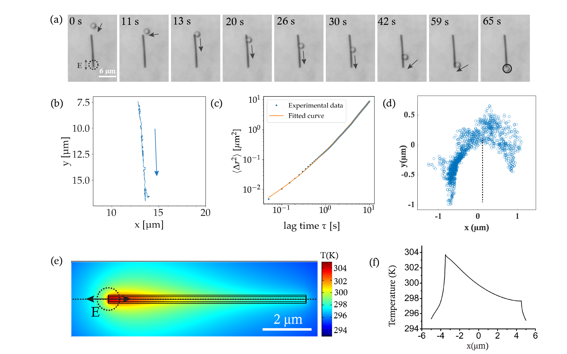
Since the particle continuously changes its micro-environment from diffusive to moving along the nanowire to permanent trapping at the excitation point, it is crucial to analyze the sections separately and not mix the resulting dynamics. Consequently,the particle is tracked from the moment it makes contact with the wire till it is transported to the other end before it gets trapped at the excitation location using Trackpy.55 The associated trajectory of the particle is plotted in figure 2(b). The trajectory plot shows that the motion of the particle is highly directional. The blue arrow indicates the direction of the movement of the particle. To further investigate the nature of the colloidal transport, we plot the MSD of the colloid on a log-log scale for the same time interval, as shown in figure 2(c). The experimentally obtained data is plotted in dotted blue. The motion of the colloid on the nanowire can be considered as a combination of 1D Brownian motion and ballistic motion. The MSD for such a system can be described as56:
| (2) |
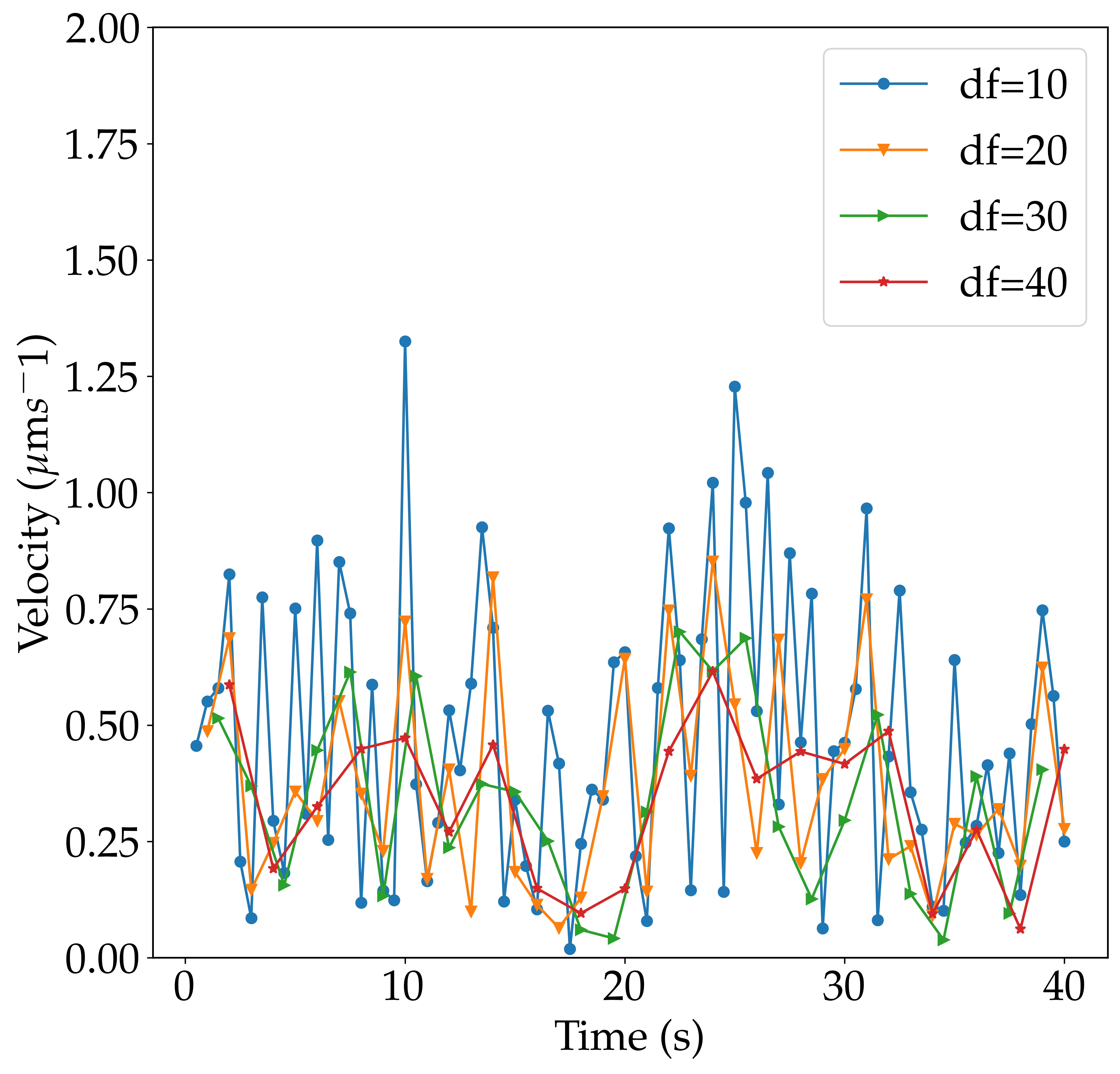
where, v is the terminal velocity of the colloid due to the external force which pulls it towards the nanowire and D is the diffusion coefficient. An analysis of the velocity of silica colloid while it is being transported on nanowire over time (SI figure 1) shows that its velocity fluctuates roughly around a constant value. By fitting equation 2 to the experimentally observed data in figure 2(c), we get v as 0.27ms-1 and a diffusion coefficient of 0.07 m2s-1. A comparative study for the Brownian motion of the ensemble of colloids which are under no laser excitation is given in SI figure 1(see supplementary movie S2). Other examples of particle-wire transport system are given in SI figure 3.
A previous study reported the transport of a much smaller TiO2 particle along the nanowire, but it is not permanently trapped and is pushed away from the excitation point into the solution. 49 In our case, once the silica colloid reaches the excitation point, it is permanently trapped there as long as the excitation laser is on. The trajectory of the trapped particle is shown in figure 2(d). By turning off the laser, the particle can again be released into the solution.
The efficient excitation of SPPs is crucial for the transport of the colloid. When a metallic structure is irradiated by an electromagnetic field, there is an associated heat generated in the system due to Joule heating.51 After plasmons are launched along the nanowire, heat is generated at the excitation point as well as along the length of the wire due to plasmon dissipation. Consequently, the temperature of the nanowire increases and the heat is released to the environment. The temperature distribution details were obtained through COMSOL. The numerically calculated temperature distribution in the xy plane at z=0 is shown in figure 2 (d). The system attains a total temperature increment of 10 K at the excitation point. A plot of the temperature distribution at the top of the wire at z=317 nm is plotted in figure 2 (e) which shows that the temperature is highest at the excitation point of the wire and gradually decreases along the length of the nanowire.
As reported previously34, silica colloids show thermophoretic migration from a lower temperature to a higher temperature. When the silver nanowire is excited, a temperature gradient is set up in the nanowire as well as its surroundings. Initially, the particle is moving freely in the water. When the colloid comes into the field of optothermal gradient of the system, it senses the temperature gradient and and is pulled towards the excitation point having the highest temperature.
To compare with a particle of different composition, and to emphasize the role of thermophoretic interactions, experiments were also performed at the same laser power with the polystyrene (PS) beads of size 2.2 m (See supplementary movie S3). PS beads have been previously reported to move from a higher temperature to a lower temperature.5758 In our experiments, the PS particles initially come closer to the nanowire but as they come too close, they are repelled from the excitation point. The selective trapping of the particles shows that thermophoresis has a contributing factor in trapping the particle.
3.2 Confined dynamics of multiple colloids
A silver nanowire can not only trap and transport a single colloid but can also be used to assemble multiple particles along its length. A concentrated solution of silica colloids in milli-Q water was prepared so that multiple particles can come into the field of optothermal potential simultaneously. Figure 3 (a) shows the time series images of the gradual assembly of the colloids along the length of the nanowire. At 0 s, two silica particles are trapped at the excitation point. Within 32 s, ten particles are aligned along the length of the nanowire.
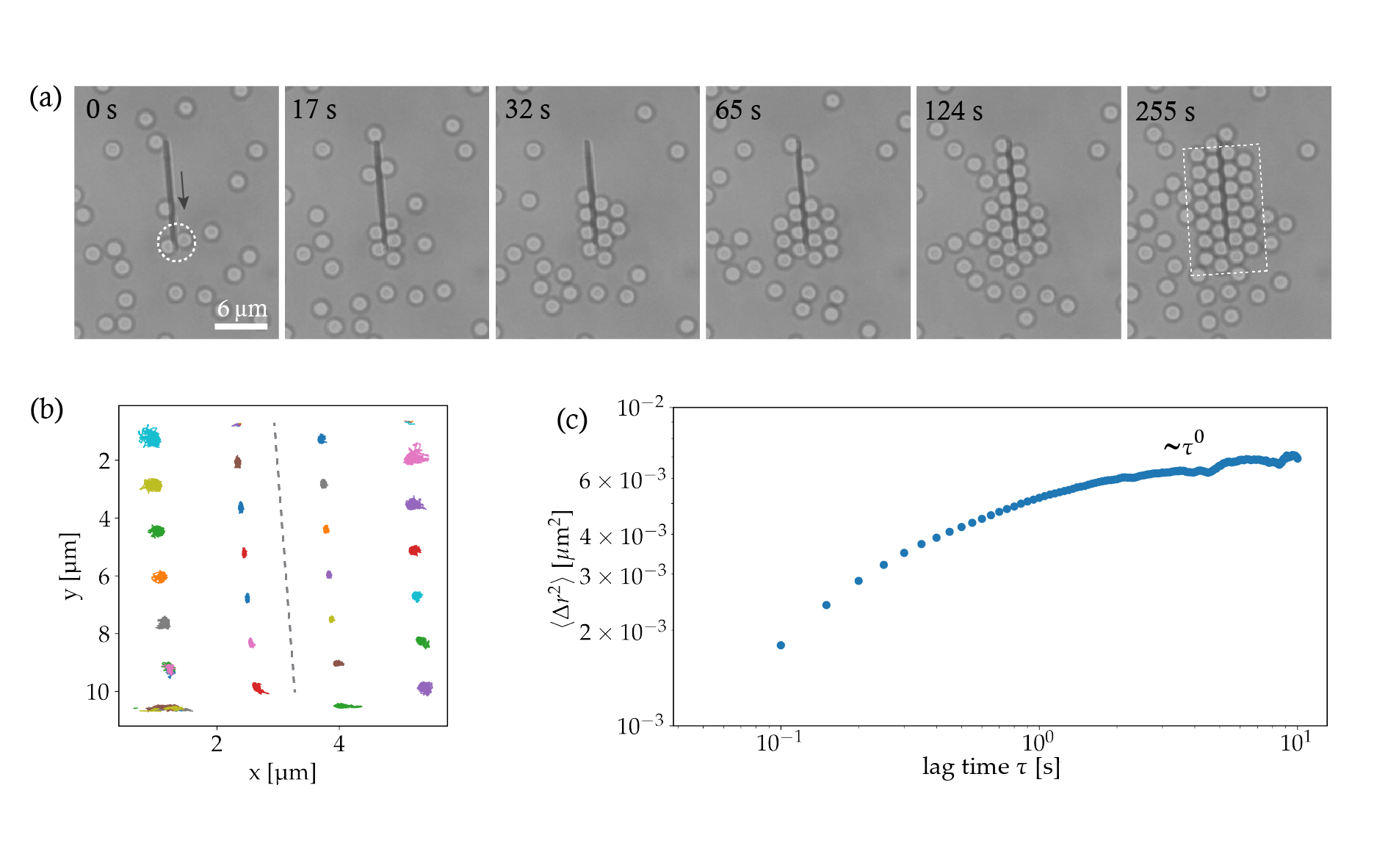
As shown in figure 2 (e), the temperature is higher towards the first half of the wire closer to the excitation point. Consequently, the particles first accumulate near the excitation end of the nanowire and start forming the second layer on both sides of the wire at 65 s. At 255 s, we observe a two layer formation of the colloids, which are stably trapped there as long as the laser excitation is on. The trajectories of the multiple particles shown in dotted white box in figure 3(a) over a period of 150 s are plotted in figure 3 (b). See supplementary movie S4 for the entire assembly process. The first layer closer to the wire exhibits stronger confinement to the nanowire as can be seen through the smaller spread of the trajectories of the individual colloids. Moreover, the trajectories are more confined perpendicular to the wire compared to along the wire. Since, the second layer of the colloids are not bounded by a third layer, they exhibit a larger spread in the trajectories perpendicular to the wire. The averaged MSD of all the particles is plotted in figure 3 (c). The curve almost falls flat and saturates at large lag times with the diffusion exponent of 0 indicating the confined motion of the trapped particles. The movement of the particles is impeded and they can not freely diffuse away from the trapped location.
3.3 Polarization dependence of the assembly
The heat generated by the silver nanowire can be contributed to two factors. One is absorption by the wire, and the other is due to the generation of SPPs59. As mentioned previously, the SPPs play a crucial role in the generation of heat and consequently trapping the colloidal particles. Keeping the polarization of the beam along the silver nanowire leads to efficient excitation of SPPs as can be seen in figure 4 (a)(ii) and consequently, should lead to faster assembly process. Figure 4(a) and 4(b) compares the extent of the assembly for the two cases, one when the polarization of the incoming beam is along the length of the nanowire and second, when it is perpendicular to it.
First the polarization is kept along the nanowire and the snapshot of the assembly is taken at 7 minutes as shown in figure 4(a)(i). Once the assembly reaches the saturation point, the laser is switched off and the colloids are free to redisperse into the solution. Now, the polarization of the laser is switched to perpendicular to the wire. In this case the efficiency of excitation of propagating plasmons is drastically reduced and no outcoupling of the light from the distal end is observed as can be seen in figure 4(b)(ii). A snapshot of the assembly after the same time of 7 minutes is shown in figure 4 (b)(i). A few particles near the excitation point and along the length of the wire can be seen but there is no significant assembly of the particles. The numerically calculated temperature distribution in the xy plane at z=0 is shown in figure 4(c). A comparative temperature distribution for the both the polarizations at the top of the wire is plotted in figure 4 (d). The assembly process for both the polarization can be found in supplementary movie S5 and movie S6.
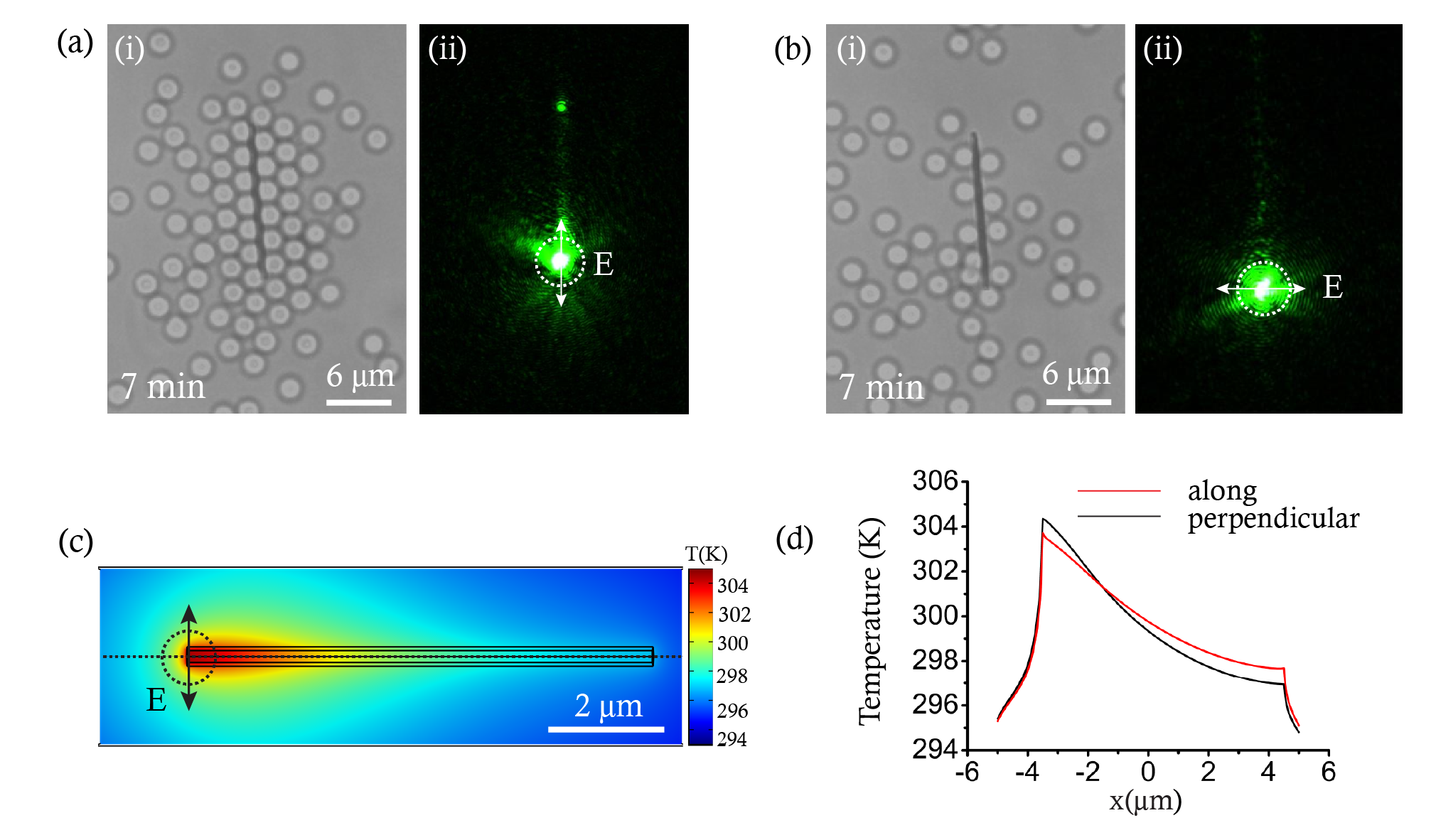
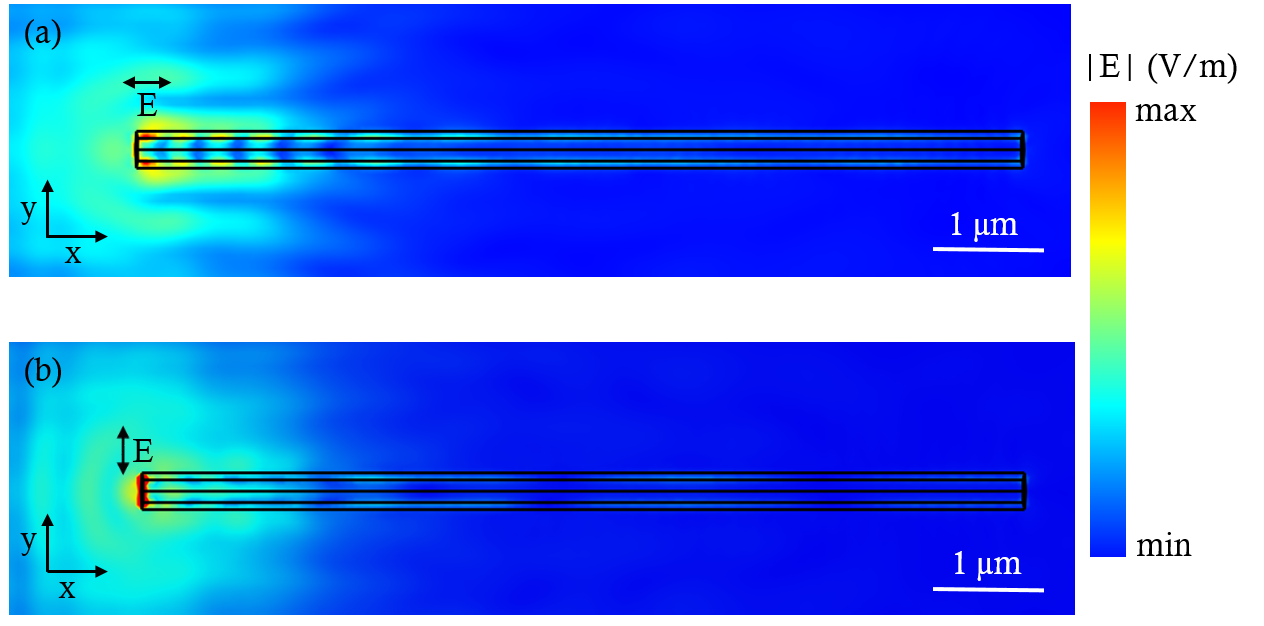
A higher temperature increase at the excitation point is found in the case of perpendicular polarization. It can be attributed to the fact that at the excitation point the electric field is confined to a smaller region along the diameter of the wire leading to more localized plasmon excitation. The near-field electric field distribution for both the cases is shown in supplementary figure S3. This enhances the temperature increment at this point, but the gradual fall in the temperature is more as there is less efficient excitation of plasmons along the nanowire and the resultant temperature increase due to plasmon dissipation is missing when there are no propagating plasmons.
4 Conclusion
We have experimentally studied the transport of the silica colloid by utilizing SPPs of a silver nanowire. Such a pulled colloid is spatio-temporally trapped at the nanowire excitation. Upon increasing the concentration of the colloids, we observed extended assemblies whose collective dynamics was studied and shown to be confined in nature. This assembly process is sensitive to the excitation polarization at nanowire input.This polarization-sensitivity was also corroborated in the simulated temperature distribution around the nanowire, thus indicating an intricate combination of optical and thermal effects facilitated by the nanowire SPPs. Given that the colloids are driven out of equilibrium by a quasi-one dimensional optothermal substrate, they may serve as interesting test-beds for active and driven matter at sub-micron scales. We envisage the utility of SPPs to create intriguing optothermal potentials to drive soft matter out of equilibrium, that can complement and improvise on conventional optical schemes.
Authors thank Pragya Kushwaha, Shailendra Kumar Chaubey and Chetna Taneja for fruitful discussions. This work was partially funded by Air Force Research Laboratory grant (FA2386-18-1-4118 R& D18IOA118) and Swarnajayanti fellowship grant (DST/SJF/PSA02/2017-18) to G V PK.
The videos can be found here
References
- Palacci et al. 2013 Palacci, J.; Sacanna, S.; Steinberg, A. P.; Pine, D. J.; Chaikin, P. M. Living crystals of light-activated colloidal surfers. Science 2013, 339, 936–940
- Lozano et al. 2016 Lozano, C.; Ten Hagen, B.; Löwen, H.; Bechinger, C. Phototaxis of synthetic microswimmers in optical landscapes. Nature communications 2016, 7, 1–10
- Buttinoni et al. 2012 Buttinoni, I.; Volpe, G.; Kümmel, F.; Volpe, G.; Bechinger, C. Active Brownian motion tunable by light. Journal of Physics: Condensed Matter 2012, 24, 284129
- Manoharan 2015 Manoharan, V. N. Colloidal matter: Packing, geometry, and entropy. Science 2015, 349, 1253751
- Zaidouny et al. 2013 Zaidouny, L.; Bohlein, T.; Roth, R.; Bechinger, C. Light-induced phase transitions of colloidal monolayers with crystalline order. Soft Matter 2013, 9, 9230–9236
- Wei et al. 1998 Wei, Q.-H.; Bechinger, C.; Rudhardt, D.; Leiderer, P. Experimental Study of Laser-Induced Melting in Two-Dimensional Colloids. Phys. Rev. Lett. 1998, 81, 2606–2609
- Schmidt et al. 2019 Schmidt, F.; Liebchen, B.; Löwen, H.; Volpe, G. Light-controlled assembly of active colloidal molecules. The Journal of chemical physics 2019, 150, 094905
- Jenkins and Egelhaaf 2008 Jenkins, M. C.; Egelhaaf, S. U. Colloidal suspensions in modulated light fields. Journal of Physics: Condensed Matter 2008, 20, 404220
- Dobnikar et al. 2013 Dobnikar, J.; Snezhko, A.; Yethiraj, A. Emergent colloidal dynamics in electromagnetic fields. Soft Matter 2013, 9, 3693–3704
- Chen et al. 2011 Chen, J.; Ng, J.; Lin, Z.; Chan, C. Optical pulling force. Nature photonics 2011, 5, 531–534
- Lu et al. 2017 Lu, J.; Yang, H.; Zhou, L.; Yang, Y.; Luo, S.; Li, Q.; Qiu, M. Light-induced pulling and pushing by the synergic effect of optical force and photophoretic force. Physical review letters 2017, 118, 043601
- Ali et al. 2020 Ali, R.; Pinheiro, F.; Dutra, R.; Neto, P. M. Tailoring optical pulling forces with composite microspheres. Physical Review A 2020, 102, 023514
- Li et al. 2020 Li, H.; Cao, Y.; Zhou, L.-M.; Xu, X.; Zhu, T.; Shi, Y.; Qiu, C.-W.; Ding, W. Optical pulling forces and their applications. Advances in Optics and Photonics 2020, 12, 288–366
- Bian et al. 2016 Bian, X.; Kim, C.; Karniadakis, G. E. 111 years of Brownian motion. Soft Matter 2016, 12, 6331–6346
- Frey and Kroy 2005 Frey, E.; Kroy, K. Brownian motion: a paradigm of soft matter and biological physics. Annalen der Physik 2005, 14, 20–50
- Lozano et al. 2018 Lozano, C.; Gomez-Solano, J. R.; Bechinger, C. Run-and-tumble-like motion of active colloids in viscoelastic media. New Journal of Physics 2018, 20, 015008
- Niese et al. 2020 Niese, L.; Wang, L.; Das, S.; Simmchen, J. Apparent phototaxis enabled by Brownian motion. Soft Matter 2020, 16, 10585–10590
- Blickle et al. 2007 Blickle, V.; Speck, T.; Lutz, C.; Seifert, U.; Bechinger, C. Einstein relation generalized to nonequilibrium. Physical review letters 2007, 98, 210601
- Ganguly and Chaudhuri 2013 Ganguly, C.; Chaudhuri, D. Stochastic thermodynamics of active Brownian particles. Physical Review E 2013, 88, 032102
- Bechinger et al. 2016 Bechinger, C.; Di Leonardo, R.; Löwen, H.; Reichhardt, C.; Volpe, G.; Volpe, G. Active particles in complex and crowded environments. Rev. Mod. Phys. 2016, 88, 045006
- Rings et al. 2010 Rings, D.; Schachoff, R.; Selmke, M.; Cichos, F.; Kroy, K. Hot brownian motion. Physical review letters 2010, 105, 090604
- Su et al. 2021 Su, X.; Fischer, A.; Cichos, F. Towards Measuring the Maxwell–Boltzmann Distribution of a Single Heated Particle. Frontiers in Physics 2021, 9, 342
- Fernandez-Rodriguez et al. 2020 Fernandez-Rodriguez, M. A.; Grillo, F.; Alvarez, L.; Rathlef, M.; Buttinoni, I.; Volpe, G.; Isa, L. Feedback-controlled active brownian colloids with space-dependent rotational dynamics. Nature communications 2020, 11, 1–10
- Caciagli et al. 2020 Caciagli, A.; Singh, R.; Joshi, D.; Adhikari, R.; Eiser, E. Controlled optofluidic crystallization of colloids tethered at interfaces. Physical Review Letters 2020, 125, 068001
- Vialetto et al. 2021 Vialetto, J.; Rudiuk, S.; Morel, M.; Baigl, D. Photothermally Reconfigurable Colloidal Crystals at a Fluid Interface, a Generic Approach for Optically Tunable Lattice Properties. Journal of the American Chemical Society 2021, 143, 11535–11543
- Arya et al. 2021 Arya, P.; Umlandt, M.; Jelken, J.; Feldmann, D.; Lomadze, N.; Asmolov, E. S.; Vinogradova, O. I.; Santer, S. Light-induced manipulation of passive and active microparticles. The European Physical Journal E 2021, 44, 1–10
- Ghosh and Ghosh 2019 Ghosh, S.; Ghosh, A. All optical dynamic nanomanipulation with active colloidal tweezers. Nature communications 2019, 10, 1–8
- Tkachenko et al. 2020 Tkachenko, G.; Toftul, I.; Esporlas, C.; Maimaiti, A.; Le Kien, F.; Truong, V. G.; Chormaic, S. N. Light-induced rotation of dielectric microparticles around an optical nanofiber. Optica 2020, 7, 59–62
- Ghosh et al. 2019 Ghosh, S.; Biswas, A.; Roy, B.; Banerjee, A. Self-assembly and complex manipulation of colloidal mesoscopic particles by active thermocapillary stress. Soft matter 2019, 15, 4703–4713
- Ghosh et al. 2020 Ghosh, S.; Ranjan, A. D.; Das, S.; Sen, R.; Roy, B.; Roy, S.; Banerjee, A. Directed self-assembly driven mesoscale lithography using laser-induced and manipulated microbubbles: Complex architectures and diverse applications. Nano letters 2020, 21, 10–25
- Ashkin et al. 1986 Ashkin, A.; Dziedzic, J. M.; Bjorkholm, J. E.; Chu, S. Observation of a single-beam gradient force optical trap for dielectric particles. Optics letters 1986, 11, 288–290
- Jones et al. 2015 Jones, P. H.; Maragò, O. M.; Volpe, G. Optical tweezers: Principles and applications; Cambridge University Press, 2015
- Melzer and McLeod 2018 Melzer, J. E.; McLeod, E. Fundamental limits of optical tweezer nanoparticle manipulation speeds. ACS nano 2018, 12, 2440–2447
- Sharma et al. 2020 Sharma, V.; Paul, D.; Chaubey, S. K.; Tiwari, S.; Kumar, G. P. Large-scale optothermal assembly of colloids mediated by a gold microplate. Journal of Physics: Condensed Matter 2020, 32, 324002
- Patra et al. 2014 Patra, P. P.; Chikkaraddy, R.; Tripathi, R. P.; Dasgupta, A.; Kumar, G. V. P. Plasmofluidic single-molecule surface-enhanced Raman scattering from dynamic assembly of plasmonic nanoparticles. Nature communications 2014, 5, 1–8
- Patra et al. 2016 Patra, P. P.; Chikkaraddy, R.; Thampi, S.; Tripathi, R. P.; Kumar, G. V. P. Large-scale dynamic assembly of metal nanostructures in plasmofluidic field. Faraday discussions 2016, 186, 95–106
- Garcés-Chávez et al. 2006 Garcés-Chávez, V.; Quidant, R.; Reece, P.; Badenes, G.; Torner, L.; Dholakia, K. Extended organization of colloidal microparticles by surface plasmon polariton excitation. Physical Review B 2006, 73, 085417
- Barnes et al. 2003 Barnes, W. L.; Dereux, A.; Ebbesen, T. W. Surface plasmon subwavelength optics. nature 2003, 424, 824–830
- Piazza and Parola 2008 Piazza, R.; Parola, A. Thermophoresis in colloidal suspensions. Journal of Physics: Condensed Matter 2008, 20, 153102
- Piazza 2008 Piazza, R. Thermophoresis: moving particles with thermal gradients. Soft Matter 2008, 4, 1740–1744
- Reichl et al. 2014 Reichl, M.; Herzog, M.; Götz, A.; Braun, D. Why charged molecules move across a temperature gradient: the role of electric fields. Physical review letters 2014, 112, 198101
- Shegai et al. 2011 Shegai, T.; Miljkovic, V. D.; Bao, K.; Xu, H.; Nordlander, P.; Johansson, P.; Kall, M. Unidirectional broadband light emission from supported plasmonic nanowires. Nano letters 2011, 11, 706–711
- Yang et al. 2016 Yang, H.; Qiu, M.; Li, Q. Identification and control of multiple leaky plasmon modes in silver nanowires. Laser & Photonics Reviews 2016, 10, 278–286
- Guo et al. 2019 Guo, Q.; Fu, T.; Tang, J.; Pan, D.; Zhang, S.; Xu, H. Routing a chiral Raman signal based on spin-orbit interaction of light. Physical review letters 2019, 123, 183903
- Johns et al. 2017 Johns, P.; Beane, G.; Yu, K.; Hartland, G. V. Dynamics of surface plasmon polaritons in metal nanowires. The Journal of Physical Chemistry C 2017, 121, 5445–5459
- Vasista et al. 2018 Vasista, A. B.; Jog, H.; Heilpern, T.; Sykes, M. E.; Tiwari, S.; Sharma, D. K.; Chaubey, S. K.; Wiederrecht, G. P.; Gray, S. K.; Kumar, G. V. P. Differential wavevector distribution of surface-enhanced Raman scattering and fluorescence in a film-coupled plasmonic nanowire cavity. Nano letters 2018, 18, 650–655
- Tiwari et al. 2020 Tiwari, S.; Taneja, C.; Sharma, V.; Vasista, A. B.; Paul, D.; Kumar, G. V. P. Dielectric microsphere coupled to a plasmonic nanowire: a self-assembled hybrid optical antenna. Advanced Optical Materials 2020, 8, 1901672
- Tiwari et al. 2021 Tiwari, S.; Vasista, A. B.; Paul, D.; Chaubey, S. K.; Kumar, G. V. P. Beaming Elastic and SERS Emission from Bent-Plasmonic Nanowire on a Mirror Cavity. The Journal of Physical Chemistry Letters 2021, 12, 6589–6595, PMID: 34242502
- Yang et al. 2016 Yang, C.; Pan, D.; Tong, L.; Xu, H. Guided transport of nanoparticles by plasmonic nanowires. Nanoscale 2016, 8, 19195–19199
- Nan and Yan 2019 Nan, F.; Yan, Z. Silver-nanowire-based interferometric optical tweezers for enhanced optical trapping and binding of nanoparticles. Advanced Functional Materials 2019, 29, 1808258
- Baffou 2017 Baffou, G. Thermoplasmonics: Heating Metal Nanoparticles Using Light; Cambridge University Press, 2017
- Baffou and Quidant 2013 Baffou, G.; Quidant, R. Thermo-plasmonics: using metallic nanostructures as nano-sources of heat. Laser & Photonics Reviews 2013, 7, 171–187
- Möller et al. 2018 Möller, T. B.; Ganser, A.; Kratt, M.; Dickreuter, S.; Waitz, R.; Scheer, E.; Boneberg, J.; Leiderer, P. Fast quantitative optical detection of heat dissipation by surface plasmon polaritons. Nanoscale 2018, 10, 11894–11900
- Sun et al. 2003 Sun, Y.; Mayers, B.; Herricks, T.; Xia, Y. Polyol synthesis of uniform silver nanowires: a plausible growth mechanism and the supporting evidence. Nano letters 2003, 3, 955–960
- Allan et al. 2021 Allan, D. B.; Caswell, T.; Keim, N. C.; van der Wel, C. M.; Verweij, R. W. soft-matter/trackpy: Trackpy v0.5.0. 2021; https://doi.org/10.5281/zenodo.4682814
- Howse et al. 2007 Howse, J. R.; Jones, R. A.; Ryan, A. J.; Gough, T.; Vafabakhsh, R.; Golestanian, R. Self-motile colloidal particles: from directed propulsion to random walk. Physical review letters 2007, 99, 048102
- Duhr and Braun 2005 Duhr, S.; Braun, D. Two-dimensional colloidal crystals formed by thermophoresis and convection. Applied physics letters 2005, 86, 131921
- Braun et al. 2014 Braun, M.; Würger, A.; Cichos, F. Trapping of single nano-objects in dynamic temperature fields. Physical Chemistry Chemical Physics 2014, 16, 15207–15213
- Li et al. 2019 Li, Q.; Chen, L.; Xu, H.; Liu, Z.; Wei, H. Photothermal modulation of propagating surface plasmons on silver nanowires. ACS Photonics 2019, 6, 2133–2140
Supplementary Material
Optothermal pulling, trapping, and assembly of colloids using nanowire plasmons
Vandana Sharma,∗ Sunny Tiwari, Diptabrata Paul, Ratimanasee Sahu, Vijayakumar Chikkadi and G. V. Pavan Kumar∗
Department of Physics, Indian Institute of Science Education and Research, Pune-411008, India
∗Email: vandana.sharma@students.iiserpune.ac.in, pavan@iiserpune.ac.in
Appendix S1
S1.1 S1: Velocity of the colloid at different frame differences (df)
S1.2 S2: Brownian motion of particles
S1.3 S3: Single particle transport along the nanowire
S1.4 S4: Near-field electric field distribution
S1.5 S5: Details of supplementary movies
Appendix S2 S1: Velocity of the colloid at different frame differences (df)
Appendix S3 S2: Brownian motion of particles
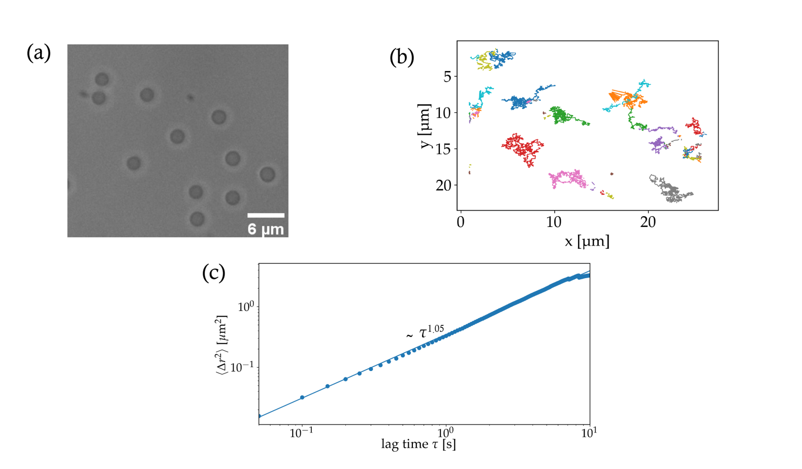
2 m Silica colloids in milli-Q water are dropcast on a glass substrate and sealed in a 120 m spacer. The Brownian motion of the particles is recorded over a period of 50 s in the absence of the nanowire and laser excitation.
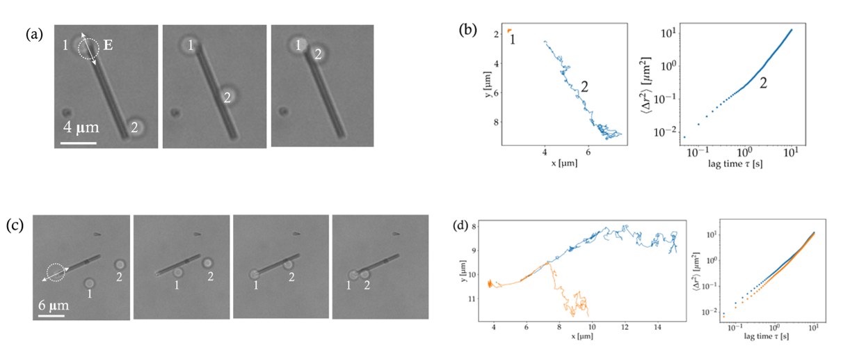
Appendix S4 S3: Single particle transport along the nanowire
Appendix S5 S4: Near-field electric field distribution
Appendix S6 S4: Details on Supporting Movies
-
1.
Supporting Movie 1: Transport of a single 2 m silica colloid from the distal end of the nanowire to the excitation point
-
2.
Supporting Movie 2: 2 m silica colloids undergoing Brownian motion.
-
3.
Supporting Movie 3: No trapping of 2.2 m polystyrene beads is observed.
-
4.
Supporting Movie 4: Assembly process of 2 m silica colloids.
-
5.
Supporting Movie 5: Assembly of silica beads when the polarization is along the nanowire.
-
6.
Supporting Movie 6: Assembly of silica beads when the polarization is perpendicular to the nanowire.