Enhanced Control of Quantum Dot Photoluminescence in Hybrid Assemblies
Abstract
The distance-dependent interaction of an emitter with a plasmonic nanoparticle or surface forms the basis of the field of plexitonics. Semiconductor quantum dots (QDs) are robust emitters due to their photostability, and, as such, offer the possibility of understanding the fundamental photophysics between one emitter and one metal nanoparticle. Hence, a key enabling challenge is the formation of such structures (i.e. systems containing both QDs and plasmonic nanoparticles) in high purity. Here, we present the translation of DNA-based self-assembly techniques to assemble metal and semiconductor nanocrystals into discrete hybrid structures, including dimers, of high purity. This method gives control over the interparticle separation, geometry, and ratio of QD:metal nanoparticle, as well as the spectral properties of the metal/QD components in the assembly to allow detailed investigation of plasmon-exciton interaction. The hybrid assemblies show the expected enhancement in steady-state photoluminescence accompanied by an increase in the QD emission rate for assemblies with a strong overlap between the QD emission and localised surface plasmon resonance. In contrast, lengthening of the QD emission lifetime (a reduction of the emission rate) of up to 1.7-fold, along with an enhancement in steady-state PL of 15 - 75 % is observed upon detuning of the QD and metal nanoparticle spectral properties. These results are understood in terms of the Purcell effect, where the gold nanoparticle acts as a damped, nanoscale cavity. Considering the metal nanoparticle using generalised nonlocal optical response theory (GNOR) and the QD as an open quantum system, we show that the response is driven by the interference experienced by the emitter for parallel and perpendicular field orientations. This understanding provides a mechanism for control of the emission rate of a QD by a metal nanoparticle across a much wider range of lifetimes than previously understood.
keywords:
Add keywords hereMonash University] ARC Centre of Excellence in Exciton Science and School of Chemistry, Monash University, Clayton, Victoria, 3800, Australia RMIT] ARC Centre of Excellence in Exciton Science and Chemical and Quantum Physics, School of Science, RMIT University, Melbourne, 3001, Australia Monash University] ARC Centre of Excellence in Exciton Science and School of Chemistry, Monash University, Clayton, Victoria, 3800, Australia \altaffiliationCurrent address: Université de Lyon, CNRS, École Normale Supérieure de Lyon, Laboratoire de Chimie UMR 5182, 46 allée d’Italie, F-69007 Lyon, France. RMIT] ARC Centre of Excellence in Exciton Science and Chemical and Quantum Physics, School of Science, RMIT University, Melbourne, 3001, Australia Monash University] ARC Centre of Excellence in Exciton Science and School of Chemistry, Monash University, Clayton, Victoria, 3800, Australia
Some journals require a graphical entry for the Table of Contents. This should be laid out “print ready” so that the sizing of the text is correct.
Inside the tocentry environment, the font used is Helvetica 8 pt, as required by Journal of the American Chemical Society.
The surrounding frame is 9 cm by 3.5 cm, which is the maximum permitted for Journal of the American Chemical Society graphical table of content entries. The box will not resize if the content is too big: instead it will overflow the edge of the box.
This box and the associated title will always be printed on a separate page at the end of the document.
1 Introduction
The excitonic states and thus the optical properties of light emitters can be controlled by judicious engineering of their local environment.1 One method to modify the electromagnetic properties of emitters is via the Purcell effect, whereby the photonic environment (i.e.the dielectric environment) of the emitter is controlled or changed. In this vein, optical microcavities, photonic crystals, interfaces, and gratings have all been used to control the emission rate of the fluorophores.2, 3 However, these approaches require precise control of the position and orientation of optically active materials at the nanoscale.
Metal nanoparticles that are much smaller than the wavelength of incident light () exhibit strong resonant excitations known as localized surface plasmon resonances (LSPRs) 4, 5. LSPRs are non-propagating modes of excitation of the conduction band electrons arising naturally from the scattering problem of a subwavelength metal nanoparticle in an oscillating electromagnetic field 6. These excitations enable metal nanoparticles to act as nanoscale optical cavities, able to focus electromagnetic energy to spots much smaller than overcoming the half-wavelength size limitation of conventional optical cavities 7, 8, 9, 10. Thus, metal nanoparticles can significantly influence the optical properties of quantum emitters, such as nanocrystal quantum dots, placed in nanoscale proximity 11.
Interaction between an emitter and a plasmonic metal nanoparticle gives rise to phenomena including plasmon-enhanced fluorescence,12, 13 surface-enhanced Raman scattering,14 energy transfer,15, 16 and coupling between plasmon and molecular resonances.17 The range of potential applications within which the phenomenon of plasmon-exciton resonance coupling may be exploited is vast and diverse, encompassing plasmon-enhanced catalytic reactions,18, 19 plasmon-enhanced optical signals,20, 21 solar cells,22, 23 and biotechnological applications.24 Various factors affect the plasmon-exciton coupling. Metal nanoparticles modify the electric field at their surface, thereby influencing the dielectric environment of nearby emitters and consequently their emission (intensity and emission rate). Another mode of interaction is damping which occurs due to energy transfer from the emitter to the metal particle acceptor. The competition between field enhancement and nonradiative damping due to energy transfer to the LSPR controls the optical response.25 Effective plasmon-fluorophore coupling requires the close proximity of the plasmon and fluorophore and significant overlap of the emission spectrum of the fluorophore with the LSPR of the metal nanocrystals.25 Dynamic control of this exciton-plasmon system opens up new avenues for novel and efficient exciton-based optoelectronic applications.
In these coupled exciton-plasmon systems, semiconductor quantum dots (QDs) offer significant advantages compared to organic fluorophores, such as photostability, high quantum yield, high absorption cross-section, and narrow, tunable emission.26, 27 Consequently, hybrid structures incorporating quantum emitters and metal nanoparticles are promising candidates for use across the range of potential applications exploiting their interactions.
Hybrid plasmon-exciton structures have been realised by the fabrication of QD-Au films, consisting of a superposition of Au or QD thin films separated by spacer layers. Within these, both quenching and enhancement of the PL intensity were achieved upon tuning the spectral overlap and variation of the polymer/polyelectrolyte layers between the films.28, 29, 30. A major concern for NP-based films is the aggregation of NPs upon drying. This causes changes in the LSPR of the metal NP and the emission of the fluorophore, thereby modifying the overall optical response of the hybrid films compared to their counterparts in the solution.31 Electron beam lithography approaches have also been widely employed to prepare two-dimensional hybrid structures, although gap variation and surface roughness lead to variations in the optical response of the coupled nanoparticles.32, 33, 34
The strong exciton-plasmon coupling has been observed in controlled, discrete systems with a one-to-one stoichiometry for the QD and metal nanoparticle. Precise profiling of distance-dependent fluorescence quenching and enhancement between a single gold nanoparticle and a quantum dot has been carried out via AFM-based micromanipulation35, 36 and by the incorporation of a single QD in a plasmonic cavity37, 38 in proof-of-principle experiments. In contrast, colloidal methods have the potential for the development of three-dimensional structures in scalable quantities. Energy transfer in electrostatically assembled clusters containing multiple dyes or quantum dots around metal nanoparticles showed a 2 - 3-fold reduction in the lifetime of the fluorophore, whilst the transfer efficiency was able to be modulated from 30% to 55% via a change in the concentration of either gold nanoparticles or the fluorophore39, 40 However, such approaches do not provide control over the number of emitters or interparticle separation. To overcome this, chemical coupling of the metal and fluorophore with short biological or chemical linkers, or silica encapsulation have been employed to develop hybrid structures, 41, 42, 43, 44 although generally, these methods provide only limited control over metal-fluorophore distances and stoichiometry, limiting their use. Consequently, a key requirement in this field is the development of assembly methods with the ability to fully control the relative location, orientation, and interparticle separation in a coupled plasmon-exciton systems.
DNA has been exploited to achieve self-assembly of gold nanoparticles by hybridisation of single-stranded DNA attached to metal nanoparticles as well as sophisticated DNA origami scaffolds.45, 46, 47, 48 The variable length of the DNA strands offers flexibility in the interparticle separation, and purification techniques enable control over the number of particles in the assembly.49, 50 Alivisatos pioneered the DNA-based self-assembly of gold nanoparticles into discrete structures.51 Subsequently, improved protocols for DNA-based gold nanoparticle assembly have been developed.52 In contrast, DNA-based QD assembly protocols are relatively underdeveloped,53, 54 with a key enabling capability, the purification of particles with one (or a given number) of attached DNA strands per particle lacking. Assembly of DNA functionalized CdTe QDs into nanoassemblies has been shown, although the purification technique (size-selective filtration) does not allow purification of QDs with only one or two DNA strands per particle.53 Gel electrophoresis approaches have also been used to purify DNA based hybrid Au/QD assemblies52, 54, 55. To date, visualisation of QDs in DNA-based QD assemblies (lacking defined numbers of DNA strands/particle) has been achieved using fluorescent straining which interferes with the optical signal of the QDs. The formation of nanostructures that show strong exciton-plasmon coupling requires two different materials, with different surface chemistries and sizes, to be assembled together in high yield, with control over geometry. The additional challenges associated with the incorporation of different materials in the assembly are significant.
The optical properties of metal-fluorophore systems are somewhat incongruous and vary between quenching56, 57 and enhancement58, 59 of the photoluminescence intensity of the emitter. Within discrete hybrid assemblies with a good overlap of the metal LSPR and fluorophore emission, strong quenching of the QD PL intensity is observed,57, 60 but at a given separation distance and with a decrease in the spectral overlap, a cross-over from quenching to enhancement in the PL intensity occurs.61, 62, 63, 64 Photoluminescence enhancement has, however, also been observed in highly resonant Au-QD systems. For example, 2.5 to 8-fold photoluminescence enhancements, with little to no change in photoluminescence lifetimes, for on-resonant Au-QD assemblies with interparticle separations in the range of have been reported.65, 66, 67 Higher degrees of steady-state photoluminescent enhancement have been observed for QD-Au hybrid films where increases of up to 50-fold have been demonstrated.68, 69 In contrast to the ambiguity in the PL intensity of hybrid systems, PL decay measurements show a reduction in the lifetime of the emitter close to the metal for all the reported hybrid systems, irrespective of quenching and enhancement in the PL intensity 70, 33. This will be further discussed in our theoretical insights section.
Here, we report an approach that overcomes many synthetic challenges to allow the controlled organization of quantum emitters near gold nanoparticles with control over emitter wavelength, metal LSPR, interparticle spacing ( and ), the number of emitters and their location around the metal nanoparticle. The assemblies were achieved with high colloidal stability, reproducibility, and purity. We demonstrate the modulation of the CdSe QD lifetime by the AuNP via the tuning of different parameters. The hybrid assemblies show increased control over the QD lifetime, with an increase in the lifetime observed when the QDs and LSPR of the metal are highly detuned, along with the expected shortening of the lifetime when these are resonant. The photoluminescence across this range consistently shows an increase in intensity. We theoretically demonstrate that the observed decay rate modifications are likely to stem from the cavity-like regulation of the QD local electromagnetic environment by gold nanoparticles. Furthermore, by modelling single metal-QD dimers using an open quantum systems framework, we demonstrate the high orientation dependence of their photoluminescence. The origin of PL intensity enhancements observed experimentally in our colloidal ensembles can be attributed to the dominance of axial field enhancements.
2 Results and Discussion
In this work, we synthesise discrete assemblies of AuNPs and CdSe/CdS QDs using a DNA-driven self-assembly approach. Assemblies are formed by mixing two sets of particles functionalised with complementary single-stranded DNA as shown in Scheme 2.2. The desired assemblies are made of gold nanospheres (10 nm diameter - AuNS10) or nanorods surrounded by one (hybrid dimers) or several QDs (core-satellite). To obtain the hybrid nanostructures in high yield, an electrophoretic method to obtain QDs attached to exactly one DNA strand per particle, while retaining the fluorescence quantum yield is developed. This ability expands the currently available library of assembled nanostructures able to be achieved, with the possibility for the selective incorporation of semiconductor materials into already established DNA-based structures.
The number of QDs attached to the gold core is controlled by varying the number of DNA strands adsorbed onto the AuNPs. The distance between the AuNPs and QDs is given by the DNA length. Two different sets of DNA strands which are 34 and 100 bases long (theoretical distances of 10 and 34 nm) are used, as is a final purification step, again with electrophoresis, to obtain the desired products in high purity. Overall, this DNA-based electrophoretic approach allows control over the number of emitters in the hybrid assembly, the interparticle separation between AuNP and QDs and the nanoparticle size and shape.
2.1 Building Blocks for Self-Assembly
The transferable nature of the DNA functionalization across different sized nanoparticles of the same material (and therefore with the same surface chemistry54) allows the incorporation of quantum dots of different sizes, and therefore different energy band gaps, into the assemblies using the same protocols. The QDs used have diameters of 3.5 nm (Q550), 4.5 nm (Q570), 6.0 nm (Q610), and 8.0 nm(Q650), with the wavelength of their lowest energy absorption 545 nm (Q550), 548 nm (Q570), 589 nm (Q610), and 622 nm (Q650).
The metal nanoparticles and quantum dots are synthesized according to standard protocols71, 72, 73, 74 and electron microscopy images of these are shown in Figure S1. The synthesis of quantum dots was carried out in organic solvent to achieve narrow particle size distribution and high photoluminescence. The former is required to achieve well-separated, discrete bands on the agarose gel employed in the DNA-based assembly approach, while the latter is required for visualisation of the separated particles. Phase transfer of the QDs into an aqueous solution is achieved with PsAA allowing good retention of the QD PL. Metal nanoparticles are ligand exchanged with bis(p-sulfonatophenyl)phenylphosphine (BSPP). We refer to the gold nanospheres as AuNS10 and gold nanorods as AuNR2.7.
2.2 Functionalisation of QDs and Au Nanoparticles with DNA
To enable the formation of the desired discrete assemblies in high purity, QDs and gold nanoparticles attached to single complementary DNA strands are prepared and mixed as shown in Scheme 2.2.
[!ht]
![[Uncaptioned image]](/html/2109.08537/assets/Hybridization_Scheme1.png) Formation of hybrid structures with different strand lengths of DNA. The wiggly lines and straight lines represent 100 and 30 bp DNA, respectively. The orange sphere represents a quantum dot while the golden sphere and rod are representing gold nanosphere and nanorod. (a) Hybrid dimers with long DNA (100 bp), interparticle separation. (b) Hybrid COSA with long DNA (100 bp), interparticle separation. (c) Hybrid dimers with short DNA (30 bp), interparticle separation. (d) Hybrid COSA with short DNA (30 bp), interparticle separation.
Formation of hybrid structures with different strand lengths of DNA. The wiggly lines and straight lines represent 100 and 30 bp DNA, respectively. The orange sphere represents a quantum dot while the golden sphere and rod are representing gold nanosphere and nanorod. (a) Hybrid dimers with long DNA (100 bp), interparticle separation. (b) Hybrid COSA with long DNA (100 bp), interparticle separation. (c) Hybrid dimers with short DNA (30 bp), interparticle separation. (d) Hybrid COSA with short DNA (30 bp), interparticle separation.
To perform DNA functionalisation of the QDs with 100-mer ssDNA, a concentrated solution of ligand-exchanged QDs (25 M) were incubated with 100 bp A1-comp tri-thiolated DNA strands, at a concentration optimized for attachment of one strand of DNA per particle (10-40 M for QDs) in salt for 24 hours to allow adsorption of the thiol groups to the nanoparticle surface. The concentration of the QDs used here is higher than that of the gold nanoparticles to allow their visualisation in the gel, however, the relative ratio of DNA to nanoparticles required for the QDs is lower compared to that for the gold nanoparticles. The presence of salt reduces the electrostatic repulsion between the particle and DNA strands, allowing the DNA to bind to the nanoparticle surface.75 Following this, to improve the colloidal stability and photoluminescence quantum yield, QDs were passivated with mercapto(ethylene glycol)6 carboxylate. PEGylation also promotes the dispersion of QDs in the high salt-containing buffer media required for both electrophoresis and DNA hybridisation.
Electrophoretic separation of QDs attached to different numbers of DNA strands is carried out by optimisation of the salt concentration and gel density, and using different DNA concentrations (Figure 1A). Well-separated, clear bands were observed upon electrophoretic separation of the DNA functionalised QDs, indicating separation of samples containing discrete numbers of DNA strands per particle. The number of DNA strands attached to the nanoparticles is determined by a comparison of each band position with respect to the reference band of the nanoparticles without DNA. An increase in DNA concentration leads to the monomer band (without DNA) disappearing, along with the appearance of higher bands indicative of QDs with multiple DNA strands per particle, including one, two, and three strands/particle. The agarose gel concentration ranges from 3.2% - 4.1% depending on the size of the QDs. This is relatively high compared to that used for gold nanoparticles (2.75%) due to the small size of the QDs compared to the gold nanoparticles. The DNA-functionalised QDs were bright enough to be visualised in the gel from their luminescence (Figure 1A) due to the relatively good retention of their quantum yield, without the use of staining agents such as ethidium bromide. Extraction of particles functionalized with one DNA strand per particle was achieved by slicing out the separated band and soaking the gel slice in TBE buffer overnight.
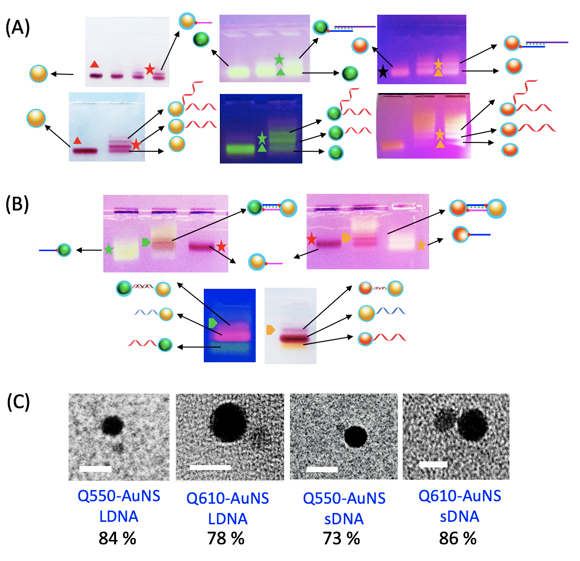
To form assemblies with short DNA strands to realise stronger interaction between the Au and QD, the challenge is to achieve well-resolved gel bands containing one DNA strand per particle on purification via electrophoresis. The short-chain length of the 30 bp DNA does not allow a significant size increase of the DNA-nanoparticle conjugate (compared to the nanoparticle itself), which makes it difficult to purify via electrophoresis. A toehold-mediated DNA displacement approach was thus used to achieve this goal75 as outlined in Scheme 2.2B. Different volumes of 30 bp B1 DNA (100 M) are added to a concentrated solution of the QDs, along with an equimolar concentration of the 100 bp long non-functionalized lengthening DNA strand (L1). The L1 DNA has 15 bp complementary to the B1 DNA. The B1 DNA attaches the AuNP via trithiol moiety while its overhanging strand hybridises with the L1 DNA. This conjugation increases the effective length/bulk of the DNA, therefore facilitating the separation of the nanoparticles in the gel (Figure 1A).75
The DNA functionalization and separation of products to isolate nanoparticles containing one DNA strand per particle of small gold nanospheres () is well-known and was carried out according to literature procedures54, 76, 77, 78 by incubating gold nanoparticles (1-5 M) with 100 bp A1 DNA (5-30 M) and their subsequent purification via gel electrophoresis (shown in Figure 1A). The procedure outlined above was repeated for 30 bp DNA with gold nanoparticles to achieve one strand of B1-comp and L2 DNA per gold nanoparticle.75 The nanoparticles were then used for the formation of the assemblies.
2.3 Self-Assembly of Au-QD Dimers
To form discrete QD:Au hybrid dimers in high yield with the 100 bp DNA linker (Scheme 2.2A), the AuNP and QDs (containing one A1-DNA and one complementary A1-DNA per particle, respectively) are incubated in the presence of salt (Scheme 2.2A). Hybrid Au-QD dimer assemblies with 30 bp linkers are formed by mixing B1 and L1 DNA functionalized QDs with B1-comp and L2 DNA functionalized gold nanoparticles along with L1-comp and L2-comp lengthening DNA strands. These were hybridised overnight, resulting in a branch migration which displaces the stable final duplex (consisting of the non-thiolated 100-mer DNA pairs of L1 - L1-comp, L2 - L2-comp), along with the short DNA bound Au-QD assembly.
Following incubation of the DNA functionalised nanoparticles, the assemblies were further purified via electrophoresis to remove unhybridised nanoparticles and higher-order assemblies. Figure 1B presents the gels (2.5%) of the electrophoretic purification of the short and long DNA assembled hybrid dimers Q550-AuNS10 and Q610-AuNS10. The first and second bands (from the bottom) in the gels correspond to the monomer QD/AuNPs functionalized with one DNA strand, respectively, whilst the third band corresponds to the hybrid dimer assembly. No slower bands, indicative of the formation of higher-order assemblies, are observed. The bands containing the assemblies were eluted from the gel and characterized by transmission electron microscopy (TEM) as shown in Figure 1C. The high contrast particles in the TEM images are gold nanoparticles, while the nanoparticles with lower contrast are the semiconductor QDs. The dimer yields range from 73% to 86% (Figure S3) following deposition on the substrate. The distance between the Au nanoparticle and QD, as calculated from the TEM images, is with 100 bp DNA and with 30 bp DNA. The theoretically calculated interparticle separation between the metal NP and QD with fully-extended 30 bp and 100 bp DNA, as expected in solution phase, is and , respectively. The smaller interparticle separation achieved on deposition compared to the theoretically predicted distance based on the length of the DNA double-stranded helix linker is likely due to the drying effects upon deposition, including van der Waals interactions between the particles and capillary drying effects.
2.4 Self-Assembly of Au-QD Core-Satellite (COSA) Structures
The versatility of the DNA-based approach following isolation of QDs containing one DNA strand per particle is highlighted by the adaptation of the DNA hybridisation scheme to form Au-QD core-satellite structures. The fabrication of core-satellite hybrid structures (Scheme 2.2C) containing a metal core and multiple monofunctionalised QD satellites were formed by initially conjugating the central gold nanoparticle with an excess of DNA prior to the self-assembly. The higher DNA density is achieved by mixing the particles with higher DNA (40 - 90 M) and salt concentrations (50 - 100 M), and is confirmed by lower electrophoretic mobility of the particles on the agarose gel (Figure 2A). The high salt concentration reduces the electrostatic interaction between the negatively charged ssDNA and thus allows a higher DNA density to be functionalised onto the AuNP surface. Additionally, running the cores on the gel separates the DNA-coated particles from excess (non-binding) DNA strands and is a necessary step prior to assembly. It also has the benefit of purifying the desired nanoparticles from side-products (different shapes for example) as can be seen in the gel images for the purification of AuNR, which are separated from nanosphere byproducts, the blue and red bands respectively in the gels in Figure 2A ( AuNS50).
To form hybrid QD-AuNS10 COSA and QD-AuNR2.7 COSA assemblies with 100 bp DNA, the fully A1 DNA functionalised AuNS10 and AuNR2.7 core particles are hybridised with QD satellite particles containing one complementary A1-comp DNA strand per particle. The hybrid core-satellite assemblies were again purified via a second gel electrophoresis step on a diluted gel (1.5%) to separate the hybrid COSA assemblies from the unbound satellites (Figure 2B and S8). The low mobility of the COSA assembly compared to single unconjugated nanoparticles results in separate bands for these species as shown in Figure 2A (lower panel). Similarly, to form 30 bp DNA-based COSA structures, a high concentration (40-90 M) of B1-DNA and L1 lengthening strand are mixed with the core AuNP, which, after purification, is hybridized with QDs functionalised with one complementary B1-comp and L1-comp DNA duplex per particle as outlined in (Scheme 2.2D).
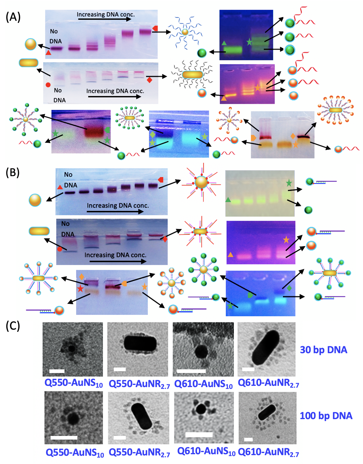
The TEM images of the COSA structures in Figure 2C and S3 show the distribution of the number of satellites per metal core ranges from 1 - 10 for AuNS10 and 1 - 20 for AuNR2.7 hybrid assemblies (Fig S4, Table 1). As expected, the number of satellites around the metal core decreases as the size of the QD increases from Q550 to Q650. The coverage of the satellites around the core NP also depends upon the available surface area of the core NP. Therefore, a higher DNA valency, and therefore a higher number of QD satellites, is observed for gold nanorod cores AuNR2.7 relative to gold nanospheres of 10 nm diameter.
2.5 Photochemistry
The 1:1 metal:QD assemblies allow the effect of the presence of just one metal nanoparticle on a QD to be quantified, and the experimentally observed optical response accurately modelled to understand the fundamental theoretical basis of the observed photophysics.
The series of QD sizes incorporated into the assemblies facilitates comparison of the response as a function of the spectral overlap between the gold nanoparticle LSPR energy and emission of the QDs, as shown in Figure S2. There is a higher degree of spectral overlap between Q550 and the gold nanospheres (AuNS10) relative to these with Q570, Q610 and Q650 () respectively.
2.5.1 Hybrid Dimers
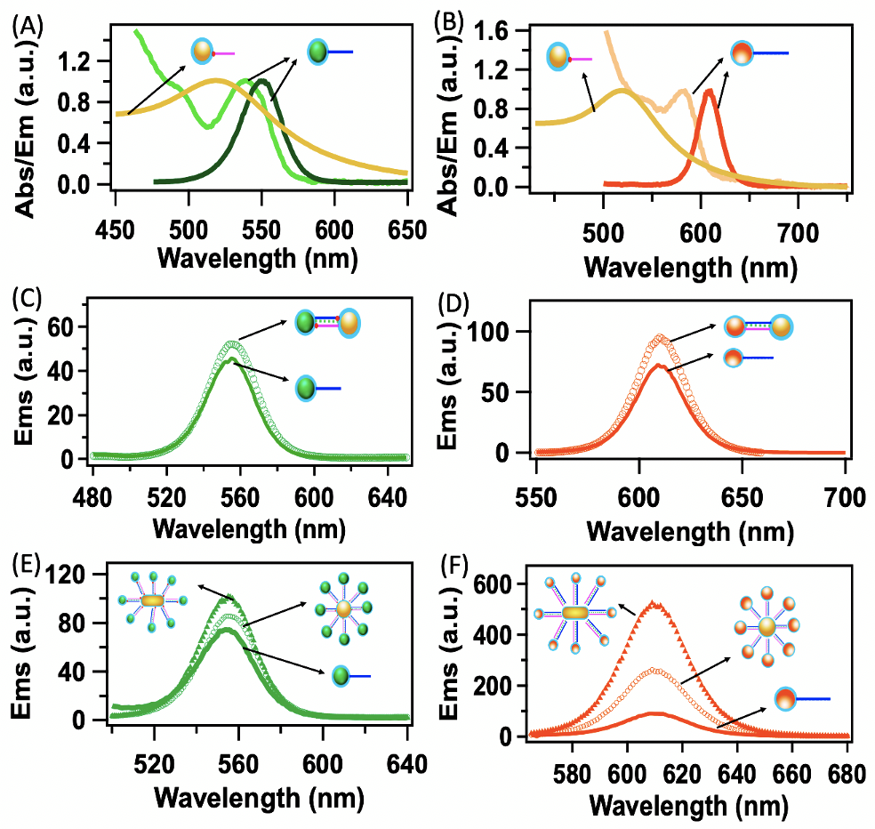
The emission spectra of the Q550-AuNS10 dimers with 30 bp DNA are shown in Figure 3C. The photoluminescence intensity in the Q550-AuNS10 dimer increases by 19% compared to the monomer (Figure 3A) before hybridisation (24% for the 100 bp dimer). The observed enhancement is a consequence of the balance between the enhancement of the electric field and quenching of the QD PL, both of which are distance-dependent, at 10 and 34 nm from the metal particle.79, 66
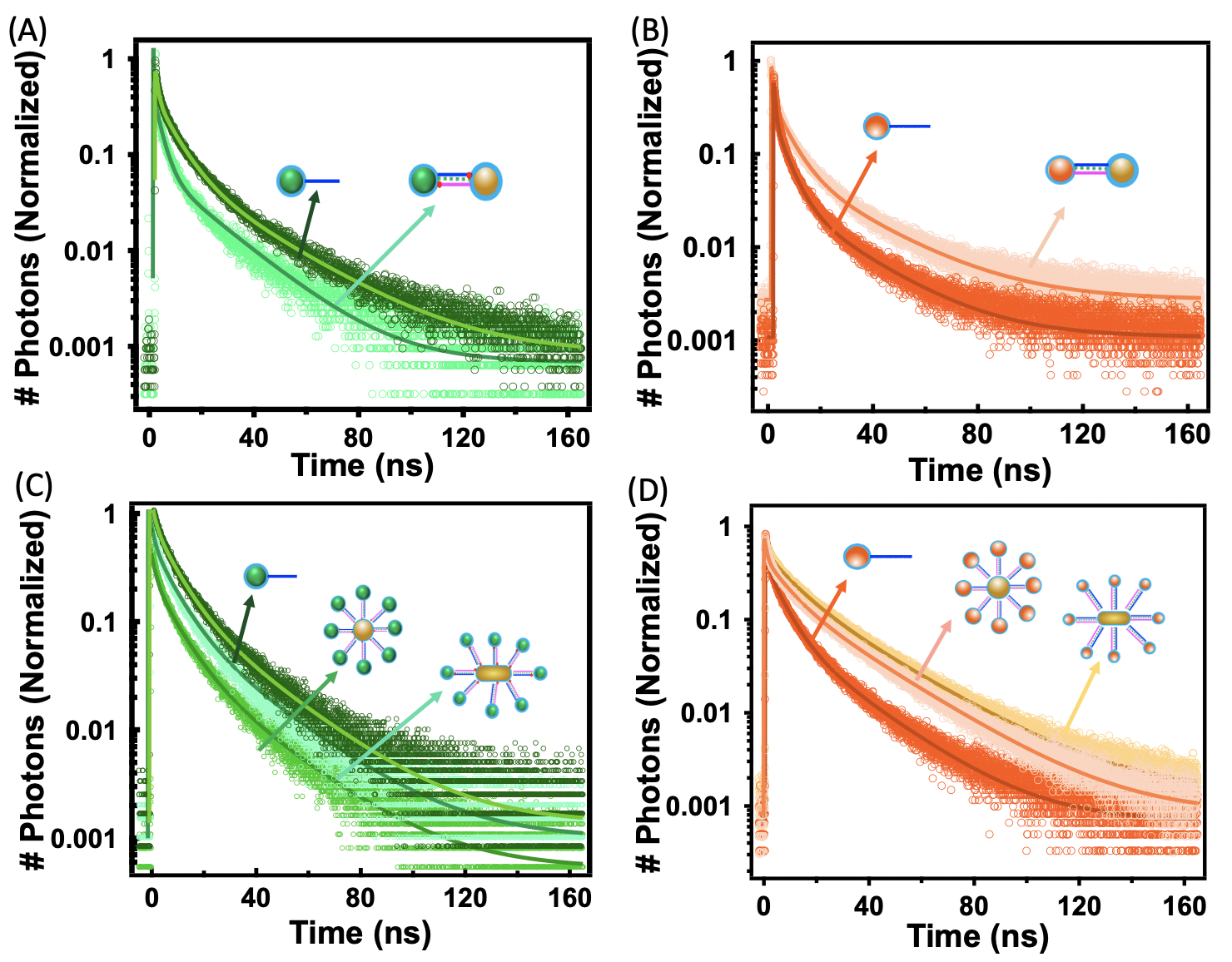
The PL decays were fit using a tri-exponential model, reflecting the broad distribution of emission rates of the QD, and are tabulated in Table 1 for dimers linked by 30 bp DNA linkers and Table S3 for 100 bp DNA linkers. The decay histogram for Q550-AuNS10 (30 bp DNA linker) is shown in Figure 4A. The average lifetime of Q550 is reduced from that of the reference QD, , to upon incorporation of the QD into the dimer (Table 1), a reduction of . The increase in the steady-state emission intensity (Figure 3C) and decrease in lifetime for Q550-AuNS10 is consistent with literature reports,80, 81, 69, 82 and attributed to coupling of the QD excited state to the AuNP LSPR.83 The reduction in the degree of quenching of the lifetime of the 100 bp DNA-based Q550-AuNS10 hybrid dimer (Figure S6C, Table S3) compared with that of the shorter linker, along with the greater enhancement in the steady-state PL intensity for this larger separation between the QD and gold nanosphere, is also consistent with literature reports.80
| Assembly | NP 1 | NP 2 | NP 2 | () | () | |
|---|---|---|---|---|---|---|
| Type | ( | QD-ref | Assembly | |||
| ) | ||||||
| Dimer | AuNS10 | Q550 | 1 | 4.78 | 9.9 | 7.8 |
| Dimer | AuNS10 | Q610 | 1 | 2.78 | 12.9 | 16.3 |
| COSA | AuNS10 | Q550 | 9 | 4.78 | 8.4 | 6.5 |
| COSA | AuNS10 | Q570 | 7 | 3.89 | 13.1 | 14.4 |
| COSA | AuNS10 | Q610 | 5 | 2.78 | 12.0 | 19.8 |
| COSA | AuNS10 | Q650 | 5 | 1.62 | 9.0 | 9.7 |
| COSA | AuNR2.7 | Q550 | 18 | 1.61 | 8.4 | 6.7 |
| COSA | AuNR2.7 | Q570 | 16 | 2.80 | 13.1 | 16.0 |
| COSA | AuNR2.7 | Q610 | 13 | 2.88 | 12.0 | 20.3 |
| COSA | AuNR2.7 | Q650 | 13 | 3.93 | 9.0 | 10.7 |
In contrast to the results for the Q550 dimer, those for Q610-AuNS10, within which the LSPR and QD emission have little spectral overlap (Figure 3B), show both an increase in both the steady-state PL intensity and the PL lifetime of the QD, that is, the photoluminescence lifetime of the QD is not quenched, but extended (a decrease in the emission rate). The steady-state photoluminescence intensity of Q610-AuNS10 dimer linked with a 30 bp DNA linker increases by 23% compared to the reference (Figure 3D), a slightly larger enhancement compared to that for Q550-AuNS10. A lengthening of the QD lifetime from to upon incorporation of the QD into the dimer (30 bp linker length), equating to an increase of is apparent (Figure 4B). For Q610-AuNS10 assemblies linked via a 100 bp DNA linker, steady-state PL intensities increased by 13% (Figure S6B), and the PL lifetime increased by 1.2 (Figure S6D) relative to the reference. Hence, also in contrast to those for the Q550-AuNS10 dimer, the changes in PL intensity and lifetime are both more pronounced for the shorter interparticle separation.
The reduction of lifetimes at one end of the spectral overlap scale (i.e. high spectral overlap), combined with the increase in the lifetime for low spectral overlap provides the opportunity for full control of the emission decay rate of the QD in metal-semiconductor hybrid systems. The level of control extends across a time period greater than previously appreciated as a consequence of the increase in the lifetime when the spectral overlap between the nanoparticles is detuned. For all systems, irrespective of the change in emission rate, an increase in the steady-state photoluminescence intensity was observed, although the relative trends with respect to their interparticle separation are different due to the competing factors.
2.5.2 Hybrid Core-satellite Assemblies
The core-satellite assemblies contain multiple QD particles around a metal nanoparticle, correlating with many donor/single acceptor systems in the framework of energy transfer systems. Relatively little change in the degree of enhancement of the steady-state PL intensity (increase by 29%, Figure 3E)) or lifetime quenching (0.79, Table 1) is observed upon increasing the number of QDs around Q550-AuNS10-COSA with 30 bp linking DNA (Figure 2C) compared to the dimer.
Hybrid Q610-AuNS10 COSA assemblies with 30 bp linker length, however, have a greater degree of enhancement in the PL intensity (48% enhancement), shown in Figure 3F, compared to the dimer assembly. Similarly, multiple Q610 quantum dots around a single gold nanoparticle also results in a large increase, (/, in the PL lifetime relative to the monomer (Figure 3F, Table 1), and 1.2 relative to the dimer. Enhancement in PL intensity and lengthening of the average lifetime for hybrid assemblies incorporating Q570 and Q650, which have varying degrees of de-tuning of their emission with the LSPR of AuNS10 and 30 bp linkers, is also observed (Figure S9). The degree of spectral overlap for the Q650 hybrid assembly is lower than Q610. A lower enhancement in the magnitude of PL intensity and average lifetime for the Q650 than Q610 hybrid assemblies is however observed. This is attributed to an increase in the center to center spacing between the AuNP and Q650 owing to the bigger size of Q650, with potentially some effect from the low quantum yield of Q650. The quenching of the quantum emitter lifetime is more pronounced for the QD with a higher spectral overlap with AuNS10, Q550-AuNS10-COSA, for which the quenching dominates the optical response at short interparticle separations. For all other assemblies, a trade-off exists between the degree of spectral overlap and interparticle separation.
Consistent with the results for dimers, increasing the interparticle separation within the core-satellite structures based on AuNS10 led to an increase in the steady-state photoluminescence intensity to 31% and 35% for Q550-AuNS10-COSA and Q610-AuNS10-COSA assemblies, respectively. The lifetime decreased by for Q550-AuNS10-COSA and increased by for Q610-AuNS10-COSA assemblies (Figure S7, Table S4). Control of the assembly structure thus allows tuning of the degree of photoluminescence enhancement and emission rate.
The enhancement effect in the PL intensity and the change in the lifetime values in the hybrid assemblies are more pronounced upon the incorporation of larger, asymmetric plasmonic particles such as gold nanorods. For nanorod assemblies with 30 bp DNA linkers, the PL intensity increased from the reference PL values to 45% for Q550-AuNR2.7-COSA assemblies and 75% for Q610-AuNR2.7-COSA assemblies (Figure 3E and F). The lifetime of Q550 decreased by (/ in Q550-AuNR2.7-COSA assemblies and increased by (/ for Q610-AuNR2.7-COSA assemblies (Figure 4C and D). With 100 bp DNA linkers, the same assemblies showed an increase in the intensity of the PL, although not to the same degree as observed for the shorter linkers, with the lifetime for the Q550-AuNR2.7-COSA assembly showing a similar decrease and a smaller increase in the lifetime for Q610-AuNR2.7-COSA (Figure S7). The photophysical properties of the AuNR2.7-COSA are thus consistent with both AuNS10-COSA and hybrid dimers assemblies, although of a greater magnitude. This is presumably due to the strong localisation of the near-field of the nanorod at the tips.
Previous literature shows that the local refractive index of the metal nanoparticle has a large impact on the decay kinetics of QDs in metal-semiconductor assemblies 84. In the next section, we model the optical properties of these systems to fully understand their origin.
Theoretical insights
Impact of the local electromagnetic environment on spontaneous emission
The transition rate (or the transition probability per unit time) of a quantum emitter from an excited state to a ground state follows the Fermi’s golden rule given by 85,
| (1) |
The density of states of the electromagnetic environment of the emitter is defined such that is the number of photon states per unit volume that fall within the angular frequency range to 85, 86. The transition matrix element is obtained as,
| (2) |
where is the perturbation Hamiltonian caused by light, is the electric dipole moment of the transition and is the electric field amplitude experienced by the quantum emitter. It is evident that the transition rate is determined by the interplay between the electric field experienced by the emitter and the density of states.
Damped nanoscale cavity analogy for gold nanoparticles
Let us first analyse how an MNP placed in nanoscale proximity alters the electromagnetic environment of a coupled QD using a cavity analogy. The total cavity decay rate (should not be confused with the emitter decay rate ), the quality factor and the effective mode volume of the cavity-like plasmonic mode of a spherical metal nanoparticle with radius can be estimated using the Drude approximation as and where 84,
| (3) | ||||
| (4) | ||||
| (5) |
In the above equations, and represent the bulk plasma frequency and the ohmic loss rate of the metal, is the radiative scattering rate, is the relative permittivity of the non-magnetic submerging medium (where is the background refractive index), is the contribution of bound electrons in the metal dielectric constant and is the wavenumber in the background medium for free space wavelength (and free space wavenumber ).
We estimate a dipole () mode volume , and the plasmonic decay rate and quality factor variations shown in Figure 5(a), for a gold nanoparticle of radius submerged in water, using the above equations and the following parameters from literature: , 87, and 88.
It has been shown that rather low-quality factors ranging from about 10 to 100 such as those in Figure 5(a), accompanied by small effective volumes in the nanometer scale result in high-quality factor/effective volume ratios that indicate efficient coupling to quantum emitters generally 84. This is in contrast to conventional diffraction-limited micro-cavities with comparatively much higher volumes that efficiently couple to emitters thanks to their very high-quality factors 84. Thus, our gold nanoparticle submerged in water is expected to behave as a damped, low-quality factor nanoscale cavity capable of efficiently coupling to emitters, while altering their electromagnetic environments with reasonably high bandwidths.

Using the aforementioned cavity analogy, we can now estimate the density of states of the MNP cavity for a given single mode with half-width and cavity resonance as: 85. The qualitatively estimated density of states variation for the dipole mode of the earlier MNP obtained using and is depicted in Figure 5(b). This results in a Lorentzian-shaped density of states variation suggesting that an MNP seems more likely to accept a photon emitted by a nearly resonant QD due to the high density of states, whereas this ability depletes as the QD becomes off-resonant. In essence, we can interpret the experimentally observed QD decay rate modification as a manifestation of the cavity nature of gold nanoparticles, where the emission is enhanced when the “cavity” is tuned (in Figure 4(A) for Q550-AuNS10) and the emission is suppressed when it is detuned (in Figure 4(B) for Q610-AuNS10), as suggested by equation (1).
In conventional resonant low Q cavities, the rapidity of emitter decay has been reported to increase with the number of emitters 89. This behavior is marginally mimicked by Q550-AuNS10 core-satellite assemblies with average normalized lifetime (see Table 1), in comparison to Q550-AuNS10 dimers with average normalized lifetime . Note that the observed difference between the dimer and core-satellite assemblies stems not just from the number of surrounding emitters but also from the (unquantified) increase in the local refractive index of the MNP due to the larger number of DNA strands in the latter case. It has also been reported in the context of conventional cavities that collective emission effects can be appreciably suppressed by increasing the detuning of the cavity from the emitter resonance for multiple emitter assemblies 89. This is qualitatively mimicked by our experimental observations for Q610-AuNS10 Dimer (average normalized lifetime ) and core-satellite assemblies (average normalized lifetime ). While reporting these qualitative similarities, we also emphasize that MNPs are not expected to ideally mimic conventional, diffraction-limited linear optical cavities. MNPs form feedback dipoles in response to all proximal dipoles 90, 91, which causes emitter-AuNP assemblies to behave as non-linear systems.
MNP-QD dimers as semi-classical open quantum systems
Adopting an alternative viewpoint where we can account for the aforementioned non-linearities, we now model the MNP using the generalized nonlocal optical response theory (GNOR) 87 and the QD excitons as open quantum systems.
Incorporating GNOR based non-locality corrections to the dipolar mode of the Mie expansion, the normalized decay rate for a generic dipolar emitter placed at a small centre separation from a spherical metal nanoparticle can be estimated as,
| (6a) | ||||
| (6b) | ||||
where , and denote the spontaneous emission rates of dipole emitters oriented perpendicularly and parallelly to the MNP surface, and free-space spontaneous decay rate, respectively. The decay rate of the isolated emitter in a medium of refractive index is obtainable as 92, and (with each respective subscript/superscript) denotes the emitter excitation lifetime. See the supplementary material for details, including the full form of the GNOR based MNP dipolar polarizability . The decay rate of a randomly oriented emitter in a hybrid dimer can be estimated as 84,
| (7) |
Figure 6(a) shows that for a generic emitter, the normalized excitation lifetime is expected to increase as the emission frequency moves away from the MNP resonance (and vice versa) as per equations (6a) and (6b), in line with our earlier observations and claims. It can also be seen from Figure 6(b) and (c) that the PL lifetimes of both Q550 and Q610 are theoretically expected to decrease in response to a gold nanoparticle approaching closer (decreasing ), in line with the previous single dimer based observations in literature 33. The lifetimes of QDs we reported experimentally (for dimers in Table 1) are averages resulting from the cohort of dimers present in colloidal samples, where the centre separations may vary around .

We model the exciton that forms in the QD as a coherently illuminated two-level-atom (TLA) coupled to the plasmonic field of the MNP at one of the two extreme orientations (QD transition dipole parallel or perpendicular to the MNP surface as portrayed in Figure 7). The TLA (both in isolation and in the presence of the MNP) is assumed to behave as an open quantum system that undergoes Markovian interactions with the submerging environment. This TLA model, where we equate the excitonic energy gap to the experimentally observed emission spectral peak of a given QD is only expected to approximate the emission behaviour, which is the focus of our study. It is not expected to sufficiently capture the phonon-assisted absorption mechanism of the QD which results in an absorption peak that does not overlap with the emission peak (unlike for an ideal TLA). Following the detailed formalism summarized in the supplementary material, we arrive at the following optical Bloch equations for the density matrix elements of the TLA, under the influence of an external electric field and the MNP,
| (8a) | ||||
| (8b) | ||||
| (8c) | ||||
where is the density matrix element of the TLA, considering the basis states , , , () is the corresponding emitter decay time (lifetime), is the corresponding dephasing time, is the emitter resonance and is the angular frequency of the incoming radiation. The effective Rabi frequency in the presence of the MNP () is related to the slowly varying positive frequency amplitude () of the total field experienced by the QD exciton () via the QD dipole moment element as . is the sum of three field components incident on the QD exciton: (external field , screened by the QD material), (field emanated by the dipole formed in the MNP in response to ), and (field emanated by the dipole formed in the MNP in response to the QD dipole). A detailed discussion on dipole formation in nanohybrids can be found in reference 91. The total power emitted by the TLA takes the form for the extreme dimer orientations 93, 90. For the isolated QD, , where denotes the isolated QD excited state population (see supplementary material for details).
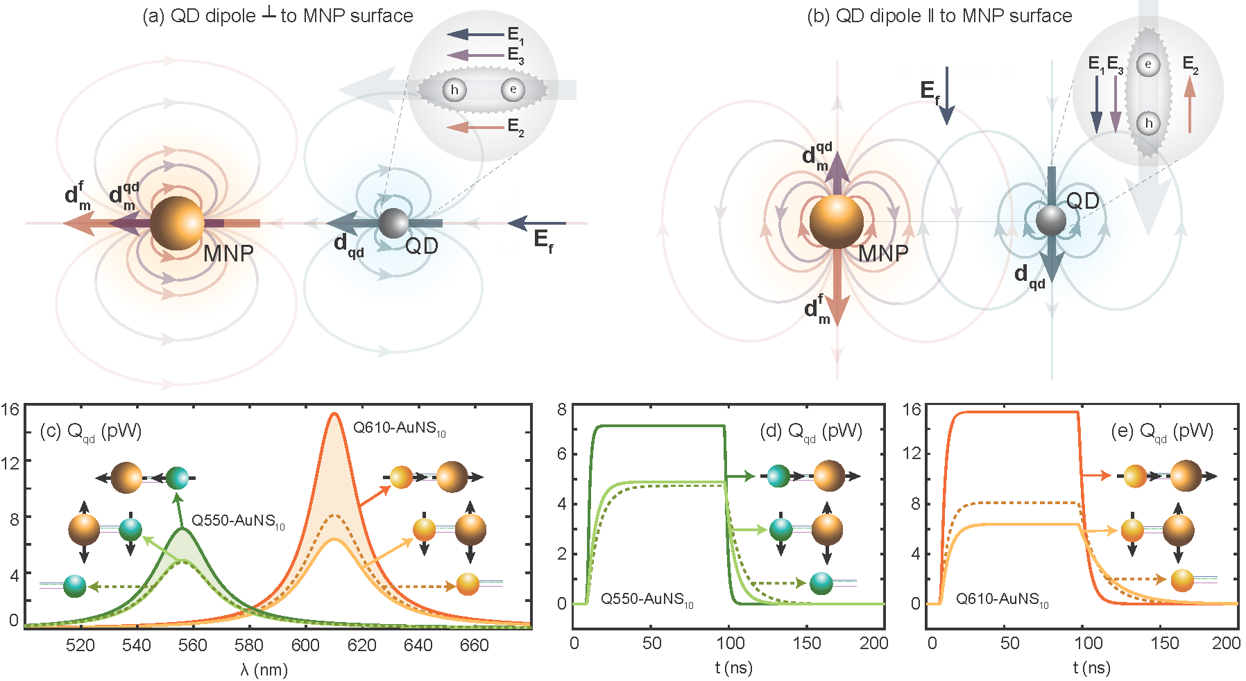
We used the above TLA-based open quantum system formalism to theoretically predict the behaviour of Q550-AuNS10 and Q610-AuNS10 dimers in extreme orientations. We assume that QDs are isotropic and form dipoles along the external field. We can see from Figure 7(a) that all fields experienced by a QD exciton constructively add in the same direction when the QD dipole is to the MNP surface (where all fields form along the axis of the hybrid), always resulting in a stronger field compared to the isolated QD (reference). This results in larger emission from the case compared to the reference for both Q550-AuNS10 and Q610-AuNS10 as observed in Figure 7(c) (where the darker solid lines for both dimers are larger than the reference). However, in the orientation (see Figure 7(b) for the schematic), there can be constructive and destructive interference effects between the field components incident on the QD exciton, resulting in the emission of the dimer being enhanced or suppressed compared to the isolated QD depending on parameter variations. It can be observed from Figure 7(c) that Q610-AuNS10 shows a large steady-state emission enhancement in the (axial) orientation compared to Q550-AuNS10. This suggests that the larger steady-state emission enhancement we experimentally observed for the Q610-AuNS10 colloidal ensemble in Figure 3(D), compared to Q550-AuNS10 in Figure 3(C), is likely to be attributable to the dominant contribution of axial field enhancement.
Temporal PL decay plots theoretically obtained for (single molecules of) both Q550-AuNS10 and Q610-AuNS10 dimers are depicted in Figure 7(d) and (e). We only focus on the emission phase of these TLA-based time-domain plots, which occurs after the electric field input is switched off near . Comparison of Q610’s steady-state isolated and dimer spectra in Figure 7(c) to its temporal plots in (e) reveals that the plasmonically enhanced case in the former subplot maps to an enhanced temporal decay while the plasmonically suppressed case maps to a suppressed temporal decay (or an enhanced lifetime, as is also observable from Figure 6). The larger decay rate observed for the orientation is expected to be caused by the larger electric field experienced by the QD in the orientation compared to the case as suggested by the Fermi’s golden rule in (1). Thus, the suppressed QD decay rate experimentally observed for the colloidal Q610-AuNS10 dimer ensemble in Figure 4(B) is likely to be attributable to the dominant contribution of the components, as suggested by equation (7).
2.6 Tunability of Surface Plasmon Resonance
To obtain a complete picture of the QD-AuNP interaction, we investigated the coupling in hybrid assemblies with fixed QD emission and variable localized surface plasmon of the AuNP. For this purpose, hybrid assemblies of Q610 with AuNS10 and different aspect ratios of gold nanorods i.e. 2.7, 3.9, and 4.1 were synthesised. The time-resolved measurements in Figure 8 compare the emission lifetime of Q610-Ref and Q610-AuNR COSA assemblies with the different aspect ratios of the metal nanorod (fit parameters in Table S5). While a lengthening of the photoluminescence lifetime for all metal hybrid systems relative to the QD alone was observed, the emission rate of Q610-AuNR2.7 (20.3 ns) is lower compared to that of Q610-AuNR3.9 and Q610-AuNR4.1 (Table 2). The spectral overlaps for these systems are not significantly different (Table 2). Nanorods are well known to have a larger near field enhancement at the tips of the nanorod, due to the longitudinal LSPR mode.94 Of the nanorods here, AuNR2.7 has the greatest width (AuNR2.7 = 20 nm, AuNR3.9 = 10 nm, AuNR4.1 = 7 nm). The wider the rod, the more QDs able to attach the tip, as apparent in the TEM images in Figure 8 and also the higher probability that QDs will be present at the nanorod tip, that is, in the position of maximum field enhancement. Consequently, the greater lengthening of the average lifetime of Q610-AuNR2.7 is attributed to the nanorod width.
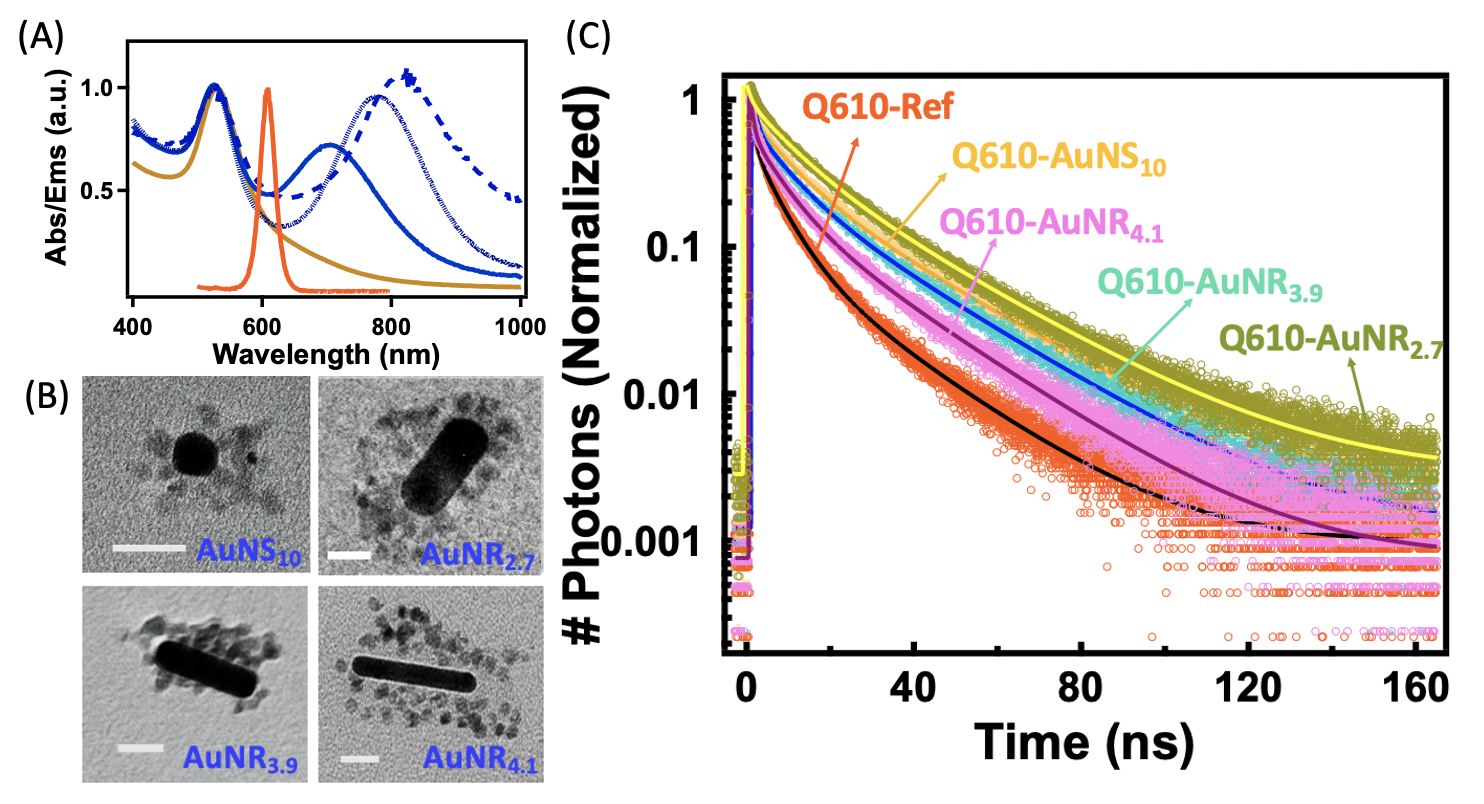
| Sample | NP 1 | NP 2 | NP 2 | ||
|---|---|---|---|---|---|
| ( | |||||
| ) | |||||
| Q610-Ref | Q610 | - | - | - | 11.8 |
| COSA | AuNS10 | Q610 | 5 | 2.78 | 19.8 |
| COSA | AuNR2.7 | Q610 | 13 | 2.88 | 20.3 |
| COSA | AuNR3.9 | Q610 | 11 | 2.56 | 18.8 |
| COSA | AuNR4.1 | Q610 | 10 | 2.11 | 15.8 |
3 Conclusion
We combine DNA-based self-assembly methods, optical spectroscopy, and theory to determine the fundamental interactions between plasmonic nanoparticles and quantum dots. Discrete assemblies incorporating quantum dots and gold nanoparticles are synthesised by translation of DNA-based assembly methods to CdSe QDs. The isolation of QDs containing only one strand of DNA per particle allows the formation of QD:AuNP dimers and core-satellite structures in high purity. The tight control of the nanoparticle sizes, stoichiometry, and interparticle separation (via DNA linker length) in the assemblies allows the interrogation of these precise structures using ensemble optical spectroscopy and theory.
A gold nanoparticle immersed in water behaves as a nanoscale damped optical cavity that mimics the qualitative behaviour of conventional diffraction-limited optical cavities when modifying the emission characteristics of quantum emitters. We show experimentally and theoretically that the detuning between a metal nanoparticle and emitter can be effectively used for the regulation (enhancement/suppression) of spontaneous emission from quantum dots. The observed lowering of the QD emission rate (by a factor of up to 1.7) gives a mechanism for greater control over the QD emission lifetime by a plasmonic nanoparticle than previously appreciated.
We show that the luminescence of QDs in the presence of a gold nanoparticle is enhanced. The steady-state emission is increased for the off-resonant dimer compared to the closely resonant dimer. Using an open quantum system-based analysis on extreme metal-QD dimer orientations, this is attributed to the dominance of axial enhancement of the electric field, and consequently the steady-state QD spectra.
4 Experimental
4.1 Materials
Cadmium oxide (CdO) (99%), octadecene (ODE) (technical grade, 90%), oleylamine (technical grade, 70%), trioctylphosphine (90%), oleic acid (technical grade, 90%), octane thiol (≥ 98.5%), tetradecylphosphonic acid, selenium powder (Se) (99.00%), sulfur (≥ 99.99%), sodium borohydride (NaBH4) (≥ 99%), gold(III) chloride trihydrate ( 99.9%), tannic acid, tri-sodium citrate dihydrate (≥ 98.5%), bis(p-sulfonatophenyl)phenylphosphine (BSPP) (97%), potassium carbonate, phosphonoacetic acid (98%), agarose (type I, low EEO), boric acid, Tris base, chloroform (high-pressure liquid chromatography (HPLC) grade), EDTA and Ficoll 400 were purchased from Sigma Aldrich. CTAB (98%) was purchased from Ajax Finechem, acetone (analytical reagent (AR) grade), and methanol (AR grade) were purchased from Merck. Thiolated ethylene glycol HS-C11-(EG)6-OCH2-COOH was purchased from Prochimia Surface (Poland). Tetramethylammonium hydroxide (TMAOH) (25% w/w in methanol) was purchased from Alfa Aesar.
PAGE-purified trithiolated DNA sequences were purchased from Fidelity Systems, Inc. (USA). The sequences of DNA used for DNA-based self-assembly are given below.
A1: \seqsplit5’-trithiol-TTTTCTCACTAAGATCGATAGAGCGATTGTGATATTTCAAGCGGTACTCCAGCTCTAGGTAGCTCCCTTTCCAATCAGCTTATGTGAGCGCCTGCCCATG-3’
A1-comp: \seqsplit5’-trithiol-TTTCATGGGCAGGCGCTCACATAAGCTGATTGGAAAGGGAGCTACCTAGAGCTGGAGTACCGCTTGAAATATCACAATCGCTCTATCGATCTTAGTGAGA-3’
B1: \seqsplit5’-trithiol-TATACCTGACCTCGGGACTTGACTGATTGT-3’
B1-comp: \seqsplit5’-trithiol-ACAATCAGTCAAGTCCCGAGGTCAGGTATA-3’
L1: \seqsplit5’-GCTGACTCGCTACTCTTTTTTTTTTTTTTTTTTTTTTTTTTTTTTTTTTTTTTTTTTTTTTTTTTTTTTTTTTTTGCATGCAGATACAATCAGTCAAGTC-3’
L1-comp: \seqsplit5’-GACTTGACTGATTGTATCTGCATGCTTTTTTTTTTTTTTTTTTTTTTTTTTTTTTTTTTTTTTTTTTTTTTTTTTTTTTTTTTTTGAGTAGCGAGTCAGC-3’
L2: \seqsplit5’-AGCACACAAGAGCTGTTTTTTTTTTTTTTTTTTTTTTTTTTTTTTTTTTTTTTTTTTTTTTTTTTTTTTTTTTTTAGCAGATACATATACCTGACCTCGG-3’
L2-comp: \seqsplit5’-CCGAGGTCAGGTATATGTATCTGCTTTTTTTTTTTTTTTTTTTTTTTTTTTTTTTTTTTTTTTTTTTTTTTTTTTTTTTTTTTTTCAGCTCTTGTGTGCT-3’
All chemicals were used as received without further purification. Ultrapure water from a MilliQ system, with resistivity 18 M was used for all aqueous solutions.
5 Synthesis
Gold Nanoparticles: Gold nanospheres were synthesised according to the method described by Piella et. al.71. Different aspect ratios of gold nanorods were synthesised using the method reported by Nikoobakht and El-Sayed.72
CdSe Core Nanocrystals: CdSe cores were synthesized according to the reported method.73
Shelling of CdSe Cores: The CdSe core particles were shelled according to the published protocol of Boldt et. al.74
5.1 Ligand Exchange of Nanoparticles
Quantum dots were transferred from the organic to aqueous phase by ligand exchange. For this, phosphonoacetic acid (PsAA) (0.1 M, 5 mL) solution was prepared in methanol and added to a dispersion of the QDs in chloroform (13 M, 1.00 mL). The pH of the PsAA solution was maintained using TMAOH (tetramethylaluminum hydroxide), (25% solution in methanol). The QD dispersion in chloroform was added and then the dispersion was sonicated for 1-2 minutes. Water was added and the QDs migrated to the aqueous layer following sonication, leaving a colorless organic layer. The aqueous layer containing the QDs was collected and washed twice by precipitation via the addition of acetone ( 5–10 mL). The precipitate was centrifuged and the pellet was redispersed in water to remove excess PsAA.
Colloidal gold nanoparticles with citrate ligands with a diameter of 10 nm were mixed overnight with BSPP (200.0 L, 90.0 mM). These were then purified by centrifugation at 13400 rpm for 50 min. The supernatant was removed and the pellet redispersed in BSPP buffer and washed three more times to achieve colloidally stable gold nanoparticles. After the final washing, the pellet was collected and re-dispersed in a known volume of BSPP to achieve a final concentration of nanoparticles (2 - 5 M).
5.2 NP-DNA Functionalisation
QDs-Short (30 bp) DNA Conjugation:
Thiolated short DNA (100.0 M) (0.5, 1.0, 2.0, 3.0, 4.0, and 5.0 L) were mixed with the equimolar concentration of non-functionalized lengthening DNA strands (100 bp) which was then left to incubate overnight. The DNA solution was then added to the highly concentrated quantum dot solution (25.0 M, 5 L) in the presence of BSPP (90.0 mM, 1.0 L) and NaCl (1 M, 1 mL). The samples were incubated overnight before running on the gel for purification.
QDs-Long (100 bp) DNA Conjugation:
Thiolated DNA (100.0 M) (0.5, 1.0, 2.0, 3.0, 4.0, and 5.0 L) were added to highly concentrated quantum dot solution (25.0 M, 5 L) in the presence of BSPP (90.0 mM, 1.0 L) and NaCl (1 M, 1 mL). The samples were incubated overnight before running on the gel for purification.
AuNP-Short (30 bp) DNA Conjugation:
Short (30 bp) thiolated DNA (0.5, 1.0, 2.0, 3.0, 4.0, and 5.0 L of 100.0 M) were mixed with an equimolar concentration of non-functionalized lengthening DNA strands (100 bp) and the dispersion left to incubate overnight. The DNA solution was then added to the highly concentrated gold nanoparticle solution (2-5 M, 5 L) in the presence of BSPP (90.0 mM, 1.0 L) and NaCl (1 M, 1 mL). The samples were incubated overnight before running on the gel for purification.
AuNP-Long (100 bp) DNA Conjugation:
The thiol-functionalised DNA (0.1, 1.0, 3.0 and 10.0 L of 100.0 M) were separately mixed with the highly concentrated dispersion of gold nanoparticles (AuNP) (2-5 M, 5.0 L) in the presence of salt (NaCl) (1.0 M, 1 mL) and BSPP (90.0 mM, 1.0 L). The samples were left overnight for incubation.
5.3 Purification of NP-DNA conjugates
Prior to electrophoresis (15 minutes), mPEG (100,000–500,000 molar excess with respect to the AuNP concentration, 1.0 L) was added to the Au-DNA samples and HS-C11-(EG)6-OCH2-COOH (340 excess with respect to the QDs concentration, 1.0 L) to the QD-DNA samples. Before loading the samples on the gel, Ficoll solution (20%, 4.0 L) was added as a loading buffer to increase the density of samples. The nanoparticle-DNA conjugate mixtures containing different concentrations of DNA were loaded on the agarose gel (2.7% for AuNPs, 4.1% for Q550, 3.8% for Q570, 3.4% for Q610, and 3.2% for Q650). For the electrophoresis, the voltage was set to 80 V for 35 - 45 min. The same procedure was performed for all Au and QD particles, for both pairs of complementary DNA strands. An electro-elution procedure was used to extract AuNP-DNA conjugates from the gel and the extracted solution was concentrated via centrifugation before hybridization. For the extraction of QD-DNA conjugates, gel bands were cut and soaked in 0.5X TBE buffer overnight. The resulting dispersion was then concentrated by evaporating the solution under N2 stream.
For COSA assemblies, the central AuNP was mixed with a higher DNA concentration (10-fold excess) to fully allow the DNA coverage around AuNP. The samples were left overnight for DNA conjugation and were then run on 1% (w/v) of the agarose gel for purifying it from the unbound DNA strands.
For the preparation of gel, agarose powder was mixed with 0.5x TBE buffer to give the desired concentration of gel. The mixture was heated in the microwave oven (800 W) for 2 minutes to dissolve the gel. The gel was cooled to 60-70 ∘C and poured into the electrophoresis tray. When the gel was fully solidified, the comb was removed. The gel was immersed in 0.5X TBE buffer. The sample was put into the wells created by the comb. Set the voltage to 80 V for 35 min and left the samples moved in the gel.
5.4 Self-Assembly and Purification
After achieving purified samples containing the desired number of DNA strands per particle, AuNS and QDs with one DNA and one complementary DNA strand/particle respectively were mixed together in the presence of optimized salt concentration (1 M, 1.0 L) to form the hybrid dimer assembly. Hybrid COSA assemblies were formed after mixing the central fully DNA functionalized AuNP cores with one complementary DNA containing QD satellite particles in the presence of salt (2 M, 1.0 L).
The assemblies were run in the second gel electrophoresis step on a dilute gel (1% - 2.5%) to purify the dimer assembly from the unconjugated Au and QD particles and the satellite bearing core nanoparticles from the unbound satellites. The low mobility rate of dimers and core-satellites assemblies compared to single unconjugated nanoparticles was responsible for separate bands while running on the agarose gel. Therefore, the relative mobility rate forms the basis of purification of an assembly from the unconjugated nanoparticles. The band containing the purified structures of interest was cut from the gel, and the assembly samples were extracted separately from each band by the electro-elution procedure. The assembly solution was then concentrated upon centrifugation.
5.5 Calculating Concentrations for Hybrid Assemblies
We used inductively coupled plasma mass spectrometry (ICP-MS) to determine the concentrations of QDs in the hybrid assembly. An eight-point calibration curve was constructed using the known standard solutions of various concentrations. The correlation curves showed an r2 value of 0.99. The concentration of the QDs was calculated using the isotopes of Cd(111) and Cd(112), Se (78), Se (82), Zn(66) Zn(68), and Au(197).
5.6 Sample Preparation
Samples for electron microscopy were prepared by depositing 10 L of the purified assembly on an ultra-thin TEM grid which was then soaked in a 1:1 mixture of water and ethanol for 10-15 minutes. The substrate was then rinsed with absolute ethanol and allowed to dry. Quartz cuvettes of 3 mm path length were used for all photophysical measurements. The cuvette was pre-cleaned by soaking in a nitric acid bath overnight and then washing with copious amounts of water.
6 Instrumentation
Transmission electron microscopy (TEM) was performed on FEI Tecnai T20 Twin LaB6. Scanning electron microscopy was done on FEI Magellan 400 FEG SEM.
ICP-MS measurements were carried out using a PerkinElmer Nexion 350 ICP-MS instrument.
UV-Visible spectra were collected using an Agilent Cary 60 UV-Vis Spectrophotometer.
The luminescence spectra of the samples were recorded on a Cary Eclipse Fluorescence Spectrophotometer. For measuring the photoluminescence signal, the optical density of the samples was kept below 0.1 to avoid the inner filter effect.
7 Photophysical Measurements
The excitation and emission parameters used for the steady-state PL measurements were as follows: excitation wavelength = 425 nm, excitation slit = 5 nm, emission slit = 5 nm, scan rate = 600 nm/min and integration time = 0.1 s.
7.1 Time Correlated Single Photon Counting
The fluorescence lifetime decays of both the reference and the assembled quantum dots were measured via a home-built time-resolved single-photon counting (TCSPC) setup. Excitation was provided by a picosecond pulsed supercontinuum laser (Fianium, SC 400-4-pp) with the emission wavelength of 425 nm, selected using a 10 nm bandpass filter (laser power = 1 mW). The emission signal was collected at right angles to the excitation beam which was then directed towards the Multichannel plate detector. Photon emissions were recorded by a photon counting module (PicoHarp 300, Picoquant). The instrument response function (IRF) was measured by using a scattering solution of milk powder in water to be 0.6 ns. The TCSPC data was collected at the 5 MHz frequency and 32 ps time intervals.
7.2 Data Analysis
The time-resolved emission decays were fit with convolution with the IRF to a series of exponential functions until a good fit was obtained as judged by the reduced chi-squared () value and the randomness of residuals. The fits were performed in custom-written routines in Igor Pro 6.3 software.
This work was undertaken within the ARC Centre of Excellence in Exciton Science, supported by the Australian Research Council (ARC) Grant CE170100026. The authors acknowledge the use of instruments and scientific and technical assistance at the Monash Centre for Electron Microscopy, a Node of Microscopy Australia. Computational resources were provided by the National Computational Infrastructure (NCI) at the Australian National University. HH gratefully acknowledges Roslyn Forecast and Francesco Campaioli for insightful discussions.
Electron microscopy images of colloids, steady state spectra of all QDs and gold nanoparticles, wide-field images of assemblies, quantification of number of satellites in core-satellite assemblies, lifetime and steady-state data for assemblies with 100 bp DNA linkers and incorporating Q570 and Q650, along with tabulations of fit data are available online:
References
- Bellessa et al. 2004 Bellessa, J.; Bonnand, C.; Plenet, J.; Mugnier, J. Strong coupling between surface plasmons and excitons in an organic semiconductor. Phys. Rev. Lett. 2004, 93, 036404
- Bayer et al. 2001 Bayer, M.; Reinecke, T.; Weidner, F.; Larionov, A.; McDonald, A.; Forchel, A. Inhibition and enhancement of the spontaneous emission of quantum dots in structured microresonators. Phys. Rev. Lett. 2001, 86, 3168
- Santhosh et al. 2016 Santhosh, K.; Bitton, O.; Chuntonov, L.; Haran, G. Vacuum Rabi splitting in a plasmonic cavity at the single quantum emitter limit. Nat. Commun. 2016, 7, 1–5
- Hapuarachchi et al. 2018 Hapuarachchi, H.; Mallawaarachchi, S.; Hattori, H. T.; Zhu, W.; Premaratne, M. Optoelectronic figure of merit of a metal nanoparticle—quantum dot (MNP-QD) hybrid molecule for assessing its suitability for sensing applications. J. Phys. Condens. Matter 2018, 30, 054006
- Liu et al. 2017 Liu, Y.; Dai, X.; Mallawaarachchi, S.; Hapuarachchi, H.; Shi, Q.; Dong, D.; Thang, S. H.; Premaratne, M.; Cheng, W. Poly (n-isopropylacrylamide) capped plasmonic nanoparticles as resonance intensity-based temperature sensors with linear correlation. J. Mater. Chem. C 2017, 5, 10926–10932
- Maier 2007 Maier, S. A. Plasmonics: fundamentals and applications; Springer Science & Business Media, 2007; Chapter 5
- Hapuarachchi et al. 2017 Hapuarachchi, H.; Premaratne, M.; Bao, Q.; Cheng, W.; Gunapala, S. D.; Agrawal, G. P. Cavity QED analysis of an exciton-plasmon hybrid molecule via the generalized nonlocal optical response method. Phys. Rev. B 2017, 95, 245419
- Ridolfo et al. 2010 Ridolfo, A.; Di Stefano, O.; Fina, N.; Saija, R.; Savasta, S. Quantum plasmonics with quantum dot-metal nanoparticle molecules: influence of the Fano effect on photon statistics. Phys. Rev. Lett. 2010, 105, 263601
- Waks and Sridharan 2010 Waks, E.; Sridharan, D. Cavity QED treatment of interactions between a metal nanoparticle and a dipole emitter. Phys. Rev. A 2010, 82, 043845
- Gettapola et al. 2019 Gettapola, K.; Hapuarachchi, H.; Stockman, M. I.; Premaratne, M. Control of quantum emitter-plasmon strong coupling and energy transport with external electrostatic fields. J. Phys. Condens. Matter 2019, 32, 125301
- Hapuarachchi et al. 2018 Hapuarachchi, H.; Gunapala, S. D.; Bao, Q.; Stockman, M. I.; Premaratne, M. Exciton behavior under the influence of metal nanoparticle near fields: Significance of nonlocal effects. Phys. Rev. B 2018, 98, 115430
- Lee et al. 2004 Lee, J.; Govorov, A. O.; Dulka, J.; Kotov, N. A. Bioconjugates of CdTe nanowires and Au nanoparticles: plasmon− exciton interactions, luminescence enhancement, and collective effects. Nano Lett. 2004, 4, 2323–2330
- Munechika et al. 2010 Munechika, K.; Chen, Y.; Tillack, A. F.; Kulkarni, A. P.; Plante, I. J.-L.; Munro, A. M.; Ginger, D. S. Spectral control of plasmonic emission enhancement from quantum dots near single silver nanoprisms. Nano Lett. 2010, 10, 2598–2603
- Hugall et al. 2009 Hugall, J. T.; Baumberg, J. J.; Mahajan, S. Surface-enhanced Raman spectroscopy of CdSe quantum dots on nanostructured plasmonic surfaces. Appl. Phys. Lett. 2009, 95, 141111
- Hao et al. 2011 Hao, Y.-W.; Wang, H.-Y.; Jiang, Y.; Chen, Q.-D.; Ueno, K.; Wang, W.-Q.; Misawa, H.; Sun, H.-B. Hybrid-State Dynamics of Gold Nanorods/Dye J-Aggregates under Strong Coupling. Angew. Chem. Int. Ed. 2011, 50, 7824–7828
- Quach et al. 2011 Quach, A. D.; Crivat, G.; Tarr, M. A.; Rosenzweig, Z. Gold nanoparticle− quantum dot− polystyrene microspheres as fluorescence resonance energy transfer probes for bioassays. J. Am. Chem. Soc. 2011, 133, 2028–2030
- Ureña et al. 2012 Ureña, E. B.; Kreuzer, M. P.; Itzhakov, S.; Rigneault, H.; Quidant, R.; Oron, D.; Wenger, J. Excitation enhancement of a quantum dot coupled to a plasmonic antenna. Adv. Mater. 2012, 24, OP314–OP320
- Zheng et al. 2007 Zheng, Y.; Zheng, L.; Zhan, Y.; Lin, X.; Zheng, Q.; Wei, K. Ag/ZnO heterostructure nanocrystals: synthesis, characterization, and photocatalysis. Inorg. Chem. 2007, 46, 6980–6986
- Mahanti and Basak 2014 Mahanti, M.; Basak, D. Enhanced ultraviolet photoresponse in Au/ZnO nanorods. Chem. Phys. Lett. 2014, 612, 101–105
- Mahanti and Basak 2014 Mahanti, M.; Basak, D. Cu/ZnO nanorods′ hybrid showing enhanced photoluminescence properties due to surface plasmon resonance. J. Lumin. 2014, 145, 19–24
- Peh et al. 2010 Peh, C.; Ke, L.; Ho, G. Modification of ZnO nanorods through Au nanoparticles surface coating for dye-sensitized solar cells applications. Mater. Lett. 2010, 64, 1372–1375
- Gan et al. 2013 Gan, Q.; Bartoli, F. J.; Kafafi, Z. H. Plasmonic-enhanced organic photovoltaics: Breaking the 10% efficiency barrier. Adv. Mater. 2013, 25, 2385–2396
- Schladt et al. 2010 Schladt, T. D.; Shukoor, M. I.; Schneider, K.; Tahir, M. N.; Natalio, F.; Ament, I.; Becker, J.; Jochum, F. D.; Weber, S.; Köhler, O. Au@ MnO nanoflowers: hybrid nanocomposites for selective dual functionalization and imaging. Angew. Chem. Int. Ed. 2010, 49, 3976–3980
- Zhou et al. 2012 Zhou, T.; Wu, B.; Xing, D. Bio-modified Fe 3 O 4 core/Au shell nanoparticles for targeting and multimodal imaging of cancer cells. J. Mater. Chem. 2012, 22, 470–477
- Sajanlal et al. 2011 Sajanlal, P. R.; Sreeprasad, T. S.; Samal, A. K.; Pradeep, T. Anisotropic nanomaterials: structure, growth, assembly, and functions. Nano Rev. 2011, 2, 5883
- Costa-Fernández et al. 2006 Costa-Fernández, J. M.; Pereiro, R.; Sanz-Medel, A. The use of luminescent quantum dots for optical sensing. Trends Analyt. Chem. 2006, 25, 207–218
- Baskoutas and Terzis 2006 Baskoutas, S.; Terzis, A. F. Size-dependent band gap of colloidal quantum dots. J. Appl. Phys. 2006, 99, 013708
- Kim et al. 2019 Kim, K.-S.; Zakia, M.; Yoon, J.; Yoo, S. I. Metal-enhanced fluorescence in polymer composite films with Au@ Ag@ SiO 2 nanoparticles and InP@ ZnS quantum dots. RSC Adv. 2019, 9, 224–233
- LeBlanc et al. 2013 LeBlanc, S. J.; McClanahan, M. R.; Jones, M.; Moyer, P. J. Enhancement of multiphoton emission from single CdSe quantum dots coupled to gold films. Nano Lett. 2013, 13, 1662–1669
- Kulakovich et al. 2002 Kulakovich, O.; Strekal, N.; Yaroshevich, A.; Maskevich, S.; Gaponenko, S.; Nabiev, I.; Woggon, U.; Artemyev, M. Enhanced luminescence of CdSe quantum dots on gold colloids. Nano Lett. 2002, 2, 1449–1452
- Chaikin et al. 2013 Chaikin, Y.; Kedem, O.; Raz, J.; Vaskevich, A.; Rubinstein, I. Stabilization of metal nanoparticle films on glass surfaces using ultrathin silica coating. Anal. Chem. 2013, 85, 10022–10027
- Jensen et al. 2000 Jensen, T. R.; Malinsky, M. D.; Haynes, C. L.; Van Duyne, R. P. Nanosphere lithography: tunable localized surface plasmon resonance spectra of silver nanoparticles. J. Phys. Chem. B 2000, 104, 10549–10556
- Ratchford et al. 2011 Ratchford, D.; Shafiei, F.; Kim, S.; Gray, S. K.; Li, X. Manipulating coupling between a single semiconductor quantum dot and single gold nanoparticle. Nano Lett. 2011, 11, 1049–1054
- Ratchford et al. 2011 Ratchford, D.; Shafiei, F.; Kim, S.; Gray, S. K.; Li, X. Manipulating coupling between a single semiconductor quantum dot and single gold nanoparticle. Nano Lett. 2011, 11, 1049–1054
- Eckel et al. 2007 Eckel, R.; Walhorn, V.; Pelargus, C.; Martini, J.; Enderlein, J.; Nann, T.; Anselmetti, D.; Ros, R. Fluorescence-Emission Control of Single CdSe Nanocrystals Using Gold-Modified AFM Tips. Small 2007, 3, 44–49
- Masuo et al. 2016 Masuo, S.; Kanetaka, K.; Sato, R.; Teranishi, T. Direct observation of multiphoton emission enhancement from a single quantum dot using AFM manipulation of a cubic gold nanoparticle. ACS Photonics 2016, 3, 109–116
- Chikkaraddy et al. 2016 Chikkaraddy, R.; De Nijs, B.; Benz, F.; Barrow, S. J.; Scherman, O. A.; Rosta, E.; Demetriadou, A.; Fox, P.; Hess, O.; Baumberg, J. J. Single-molecule strong coupling at room temperature in plasmonic nanocavities. Nature 2016, 535, 127–130
- Leng et al. 2018 Leng, H.; Szychowski, B.; Daniel, M.-C.; Pelton, M. Strong coupling and induced transparency at room temperature with single quantum dots and gap plasmons. Nat. Commun. 2018, 9, 1–7
- Sen et al. 2007 Sen, T.; Sadhu, S.; Patra, A. Surface energy transfer from rhodamine 6G to gold nanoparticles: A spectroscopic ruler. Appl. Phys. Lett. 2007, 91, 043104
- Wargnier et al. 2004 Wargnier, R.; Baranov, A. V.; Maslov, V. G.; Stsiapura, V.; Artemyev, M.; Pluot, M.; Sukhanova, A.; Nabiev, I. Energy transfer in aqueous solutions of oppositely charged CdSe/ZnS core/shell quantum dots and in quantum dot- nanogold assemblies. Nano Lett. 2004, 4, 451–457
- Kang and Taton 2005 Kang, Y.; Taton, T. A. Core/shell gold nanoparticles by self-assembly and crosslinking of micellar, block-copolymer shells. Angew. Chem. Int. Ed. 2005, 44, 409–412
- Ofir et al. 2008 Ofir, Y.; Samanta, B.; Rotello, V. M. Polymer and biopolymer mediated self-assembly of gold nanoparticles. Chem. Soc. Rev. 2008, 37, 1814–1825
- Quintiliani et al. 2014 Quintiliani, M.; Bassetti, M.; Pasquini, C.; Battocchio, C.; Rossi, M.; Mura, F.; Matassa, R.; Fontana, L.; Russo, M. V.; Fratoddi, I. Network assembly of gold nanoparticles linked through fluorenyl dithiol bridges. J. Mater. Chem. C 2014, 2, 2517–2527
- Lin et al. 2016 Lin, G.; Chee, S. W.; Raj, S.; Král, P.; Mirsaidov, U. Linker-mediated self-assembly dynamics of charged nanoparticles. ACS Nano 2016, 10, 7443–7450
- Li et al. 2009 Li, H.; Carter, J. D.; LaBean, T. H. Nanofabrication by DNA self-assembly. Mater. Today 2009, 12, 24–32
- Schreiber et al. 2014 Schreiber, R.; Do, J.; Roller, E.-M.; Zhang, T.; Schüller, V. J.; Nickels, P. C.; Feldmann, J.; Liedl, T. Hierarchical assembly of metal nanoparticles, quantum dots and organic dyes using DNA origami scaffolds. Nat. Nanotechnol. 2014, 9, 74–78
- Wang et al. 2012 Wang, R.; Nuckolls, C.; Wind, S. J. Assembly of heterogeneous functional nanomaterials on DNA origami scaffolds. Angew. Chem. Int. Ed. 2012, 51, 11325–11327
- Zhang and Liedl 2019 Zhang, T.; Liedl, T. DNA-Based Assembly of Quantum Dots into Dimers and Helices. Nanomaterials 2019, 9, 339
- Liu and Liedl 2018 Liu, N.; Liedl, T. DNA-assembled advanced plasmonic architectures. Chem. Rev. 2018, 118, 3032–3053
- Guerrini et al. 2012 Guerrini, L.; McKenzie, F.; Wark, A. W.; Faulds, K.; Graham, D. Tuning the interparticle distance in nanoparticle assemblies in suspension via DNA-triplex formation: correlation between plasmonic and surface-enhanced Raman scattering responses. Chem. Sci. 2012, 3, 2262–2269
- Alivisatos et al. 1996 Alivisatos, A. P.; Johnsson, K. P.; Peng, X.; Wilson, T. E.; Loweth, C. J.; Bruchez, M. P.; Schultz, P. G. Organization of’nanocrystal molecules’ using DNA. Nature 1996, 382, 609–611
- Fu et al. 2004 Fu, A.; Micheel, C. M.; Cha, J.; Chang, H.; Yang, H.; Alivisatos, A. P. Discrete nanostructures of quantum dots/Au with DNA. J. Am. Chem. Soc. 2004, 126, 10832–10833
- Tikhomirov et al. 2011 Tikhomirov, G.; Hoogland, S.; Lee, P.; Fischer, A.; Sargent, E. H.; Kelley, S. O. DNA-based programming of quantum dot valency, self-assembly and luminescence. Nat. Nanotechnol. 2011, 6, 485
- Lermusiaux et al. 2020 Lermusiaux, L.; Nisar, A.; Funston, A. M. Flexible synthesis of high-purity plasmonic assemblies. Nano Res. 2020, 14, 635–645
- Fernandez et al. 2020 Fernandez, M.; Urvoas, A.; Even-Hernandez, P.; Burel, A.; Meriadec, C.; Artzner, F.; Bouceba, T.; Minard, P.; Dujardin, E.; Marchi, V. Hybrid gold nanoparticle–quantum dot self-assembled nanostructures driven by complementary artificial proteins. Nanoscale 2020, 12, 4612–4621
- Vaishnav and Mukherjee 2018 Vaishnav, J. K.; Mukherjee, T. K. Long-range resonance coupling-induced surface energy transfer from CdTe quantum dot to plasmonic nanoparticle. J. Phys. Chem. C 2018, 122, 28324–28336
- Pons et al. 2007 Pons, T.; Medintz, I. L.; Sapsford, K. E.; Higashiya, S.; Grimes, A. F.; English, D. S.; Mattoussi, H. On the quenching of semiconductor quantum dot photoluminescence by proximal gold nanoparticles. Nano Lett. 2007, 7, 3157–3164
- Tripathi et al. 2014 Tripathi, L.; Praveena, M.; Valson, P.; Basu, J. Long range emission enhancement and anisotropy in coupled quantum dots induced by aligned gold nanoantenna. Appl. Phys. Lett. 2014, 105, 163106
- Tripathi et al. 2013 Tripathi, L.; Praveena, M.; Basu, J. Plasmonic tuning of photoluminescence from semiconducting quantum dot assemblies. Plasmonics 2013, 8, 657–664
- Nikoobakht et al. 2002 Nikoobakht, B.; Burda, C.; Braun, M.; Hun, M.; El-Sayed, M. A. The Quenching of CdSe Quantum Dots Photoluminescence by Gold Nanoparticles in Solution. Photochem. Photobiol. 2002, 75, 591–597
- Haridas et al. 2011 Haridas, M.; Tripathi, L.; Basu, J. Photoluminescence enhancement and quenching in metal-semiconductor quantum dot hybrid arrays. Appl. Phys. Lett. 2011, 98, 27
- Huang et al. 2015 Huang, Q.; Chen, J.; Zhao, J.; Pan, J.; Lei, W.; Zhang, Z. Enhanced photoluminescence property for quantum dot-gold nanoparticle hybrid. Nanoscale Res. Lett. 2015, 10, 1–6
- Liu et al. 2006 Liu, N.; Prall, B. S.; Klimov, V. I. Hybrid gold/silica/nanocrystal-quantum-dot superstructures: synthesis and analysis of semiconductor− metal interactions. J. Am. Chem. Soc. 2006, 128, 15362–15363
- Inoue et al. 2015 Inoue, A.; Fujii, M.; Sugimoto, H.; Imakita, K. Surface plasmon-enhanced luminescence of silicon quantum dots in gold nanoparticle composites. J. Phys. Chem. C 2015, 119, 25108–25113
- Kormilina et al. 2018 Kormilina, T.; Stepanidenko, E.; Cherevkov, S.; Dubavik, A.; Baranov, M.; Fedorov, A.; Baranov, A.; Gun’Ko, Y.; Ushakova, E. A highly luminescent porous metamaterial based on a mixture of gold and alloyed semiconductor nanoparticles. J Mater Chem C 2018, 6, 5278–5285
- Ribeiro et al. 2013 Ribeiro, T.; Prazeres, T.; Moffitt, M.; Farinha, J. Enhanced photoluminescence from micellar assemblies of cadmium sulfide quantum dots and gold nanoparticles. J. Phys. Chem. C 2013, 117, 3122–3133
- Nepal et al. 2013 Nepal, D.; Drummy, L. F.; Biswas, S.; Park, K.; Vaia, R. A. Large scale solution assembly of quantum dot–gold nanorod architectures with plasmon enhanced fluorescence. ACS Nano 2013, 7, 9064–9074
- Hsieh et al. 2007 Hsieh, Y.-P.; Liang, C.-T.; Chen, Y.-F.; Lai, C.-W.; Chou, P.-T. Mechanism of giant enhancement of light emission from Au/CdSe nanocomposites. Nanotechnology 2007, 18, 415707
- Song et al. 2005 Song, J.-H.; Atay, T.; Shi, S.; Urabe, H.; Nurmikko, A. V. Large enhancement of fluorescence efficiency from CdSe/ZnS quantum dots induced by resonant coupling to spatially controlled surface plasmons. Nano Lett. 2005, 5, 1557–1561
- Li et al. 2016 Li, J.; Krasavin, A. V.; Webster, L.; Segovia, P.; Zayats, A. V.; Richards, D. Spectral variation of fluorescence lifetime near single metal nanoparticles. Sci. Rep. 2016, 6, 1–10
- Piella et al. 2016 Piella, J.; Bastús, N. G.; Puntes, V. Size-Controlled Synthesis of Sub-10-nanometer Citrate-Stabilized Gold Nanoparticles and Related Optical Properties. Chem. Mater. 2016, 28, 1066–1075
- Nikoobakht and El-Sayed 2003 Nikoobakht, B.; El-Sayed, M. A. Preparation and growth mechanism of gold nanorods (NRs) using seed-mediated growth method. Chem. Mater. 2003, 15, 1957–1962
- Carbone et al. 2007 Carbone, L.; Nobile, C.; De Giorgi, M.; Sala, F. D.; Morello, G.; Pompa, P.; Hytch, M.; Snoeck, E.; Fiore, A.; Franchini, I. R., et al. Synthesis and micrometer-scale assembly of colloidal CdSe/CdS nanorods prepared by a seeded growth approach. Nano Lett. 2007, 7, 2942–2950
- Boldt et al. 2013 Boldt, K.; Kirkwood, N.; Beane, G. A.; Mulvaney, P. Synthesis of Highly Luminescent and Photo-Stable, Graded Shell CdSe/Cd x Zn1–x S Nanoparticles by In Situ Alloying. Chem. Mater. 2013, 25, 4731–4738
- Busson et al. 2011 Busson, M. P.; Rolly, B.; Stout, B.; Bonod, N.; Larquet, E.; Polman, A.; Bidault, S. Optical and topological characterization of gold nanoparticle dimers linked by a single DNA double strand. Nano Lett. 2011, 11, 5060–5065
- Yao et al. 2006 Yao, H.; Yi, C.; Tzang, C.-H.; Zhu, J.; Yang, M. DNA-directed self-assembly of gold nanoparticles into binary and ternary nanostructures. Nanotechnology 2006, 18, 015102
- Lermusiaux and Funston 2018 Lermusiaux, L.; Funston, A. M. Plasmonic isomers via DNA-based self-assembly of gold nanoparticles. Nanoscale 2018, 10, 19557–19567
- Chen et al. 2015 Chen, Z.; Lan, X.; Chiu, Y.-C.; Lu, X.; Ni, W.; Gao, H.; Wang, Q. Strong chiroptical activities in gold nanorod dimers assembled using DNA origami templates. Acs Photonics 2015, 2, 392–397
- Kulakovich et al. 2002 Kulakovich, O.; Strekal, N.; Yaroshevich, A.; Maskevich, S.; Gaponenko, S.; Nabiev, I.; Woggon, U.; Artemyev, M. Enhanced luminescence of CdSe quantum dots on gold colloids. Nano Lett. 2002, 2, 1449–1452
- Li et al. 2010 Li, X.; Kao, F.-J.; Chuang, C.-C.; He, S. Enhancing fluorescence of quantum dots by silica-coated gold nanorods under one-and two-photon excitation. Opt. Express 2010, 18, 11335–11346
- Cohen-Hoshen et al. 2012 Cohen-Hoshen, E.; Bryant, G. W.; Pinkas, I.; Sperling, J.; Bar-Joseph, I. Exciton–plasmon interactions in quantum dot–gold nanoparticle structures. Nano Lett. 2012, 12, 4260–4264
- Nepal et al. 2013 Nepal, D.; Drummy, L. F.; Biswas, S.; Park, K.; Vaia, R. A. Large scale solution assembly of quantum dot–gold nanorod architectures with plasmon enhanced fluorescence. ACS Nano 2013, 7, 9064–9074
- Vaishnav and Mukherjee 2018 Vaishnav, J. K.; Mukherjee, T. K. Long-range resonance coupling-induced surface energy transfer from CdTe quantum dot to plasmonic nanoparticle. J. Phys. Chem. C 2018, 122, 28324–28336
- Colas des Francs et al. 2012 Colas des Francs, G.; Derom, S.; Vincent, R.; Bouhelier, A.; Dereux, A. Mie plasmons: modes volumes, quality factors, and coupling strengths (purcell factor) to a dipolar emitter. Int. J. Opt. 2012, 2012
- Fox 2006 Fox, M. Quantum optics: an introduction; OUP Oxford, 2006; Vol. 15
- Premaratne and Agrawal 2021 Premaratne, M.; Agrawal, G. P. Theoretical Foundations of Nanoscale Quantum Devices; Cambridge University Press, 2021; Chapter 1
- Raza et al. 2015 Raza, S.; Bozhevolnyi, S. I.; Wubs, M.; Mortensen, N. A. Nonlocal optical response in metallic nanostructures. J. Phys. Condens. Matter 2015, 27, 183204
- Kolwas and Derkachova 2010 Kolwas, K.; Derkachova, A. Plasmonic abilities of gold and silver spherical nanoantennas in terms of size dependent multipolar resonance frequencies and plasmon damping rates. Opto-Electron. Rev. 2010, 18, 429–437
- Seke and Rattay 1987 Seke, J.; Rattay, F. N-atom spontaneous emission in a detuned damped cavity. J. Opt. Soc. Am. B 1987, 4, 380–386
- Artuso 2012 Artuso, R. D. The Optical Response of Strongly Coupled Quantum Dot-Metal Nanoparticle Hybrid Systems. Ph.D. thesis, University of Maryland, College Park, Maryland, United States, 2012
- Hapuarachchi and Cole 2020 Hapuarachchi, H.; Cole, J. H. Influence of a planar metal nanoparticle assembly on the optical response of a quantum emitter. Phys. Rev. Res. 2020, 2, 043092
- Duan and Reid 2006 Duan, C.-K.; Reid, M. F. Dependence of the spontaneous emission rates of emitters on the refractive index of the surrounding media. J. Alloys Compd. 2006, 418, 213–216
- Wrigge 2008 Wrigge, G. Coherent and incoherent light scattering in the resonance fluorescence of a single molecule. Ph.D. thesis, ETH Zurich, 2008
- Xue et al. 2017 Xue, Y.; Ding, C.; Rong, Y.; Ma, Q.; Pan, C.; Wu, E.; Wu, B.; Zeng, H. Tuning plasmonic enhancement of single nanocrystal upconversion luminescence by varying gold nanorod diameter. Small 2017, 13, 1701155