11institutetext: Computer Science Department, College of Staten Island (CUNY)
22institutetext: City University of New York Graduate Center
22email: louis.petingi@csi.cuny.edu
Graph-Theoretic Partitioning of RNAs and Classification of Pseudoknots-II
Abstract
Dual graphs have been applied to model RNA secondary structures with pseudoknots, or intertwined base pairs. In previous works, a linear-time algorithm was introduced to partition dual graphs into maximally connected components called blocks and determine whether each block contains a pseudoknot or not. As pseudoknots can not be contained into two different blocks, this characterization allow us to efficiently isolate smaller RNA fragments and classify them as pseudoknotted or pseudoknot-free regions, while keeping these sub-structures intact. Moreover we have extended the partitioning algorithm by classifying a pseudoknot as either recursive or non-recursive in order to continue with our research in the development of a library of building blocks for RNA design by fragment assembly. In this paper we present a methodology that uses our previous results and classify pseudoknots into the classical H,K,L, and M types, based upon a novel representation of RNA secondary structures as dual directed graphs (i.e., digraphs). This classification would help the systematic analysis of RNA structure and prediction as for example the implementation of more accurate folding algorithms.
Keywords:
Graph Theory RNA Secondary Structures Partitioning Bi-connectivity Pseudoknots.1 Introduction
Let be undirected graph composed of by a finite set of vertices and a set of unordered pairs of vertices. called edges, where each edge represents a relation between two vertices.
In 2003, Gan et. al [9] introduced dual graphs to model RNA secondary structures (2D). The 2D elements of RNA molecules consist of double-stranded (stem) regions defined by base pairing such as Adenine-Uracil,
Guanine-Cytosine, Guanine-Uracil, and single stranded loops; stems and loops are mapped to the vertices and edges of the corresponding dual graph, respectively (later we present an alternative definition of dual graphs).
Dual graphs can represent complex RNA structures called pseudoknots (PKs), which result when two base-paired regions intertwined. Pseudoknots have been associated with a diverse range of important RNA activities as for example in viral gene expression and genome replication (e.g., hepatitis C, and SARS-CoV viruses). Even though emphasis has been recently placed on viral translational initiation and elongation, the broader roles of pseudoknots are well-documented [3, 20].
Frameshifting is promoted by an mRNA (i.e., messenger RNA). In programmed ribosomal frameshifting, the ribosome is forced to shift one nucleotide backwards into an overlapping reading frame and to translate an entirely new sequence of amino acids. This process is essential in the replication of viral pathogens. The signal composed of two essential elements: a heptanucleotide ‘slippery’ sequence and an adjacent mRNA secondary structure, predominately a pseudoknot.
In [25, 26] a linear-time partitioning algorithm was introduced based on the dual graph representation of RNA 2D. This algorithm partitions a dual graph into connected components called blocks and then determines whether each block contains a pseudoknot or is a regular region. Thus our procedure provides a systematic approach to partition an RNA 2D, into smaller classified regions, while providing a topological perspective for the analysis of RNAs.
In [27] Pseudoknots were classified into two main groups: recursive and non-recursive pseudoknot. The former is distinguished from the latter because it contains an internal pseudoknotted or regular region that does not intertwine with external stems within the PK. In addition, if the PK is recursive, the partitioning algorithm uniquely identifies each recursive region.
In the classical literature, pseudoknots have been classified and predicted by folding algorithms into four types: H,K,L, and M [19]. Even though each type is defined in terms of how few stems intertwined, pseudoknots can be complex structures, recursive, and be comprised of several stems. In this paper we show based on dual directed graphs, each PK type can be identified through a series of reductions to an unique representative. This methodology will allow us to systematically analyze thousands of motifs and develop more precise RNA folding algorithms.
The stimulatory nature of PKs in viral replication, and specifically in
frameshifting, has been widely studied (see for example [2,4,5,7]). In a recent publication (2021), Bhatt et al. [2] presented a detailed study of programmed ribosomal frameshifting in translation of the SARS-CoV-2 virus. Also, in [5], structural properties of PKs viral replication in are discussed. Interesting enough evidences show that the simplest H-type pseudoknot (or related structures, see Section 3) are predominantly present in eukaryotic families of viral mRNAs. That is not the case in other secondary structures, as pseudoknots could be composed of several intertwining stems (see Section 2, Fig. 4). Related questions follow from this observation;
-
1.
Are there families of viruses or retroviruses in which other types of PKs (i.e., K, L, M) stimulate frameshifting.
-
2.
Are there structural differences in pseudoknots comprising the FSEs in eukaryotic cells versus prokaryotic cells.
-
3.
Are there viral mRNAs in which PKs are not present.
Our methodology will be able to shed some light on these outstanding questions.
In addition, our algorithms allow the study of biological functionality pertaining PKs in general RNA structure as, for example, to better understand catalytic steps of protein synthesis, plant viral RNAs with tRNA-like structures, and RNA splicing, among other functions.
In the next section, we present background material and definitions relevant to this paper, and we review the partitioning algorithm introduced in [25, 26], as well as its applications, as for example the development of a library of building blocks for RNA design by fragment assembly [15, 17]; we also review modified partitioning algorithm can detect recursive PKs and its recursive regions. In section 3, we show how the aforementioned PK types are identified from dual digraphs. In Section 4 we summarize the findings. An Appendix section includes computational tests performed by the modified algorithm, on some RNA’s motifs.
2 Background
2.1 Biological and Topological Definitions
In 2003, Gan et. al [9] introduced tree and dual graph-theoretic representations of RNA 2D motifs in a framework called RAG (RNA-As-Graphs) [7, 10, 14, 21]. Unlike tree graphs, dual graphs detect pseudoknots, thus all the results in this paper are based on the dual graph representation of RNAs 2D.
Topological properties of dual graphs, suggests an alternative way to look at the problem of detection and classification of PKs and of general RNAs. As base pairing in PKs is not well-nested, making the presence of PKs in RNA sequences more difficult to predict by the more classical dynamic programming [5] and context-free grammars standard methods [4].
Following some of the definitions from (Kravchenko, 2009 [22]), we define our biological variables as follows.
Definition 1
General terms:
-
a.
RNA primary sequence: a sequence of linearly ordered bases , where .
-
b.
canonical base pair: a base pair .
-
c.
RNA secondary structure without pseudoknot - or regular structure, encapsulated in the region : an RNA 2D structure in which no two base pairs , satisfy (i.e., no two base pairs intertwined).
-
d.
a base pair stem: a tuple in which form base pairs.
-
e.
segment region: is a tuple in which is not a base pair whenever .
-
f.
a pseudoknot encapsulated in the region : if such that and are base pairs (i.e., at least two base pairs intertwined).
A dual graph can be easily derived from the graphical representation of an RNA 2D structure: each stem is modeled by a vertex of the dual graph, and following the primary sequence in linear order (i.e., from left to right), a segment between stems and is represented by an edge in the dual graph (see Fig. 1).

In the next section we present our partitioning approach as of a dual graph , into subgraphs , called blocks.
2.2 Graph Partitioning Algorithm
The graph-theoretic partitioning algorithm is based on identifying articulation points of the dual graph representation of an RNA 2D. An articulation point is a vertex of a graph whose deletion disconnects a graph or an isolated vertex remains. Articulation points allow us to identify blocks (see Fig. 2); since a block is a maximally non-separable component, a pseudoknot cannot be then contained in two different blocks. Thus identification of these block components allows us to isolate pseudoknots (as well as pseudoknot-free blocks), without breaking their structural properties.

An algorithm for identifying (bi-connected) block components in a graph was introduced by John Hopcroft and Robert Tarjan (1973, [13]), and runs in linear computational time.
A hairpin loop occurs when two regions of the same strand, usually complementary in nucleotide sequence when read in opposite directions, base-pair to form a double helix that ends in an unpaired loop. A self-loop in the dual graph, i.e., an edge having the same vertex as the end-points, represents a hairpin, and as it does not connect two different vertices (i.e., stems), it is formally deleted from the dual graph.
From Definition 1-c, an RNA 2D structure is a regular-region (pseudoknot-free) and encapsulated in a region , if no two base pairs , satisfy , , otherwise the region is a pseudoknot; this definition yields the following main result.
Corollary 1
Corollary 1 yields the following algorithm,
Algorithm 2.1
Partitioning
-
1.
Partition the dual graph into blocks by application of Hopcroft and Tarjan’s algorithm.
-
2.
Analyze each block to determine whether contains a vertex of degree at least 3. If that is the case then the block contains a pseudoknot, according to Corollary 1. If not then the block represents a pseudoknot-free structure.
Consider as an example the dual graph shown in Figure 2. This graph is decomposed into 2 blocks. According to Corollary 1, block 1 is a pseudoknot as it has a vertex of degree at least 3, while block 2, a cycle, corresponds to a regular region.
Our partitioning algorithm was applied recently [15] to analyze the modular units of RNAs for a representative database of experimentally determined RNA structures and to develop a library of building blocks for RNA design by fragment assembly, as done recently for tree graphs, along with supporting chemical mapping experiments [16]. Among the 22 frequently occurring motifs we found for known RNAs up to 9 vertices, 15 contain pseudoknots [15]. Thus, further classification of the pseudoknotted RNAs could help in cataloging and applications to RNA design. Another application of the partitioning algorithm to small and large units of ribosomal RNAs of various prokaryotic and eukaryotic organisms helped identify common subgraphs and ancestry relationships [15].
In the next section we extend our algorithm to classify PKs as either recursive or non-recursive; the algorithm can also identify each recursive region.
2.3 Classification of pseudoknots as either recursive or non-recursive and identification of each recursive region
The RNA 2D dual graph and graphical representations depicted in this section are based upon New York University’s RAG-database [14], and R-Chie visualization software [23], respectively.
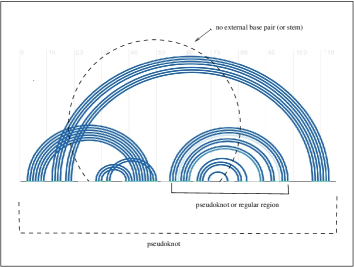
The definition of a recursive pseudoknot follows the one of Wong et al. [30]. A recursive pseudoknot is a pseudoknot in a region that contains a pseudoknotted or regular region , , and there does not exist a base pair , such that is a base of , and is a base of external to (see Fig. 3).
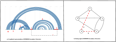
A pseudoknotted block can be classified as recursive by determining the edge-connectivity of the block. The edge-connectivity is defined as the minimum number of edges that if they are deleted then the resulting graph is disconnnected. As an example consider the Hepatitis Delta Virus Ribozyme (see Fig. 4), necessary for viral replication. The stem labeled in the graphical representation (or vertex labeled in the dual graph) is attached to the pseudoknot by the segments and in its graphical representation, or edges labeled and in the dual graph representation. As by deleting two edges in the dual graph, vertex labeled becomes disconnected, then stem is a recursive region of the PK, thus the dua graph edge-connectivity is . The proof of the following lemma was shown in [27].
Lemma 1
The dual graph representation of a pseudoknotted block is recursive if and only if the block has edge-connectivity 2.
The edge-connectivity of a graph can be determined in polynomial time in order using the max-flow min-cut theorem of network flows by Edmond and Karp [6], or Ford and Fulkerson [8].
We can also delete each pair of edges and determine if the graph is disconnected using Depth-First-Search [12] in time ; this variation allows us to find every internal recursive region of the recursive pseudoknot if such pair of edges exist.
The following is the partitioning and classification of pseudoknots algorithm.
Algorithm 2.2
Partitioning and Classification of PKs
-
i.
Input dual graph as the Adjacency Matrix, of a RNA 2D.
-
ii.
Output partitioning of the RNA 2D into recursive PK, non-recursive PK, and regular regions.
-
1.
Partition the dual graph into blocks by application of Hopcroft and Tarjan’s algorithm;
-
2.
Analyze each block to determine whether each contains a vertex of degree at least 3;
-
3.
IF the block has a vertex of degree then the block is a pseudoknot;
-
–
Apply max-flow min-cut theorem to determine edge-connectivity;
-
–
if edge-connectivity = 2 then the block is a recursive pseudoknot;
else the pseudoknot is not recursive;
-
–
-
4.
ELSE the block is a regular region;.
-
1.
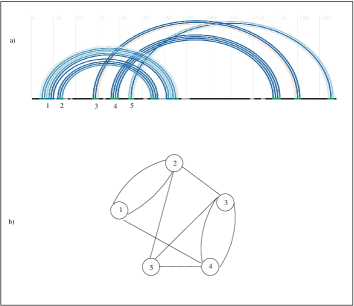
As an example of a non-recursive pseudoknot consider the Translational repression of the Escherichia coli alpha operon mRNA ([29]), illustrated in Fig. 5. The dual graph representation of this motif 2D has edge-connectivity 3, thus it is not a recursive PK.
The Appendix illustrates the output generated by the modified algorithm when is run on some of the aforementioned motifs. The algorithm is written in C++ and is archived for public use [28].
3 Classification of H, K, L, M pseudoknots types
In the classical literature, pseudoknots have been classified and predicted by folding algorithms into four types: H,K,L, and M [22]. Even though each type is defined in terms of how few stems intertwined, pseudoknots can be complex structures, recursive, and be comprised of several stems. In this paper we show based on dual directed graphs, each PK type can be identified through a series of reductions to an unique representative. This methodology will allow us to systematically analyze thousands of motifs and develop more precise RNA folding algorithms. The results and algorithms discussed in Section 2.2 and Section 2.3, based upon undirected dual graphs, can be easily extended to dual directed graphs, as the direction of a directed edge can be ignored.
In this section we follow the definitions of pseudoknots as by the papers of Kucharik et al [22] and Antczak et al. [1].
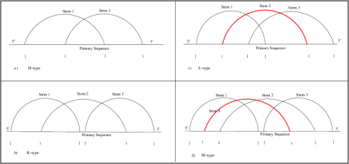
A H-type pseudoknot occurs when a nucleotide of a loop or bulge pairs with a nucleotide of a single-stranded region outside the loop; this type can be alternatively illustrated from the graphical representation (see Fig. 6-a) by the intertwining of two stems.
A K-type PK results when two nucleotides from different loops (or bulges) pair to form a double helical segment (see Fig. 6-b). Similarly we can also describe L and M types pseudoknots, derived from the H and K types, respectively, by addition of a stem (colored red) as shown in Figure 6-c and Figure 6-d.
Even though the different PK types are defined in terms of few stems, each type may be composed of several stems and also they could be recursive (see section 2.3). For example Figure 7 illustrates two H-type PKs, PKB70 (i.e., PK PK4 of Legionella pneumophilia tmRNA), causative agent of the Legionannary’s disease [21], composed of 5 stems, and recursive PKB259 (i.e., potato yellow vein virus [27]) comprising a recursive region and 3 stems. Even though two PKs of the same type of maybe structurally different, in the following section we show how using dual digraphs each motif type can be reduced to a unique digraph type-representative.


3.1 Dual digraphs and reductions to unique PK type identifiers
In this section we used dual digraphs and based upon two graph-theoretical transformations, these digraphs can be reduced to a unique representative identifying H,K,L, and M pseudoknot types.
A dual digraph can be easily derived from the graphical representation of a RNA 2D structure: each stem is modeled by a vertex of the dual digraph, and following the primary sequence in linear order (i.e., from the 5’ end to the 3’ end), a segment between stems and is represented by an directed edge in the dual digraph. Moreover each directed edge is assigned a weight corresponding to the order in which the segment is reached following the primary sequence (see Fig. 7-b); this weight is represented by .
From a RNA 2D perspective, we informally define a super-stem as a stem that completely contains another stem (sub-stem). Formally a super-stem is a -tuple , ; in which forms a base-pair (see definition 1-d), if there exist another stem (i.e., sub-stem) in the region , , and . For example in Stem shown is Figure 7-a (i.e., PKB70) is a super-stem of and , and stem is a super-stem of ; similarly stem is a super-stem with respect to . For ease of notation let represent the case when is a super-stem of . The following proposition describes how a super-stem and its corresponding sub-stem can be detected in a dual digraph,
Proposition 1
If there are exist exactly two anti-parallel directed edges and between vertices and of the dual digraph, with , then is a super-stem of .
Proof
Without loss of generality, let . Since , implies that if there is either an eternal stem , intertwining and , or, forms a self-loop; if there exist an external stem intertwining and , this stem does not intercept the primary sequence between and . If , then is not fully contained in (i.e., intertwines ).
As an example consider stems labeled and for PKB270 shown in Figure 7-a. In the corresponding dual digraph (Fig. 7-b) the vertices labeled and are connected by exactly two anti-parallel edges with . Please note that the converse it is not necessarily true, that is, if is a super-stem of , then there exist exactly two anti-parallel edges between vertices and in the dual digraph with ; for example in PKB259 (Fig. 7-e), stem is a super-stem of , however there are no anti-parallel edges between the corresponding vertices in the dual digraph (Fig. 7-f). This has to do with the fact that another stem (i.e., ) intercepts the primary sequence between and . From Proposition 1 we introduce the following graph reduction,
Reduction 3.1
Let be a dual digraph.If there are exist exactly two anti-parallel directed edges and between vertices and of , with , then transform into , where and are identified into a single vertex and any directed edge in , , , or , will result in an directed edge in , while keeping the same weights from the edges of . Similarly an edge , or in will have the corresponding edge in , while maintaining the same weights on the original edges (see Fig. 7-b-c).
A recursive PK can be classified by our partitioning algorithm and a recursive fragment can be identified (see Section 2.2). For example in the graphical representation of PKB259 (Fig. 7-e), segments and isolate the recursive fragment Stem , or equivalently, deleting exactly two edges of the dual digraph (connectivity 2), and , identified by Algorithm 2.2, disconnects the dual digraph. The following graph reduction follows from the algorithm,
Reduction 3.2
Let be a dual digraph, and be a recursive fragment adjacent to Stem by direct segment from Stem to , and by segment from to Stem (i.e., deleting segments and isolates ), then transform into , by deleting segments and , recursive fragment , and by inserting a direct segment from to (see Fig. 7-e-f).
4 Conclusions and Ongoing Work
The Covid-19 pandemic accelerated the study of viral replication and the need to develop therapeutics to control infectivity. For example, a single mutation of a single nucleotide in the frameshifting region of the SARS-CoV-2 RNA results in a concomitant obliteration of viral replication. The importance of the three-stemmed pseudoknot-dependent ribosomal frameshifting for the propagation of SARS-related coronaviruses it is well-established. We suggest that pseudoknots not only play a significant role, but a predominant one in viral transmissibility for most viruses, and our proposed techniques aim to shed some light in this area, as well to better understand the roles of PKs in general RNA functionality.
5 Appendix
Let represents an edge of a dual graph with end-vertices and .
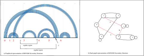
We are next illustrating the output of the partitioning algorithm tested on the tRNA-like-structure dual graph (see Fig. 9).
——————— Motif :RF01084 —————————–
===================== New Block =======
(4,5) - (4,5) -
—- this block represents a regular-region —-
===================== New Block =======
(4,0) - (3,4) - (1,3) - (6,0) - (2,6) - (1,2) - (1,2) - (0,1) -
removed edges (2,6) and (6,0), these two edges are a disconnecting set:
The block is a recursive PK.
———– Summary information for Motif :RF01084 ——————————–
———– Total number of blocks: 2
———– number of non-recursive PK blocks: 0
———– number of recursive PK blocks: 1
———– number of regular blocks : 1
————————————————————————————
Acknowledgments
The work of L. P. was supported by
PSC-CUNY Award 61249-00-49 of the City University of New York.
References
- [1] Antczak, M., Popenda, M., Zok, T., Zurkowski, M., Adamiak, R.-W, and Szachniuk, M.: New algorithms to represent complex pseudoknotted RNA structures in dot-bracket notation. Bioinformatics 34 (8): 1304-1312 (2018).
- [2] Barends, S., Rudinger-Thirion, J., Florentz, C., Giegé, R., Pleij, C., Kraal, B.: tRNA-Like Structure Regulates Translation of Brome Mosaic Virus RNA. J Virol. 78 (8): 4003–4010 (2004).
- [3] Brierley, I., Pennell, S., Gilbert, R.-J.: Viral RNA pseudoknots: versatile motifs in gene expression and replication. Nat Rev. Microbiol. 5 (8): 598-610 (2007).
- [4] Condon, A., Davy, B., Rastegari, B., Zhao, S., Tarrant, F.: Classifying RNA pseudoknotted structures. Theoretical Computer Science 320(1), 35–50 (2004).
- [5] Dirks, R.-M., and Pierce, N.-A.: A partition function algorithm for nucleic acid secondary structure including pseudoknots. J. Comput. Chem. 24(13), 1664–1677 (2003).
- [6] Edmonds, J., Karp, R.-M.: Theoretical improvements in algorithmic efficiency for network flow problems. Journal of the ACM 19 (2), 248–264 (1972).
- [7] Fera, D., Kim, N., Shiffeldrim, N., Zorn, J., Laserson, U., Gan, H.-H., Schlick, T.: RAG: RNA-As-Graphs web resource. BMC Bioinformatics 5, 88 (2004).
- [8] Ford, L. R., Fulkerson, D. R.: Maximal flow through a network. Canadian Journal of Mathematics 8, 1956, 399–404 (1956).
- [9] Gan, H.-H., Pasquali, S., Schlick, T.: Exploring the repertoire of RNA secondary motifs using graph theory; implications for RNA design. Nucleic Acids Res. 31(11), 2926–2943 (2003).
- [10] Gan, H.-H., Fera, D., Zorn, J., Shiffeldrim, N., Tang, M., Laserson, U., Kim, N., Schlick, T.: RAG: RNA-As-Graphs database–concepts, analysis, and features, Bioinformatics 20(8), 1285–1291 (2004).
- [11] Han, B., Dost, B., Bafna, V., Zhang, S.: Structural Alignment of Pseudoknotted RNA. Journal of Computational Biology 15(5), 489–504 (2008).
- [12] Harary, F.:Graph Theory, Addison-Wesley, Mass. 1969.
- [13] Hopcroft, J., Tarjan, R.: Efficient algorithms for graph manipulation. Communications of the ACM 16(6), 372–378 (1973).
- [14] Izzo, J.-A., Kim, N., Elmetwaly, S., Schlick, T.: RAG: an update to the RNA-As-Graphs resource. BMC Bioinformatics 12, 219 (2011).
- [15] Jain, S., Bayrak, C.-S., Petingi, L., Schlick, T.: Dual Graph Partitioning Highlights a Small Group of Pseudoknot-Containing RNA Submotifs. Genes 9(8), 371 (2018).
- [16] Jain, S., Ramos, S.-B., Laederach, A., Schlick, T.: A Pipeline for Computational Design of Novel RNA-Like Topologies. NAR 46(14), (2018).
- [17] Jain, S., Saju, S., Petingi, L., Schlick, T.: New Dual Graph Library for Studying RNA Graphs and Subgraphs, Accepted with minor revisions, to appear in Methods, 2019.
- [18] Jules M., Buchrieser C.: Legionella pneumophila adaptation to intracellular life and the host response: clues from genomics and transcriptomics, FEBS Letters. 581(15), 2829–2838 (2007).
- [19] Kucharik, M., Hofacker, I.-L., Stadler, P.-F., Qin, J.: Pseudoknots in RNA folding landscapes, Bioinformatics 32(2), 187–194 (2016).
- [20] Pesells, A., Serganov, A.: Structure and function of pseudknots involved in gene expression control. Wiley Interdiscip. Rev. RNA 5(6), 803–822 (2014).
- [21] Kim, N., Petingi, L., Schlick, T.: Network Theory Tools for RNA Modeling. WSEAS Transactions on Math. 12(9), 941–955 (2013).
- [22] Kravchenko, A.: Predicting RNA Secondary Structures Including Pseudoknots. University of Oxford internal report, (2009).
- [23] Lai, D., Proctor, J.-R., Zhu, J.-Y., Meyer, I.-M.:R- chie : a web server and R package for visualizing RNA secondary structures, Nucleic Acids Research 40(12), e95 (2012). e-RNA Website, https://www.e-rna.org/r-chie/rfam.cgi. Last accessed 4 January 2019.
- [24] Livieratos, I.-C. et al.: Analysis of the RNA of Potato yellow vein virus: evidence for a tripartite genome and conserved 3’-terminal structures among members of the genus Crinvirus, J. Gen. Virol. 85, 2065–2075 (2004). e-RNA Website, https://www.e-rna.org/r-chie/rfam.cgi. Last accessed 4 January 2019.
- [25] Petingi, L.: Identifying and Analyzing Pseudoknots based on Graph-Theoretical Properties of Pseudoknots: A Partitioning Approach, CUNY Graduate Center Academic Works. Internal Report (2015).
- [26] Petingi, L.: A Graph-Theoretical Approach for Partitioning RNA Secondary Structures into Pseudonotted and Pseudoknot-free Regions. Proceedings of the World Congress on Engineering and Computer Science 2016 (WCECS 2016) II, October 19-21, 2016. Best Paper Award
- [27] Petingi, L., Schlick, T.: Graph-Theoretic Partitioning of RNAs and Classification of Pseudoknots. In: Holmes I., Martín-Vide C., Vega-Rodríguez M. (eds) Algorithms for Computational Biology. AlCoB 2019. Lecture Notes in Computer Science, vol 11488. Springer, Cham
- [28] Petingi, L.: Dual Graph Partitioning Code. https://github.com/Louis-Petingi/Partition-Algorithm-3. Last accesed 3 January, 2019.
- [29] Schlax, P-J., Xavier, K.-A., Gluick, T.-C., Draper D-E.: Translational repression of the Escherichia coli alpha operon mRNA: importance of an mRNA conformational switch and a ternary entrapment complex. J Biol Chem. 276 (42), 38494–38501 (2001).
- [30] Wong, T.-K., Lam, T.-W., Sung, W.-K., Cheung, B.-W., Yiu, S.-M.: Structural alignment of RNA with complex pseudoknot structure. J Comput Biol. 18(1), 97–108 (2011).