Multi-Slice Dense-Sparse Learning for Efficient Liver and Tumor Segmentation
Abstract
Accurate automatic liver and tumor segmentation plays a vital role in treatment planning and disease monitoring.
Recently, deep convolutional neural network (DCNNs) has obtained tremendous success in 2D and 3D medical image segmentation.
However, 2D DCNNs cannot fully leverage the inter-slice information, while 3D DCNNs are computationally expensive and memory intensive.
To address these issues, we first propose a novel dense-sparse training flow from a data perspective, in which, densely adjacent slices and sparsely adjacent slices are extracted as inputs for regularizing DCNNs, thereby improving the model performance.
Moreover, we design a 2.5D light-weight nnU-Net from a network perspective, in which, depthwise separable convolutions are adopted to improve the efficiency.
Extensive experiments on the LiTS dataset have demonstrated the superiority of the proposed method.
Clinical relevance— The proposed method can effectively segment livers and tumors from CT scans with low complexity, which can be easily implemented into clinical practice.
I INTRODUCTION
Liver cancer is life-threatening and one of the most dangerous tumors to human health [1]. Computed tomography (CT) is one of the most effective non-invasive diagnostic imaging procedures to help doctors detect and characterize liver lesions [2]. Moreover, accurate localization and segmentation of liver and lesions is a crucial step for clinical diagnosis and surgical planning [3]. However, in routine clinical practice, manually segmenting liver and lesions from CT scans is time-consuming and error-prone. Therefore, automatic computer-aided segmentation methods are urgently needed.
In recent years, deep learning has advanced the development of computer-aided diagnosis [4]. In particular, fully convolutional networks (FCNs) [5, 6], e.g., 2D and 3D FCNs, have achieved promising performance for medical image segmentation [7, 8, 9, 10]. 2D FCNs have achieved good segmentation results in many medical imaging fields [11], and are broadly implemented for liver and tumor segmentation in 2D CT slices [7, 8, 12]. However, 2D FCNs ignore the inter-slice features in 3D volumetric CT scans, which limits the segmentation performance. On the other hand, replacing 2D convolutions with 3D ones, 3D FCNs are capable to explore the inter-slice correlations and learn deep 3D representations with volumetric inputs, thereby obtaining more reliable results [13]. Regardless of the accuracy, high computational complexity and cost of 3D FCNs impede the broader clinical use. To solve this issue, much research effort has been devoted to maintaining an accuracy-complexity balance [14, 15, 16], but most of them focus on constructing hybrid architectures using 2D and 3D convolutions together.
In this work, we first design a novel learning strategy from a data perspective, termed as Dense-Sparse learning, in which, two types of adjacent slices, i.e., densely adjacent slices and sparsely adjacent slice, are feed into 2D FCNs to probe the inter-slice information with different strides. Furthermore, traditional convolutions in 2D nnU-Net [17] are replaced with depthwise separable convolutions from the perspective of network to form a 2.5D light-weight nnU-Net for improving the efficiency. We extensively evaluate the proposed method on the LiTS dataset [18]. Experimental results demonstrate that the proposed method can achieve comparable performance on liver and tumor segmentation with much fewer parameters than 3D nnU-Net.
II RELATED WORK
In the past decades, deep learning has received much attention on various computer vision tasks, such as classification and segmentation [19]. A lot of methods based on deep convolutional neural network (DCNNs) have been proposed for liver and tumor segmentation [7, 12, 20, 6]. Ben-Cohen et al. [7] present an fully convolutional network (FCN) for liver and tumor segmentation. Chlebus et al. [12] propose a 2D U-Net with object-based post-processing, obtaining high performance in liver and tumor segmentation. Vorontsov et al. [20] employ two parallel U-Nets [6] for joint liver and tumor segmentation. These methods take 2D slices as inputs and thus ignore the contextual information between slices, limiting the segmentation performance, while 3D FCNs [21, 22, 23] are supposed to consider the inter-slice information, obtaining better segmentation results than 2D models. For instance, Dou et al. [21] introduce a deep supervision mechanism into a 3D FCN for boosting the segmentation performance. Nevertheless, training such 3D FCNs is time-consuming and memory-consuming, which limits the wide applications of 3D models.
To probe the inter-slice information while reducing the computational complexity, many methods from different perspectives have been proposed, including 2.5D models [24, 16] and hybrid 2D–3D models [14, 15]. For example, in [24], adjacent slices from 3D CT scans are used to train a 2.5D FCN, while Li et al. [14] design a hybrid densely connected UNet (H-DenseUNet) to jointly explore intra-slice and inter-slice features. These methods alleviate the problems of 2D and 3D models to a certain extent. Differently, we consider both data and network perspectives in our framework for more efficient liver and tumor segmentation.
III METHODOLOGY
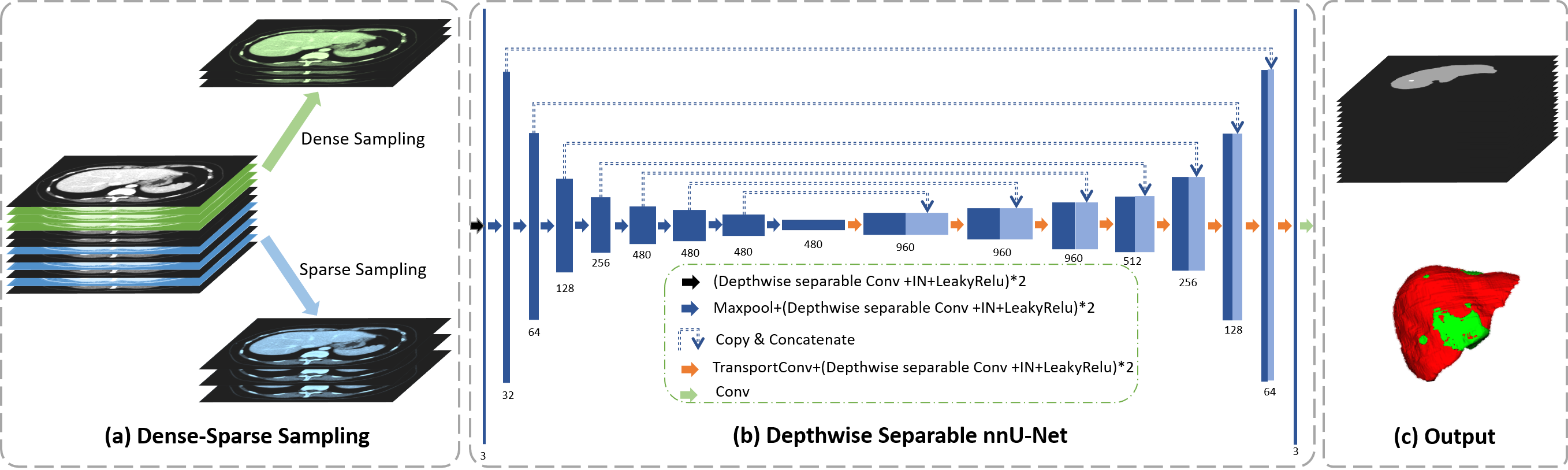
The proposed framework is presented in Fig. 1. Firstly, Dense-Sparse (DS) sampling strategy is used to generate two different inputs, i.e., densely adjacent slices and sparsely adjacent slices, for improving the model performance. Then, a 2.5D nnU-Net with depthwise separable convolutions (DS nnU-Net) is implemented as the segmentation model, in which, all convolutional layers are replaced with depthwise separable convolutions to reduce the computational complexity. We further design a two-stage dense-sparse-dense (DSD) training strategy, in which, densely adjacent slices and sparse adjacent slices are randomly selected to train the network at the first stage for fast convergence, while only densely adjacent slices are fed into the network at the second stage to improve the model robustness.
III-A Dense-Sparse Sampling
Let denote the training samples of height , width and thickness , where is the batch size. For a 2D network, only one slice () is used to generate the segmentation mask, lacking the context information for volumetric medical image segmentation. In contrast, a 3D network takes the whole volumes (all slices) as inputs, suffering from high computational costs. In common, a 2.5D network uses a stack of continuous slices during training and generates the segmentation mask for the central slice at the inference stage. For instance, if , the densely adjacent slices would be the input of the 2.5D network to produce a 2D mask for slice . The compromise can avoid high GPU memory consumption of 3D convolutions while providing inter-slice information. Beyond the densely adjacent slices, we propose a novel dense-sparse sampling method to generate densely adjacent slices and sparsely adjacent slices, as shown in Fig. 1 (a).
For the sake of simplicity, we take as an illustration. Let be the slice indexed by in a CT scan , where . In dense-sparse (DS) sampling, is a 3D input of thickness , which is described as:
| (1) |
where is integer division, and is the stride of sampling. In the case of edge slices, slices extending beyond the volume were repeated. DS sampling shares the similar spirit of the convolution kernels, in which, the stride in two dimensions is for the height and the width movement, while the stride of DS sampling is for the movement along the dimension of thickness. More specifically, when , densely adjacent slices will be generated, while is for dense sampling to produce sparsely adjacent slices . With and together, the network can learn inter-slice features efficiently. In our experiments, we set and for dense sampling and sparse sampling, respectively.
III-B Depthwise Separable nnU-Net
nnU-Net [17] is a self-adapting framework based on generic U-Net architectures, such as 2D and 3D U-Net. With various kinds of data augmentation, nnU-Net makes full use of the potentials of U-Net. Considering the memory constraints and negative impact from cropped patches for 3D network training, we select 2D nnU-Net as our backbone and design a depthwise separable nnU-Net (DS nnU-Net), which employ depthwise separable convolutions instead of standard convolution layers to further ease the computational burden, as shown in Fig. 1 (b).
Let us consider a convolutional layer with an input feature map and output feature map , where , and are spatial width, spatial height and the number of channels, respectively. The output of a standard convolution layer with kernel can be formatted as:
| (2) |
A depthwise separable (DS) convolution can be split into 2 separate kernels, i.e., the depthwise convolution and the pointwise convolution. In depthwise convolution, each kernel iterates one channel of the input feature map . The output the depthwise convolution of can be computed as
| (3) |
Following that, the pointwise convolution , also known as convolution, is applied to increase the number of channels. The mathematical formulation is
| (4) |
If we have a convolution with input channel and output channel , for standard convolution, it has parameters, while the depthwise separable convolution contains parameters, which approximately reduce the computational complexity by a factor of . We replace all standard convolutions with DS convolutions in our DS nnU-Net, which has only million (M) parameters, while 2D nnU-Net has more than M parameters.
III-C Dense-Sparse-Dense Training
Since densely adjacent slices and sparsely adjacent slices have different views of the data, which may cause negative transfer across views. To this end, we proposed a two-step progressive learning strategy, namely Dense-Sparse-Dense (DSD) training to facilitate model optimization. In the first (Dense-Sparse) step, we randomly input densely adjacent slices and sparsely adjacent slices to train and regularize the network for fast convergence. In the second (Dense) step, we retrain the network with all densely adjacent slices for increasing the model capacity without overfitting.
IV EXPERIMENTS
IV-A Dataset and Experimental Settings
We evaluate the proposed method on the Liver Tumor Segmentation (LiTS) dataset [18], which includes CT scans ( for training and for testing). Since the ground truths of testing data are not publicly available, for a fair comparison, we randomly select volumes for training and the remaining for testing in our experiments. The CT volumes are resized to multiple slices of size after resampling and normalization. Following the settings in nnU-Net, we train the network with the combination of cross-entropy loss and dice loss. We implement DS sampling with thickness . For DSD training, we set and epochs for step and step, respectively.
According to the evaluation procedures of the LiTS challenge, we evaluated the liver and tumor segmentation performance using the only golden indicator, Dice per case score, which refers to an average of Dice per volume score.
We compare our method with 2D nnU-Net and 3D nnU-Net with full resolution. Besides, to validate the effectiveness of different components of our pipeline, the following variants are evaluated.
-
•
nnU-Net-DS: 2D nnU-Net with the proposed dense-sparse sampling.
-
•
nnU-Net-DSD: 2D nnU-Net with the proposed dense-sparse-dense training strategy.
-
•
DS nnU-Net-DSD: Depthwise Separable nnU-Net with the proposed dense-sparse-dense training strategy.
| Method | Lesion | Liver | Params (M) |
| Dice per case (mean (std), %) | |||
| 2D nnU-Net | 0.801 (0.024) | 0.960 (0.004) | 41 |
| 3D nnU-Net | 0.827 (0.055) | 0.965 (0.004) | 37 |
| nnU-Net-DS | 0.804 (0.041) | 0.962 (0.002) | 41 |
| nnU-Net-DSD | 0.815 (0.032) | 0.963 (0.003) | 41 |
| DS nnU-Net-DSD | 0.814 (0.025) | 0.962 (0.003) | 7 |
IV-B Results and Discussions
The experimental results are shown in Table I. For a fair comparison, we train all models with epochs. Apparently, 3D nnU-Net outperforms 2D nnU-Net on liver and tumor segmentation because it randomly samples 3D patches from CT volumes, which is capable to capture 3D contextual information for improved performance. 3D nnU-Net has fewer layers and parameters than 2D nnU-Net, but more training and inference time is required. Although 3D networks can effectively improve segmentation performance, it is also important to pay attention to model training. It is well noted that the proposed dense-sparse sampling can help improve the performance of 2D nnU-Net on liver and tumor segmentation. With DS sampling and DSD training strategy together, 2D nnU-Net achieved comparable performance with 3D nnU-Net. The results have demonstrated the effectiveness of the proposed DS sampling and DSD training strategy for improving segmentation performance without carefully modified architectures. On the other hand, we implemented DS nnU-Net with the proposed sampling and training strategies. It is observed that there is no significant performance degradation on liver and tumor segmentation with much fewer parameters, which is around of 2D nnU-Net. Furthermore, the training time is shortened by around on a single RTX 2020ti GPU.
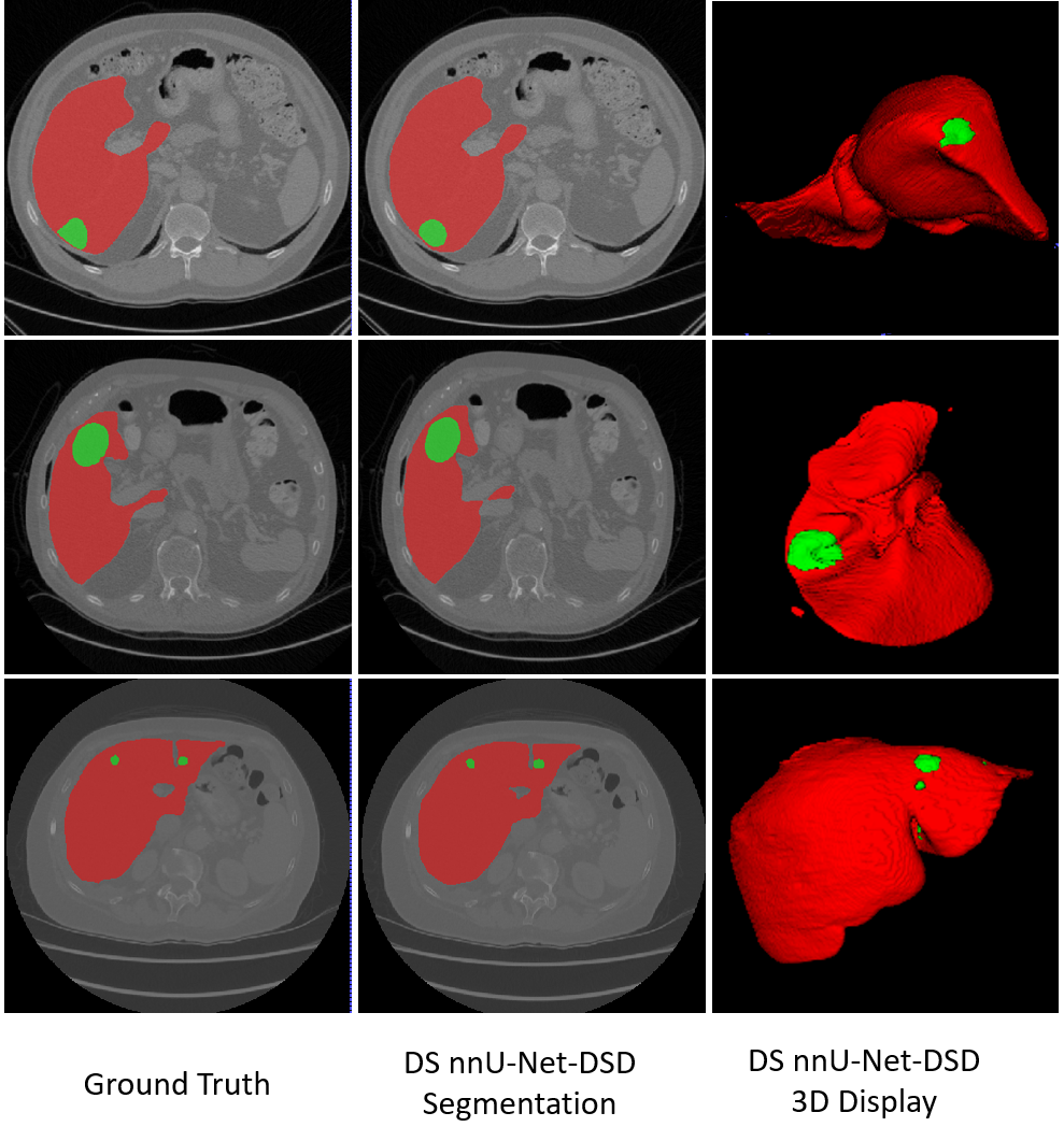
Fig. 2 shows the visualization results of our method DS nnU-Net-DSD. We can see that the masks of our method are close to the ground truth labels, which further shows the feasibility of the proposed method for efficient liver and lesion segmentation.
V CONCLUSIONS
In this work, we design a novel end-to-end deep learning framework from both perspectives of data and network for liver and tumor segmentation. Extensive experiments show that the proposed approach can obtain accurate segmentation results, as well as speed up the training and inference process with only M parameters. Moreover, ablation studies demonstrate the effectiveness of different components in our framework. In the future, we will implement the proposed method in other medical image segmentation tasks to evaluate the generalization capability.
References
- [1] Forner and Alejandro, “Hepatocellular carcinoma surveillance with mirnas,” Lancet Oncology, vol. 16, no. 7, pp. 743–745, 2015.
- [2] Kaixin Xu, Ziyuan Zhao, Jiapan Gu, Zeng Zeng, Chan Wan Ying, Lim Kheng Choon, Thng Choon Hua, and Pierce KH Chow, “Multi-instance multi-label learning for gene mutation prediction in hepatocellular carcinoma,” in 2020 42nd Annual International Conference of the IEEE Engineering in Medicine Biology Society (EMBC), 2020, pp. 6095–6098.
- [3] Jiapan Gu, Ziyuan Zhao, Zeng Zeng, Yuzhe Wang, Zhengyiren Qiu, Bharadwaj Veeravalli, Brian Kim Poh Goh, Glenn Kunnath Bonney, Krishnakumar Madhavan, Chan Wan Ying, Lim Kheng Choon, Thng Choon Hua, and Pierce K. H. Chow, “Multi-phase cross-modal learning for noninvasive gene mutation prediction in hepatocellular carcinoma,” in 2020 42nd Annual International Conference of the IEEE Engineering in Medicine Biology Society (EMBC), 2020, pp. 5814–5817.
- [4] Mohammad Hesam Hesamian, Wenjing Jia, Xiangjian He, and Paul Kennedy, “Deep learning techniques for medical image segmentation: achievements and challenges,” Journal of digital imaging, vol. 32, no. 4, pp. 582–596, 2019.
- [5] Jonathan Long, Evan Shelhamer, and Trevor Darrell, “Fully convolutional networks for semantic segmentation,” IEEE Transactions on Pattern Analysis and Machine Intelligence, vol. 39, no. 4, pp. 640–651, 2015.
- [6] Olaf Ronneberger, Philipp Fischer, and Thomas Brox, “U-net: Convolutional networks for biomedical image segmentation,” in International Conference on Medical Image Computing and Computer-Assisted Intervention, 2015.
- [7] Avi Ben-Cohen, Idit Diamant, Eyal Klang, Michal Amitai, and Hayit Greenspan, “Fully convolutional network for liver segmentation and lesions detection,” in Deep learning and data labeling for medical applications, pp. 77–85. Springer, 2016.
- [8] Patrick Ferdinand Christ, Mohamed Ezzeldin A Elshaer, Florian Ettlinger, Sunil Tatavarty, Marc Bickel, Patrick Bilic, Markus Rempfler, Marco Armbruster, Felix Hofmann, Melvin D’Anastasi, et al., “Automatic liver and lesion segmentation in ct using cascaded fully convolutional neural networks and 3d conditional random fields,” in International Conference on Medical Image Computing and Computer-Assisted Intervention. Springer, 2016, pp. 415–423.
- [9] Yuwei Pang, Dong Hu, and Min Sun, “A modified scheme for liver tumor segmentation based on cascaded fcns,” in Proceedings of the International Conference on Artificial Intelligence, Information Processing and Cloud Computing, 2019, pp. 1–6.
- [10] Shenhai Zheng, Bin Fang, Laquan Li, Mingqi Gao, Yi Wang, and Kaiyi Peng, “Automatic liver tumour segmentation in ct combining fcn and nmf-based deformable model,” Computer Methods in Biomechanics and Biomedical Engineering: Imaging & Visualization, vol. 8, no. 5, pp. 468–477, 2020.
- [11] Zaiwang Gu, Jun Cheng, Huazhu Fu, Kang Zhou, Huaying Hao, Yitian Zhao, Tianyang Zhang, Shenghua Gao, and Jiang Liu, “Ce-net: Context encoder network for 2d medical image segmentation,” IEEE transactions on medical imaging, vol. 38, no. 10, pp. 2281–2292, 2019.
- [12] Grzegorz Chlebus, Andrea Schenk, Jan Hendrik Moltz, Bram van Ginneken, Horst Karl Hahn, and Hans Meine, “Automatic liver tumor segmentation in ct with fully convolutional neural networks and object-based postprocessing,” Scientific reports, vol. 8, no. 1, pp. 1–7, 2018.
- [13] Qing Huang, Jinfeng Sun, Hui Ding, Xiaodong Wang, and Guangzhi Wang, “Robust liver vessel extraction using 3d u-net with variant dice loss function,” Computers in biology and medicine, vol. 101, pp. 153–162, 2018.
- [14] Li Xiaomeng, Chen Hao, Qi Xiaojuan, Dou Qi, Fu Chi-Wing, and Heng Pheng-Ann, “H-denseunet: Hybrid densely connected unet for liver and liver tumor segmentation from ct volumes,” IEEE Transactions on Medical Imaging, pp. 1–1, 2017.
- [15] Jianpeng Zhang, Yutong Xie, Pingping Zhang, Hao Chen, Yong Xia, and Chunhua Shen, “Light-weight hybrid convolutional network for liver tumor segmentation.,” in IJCAI, 2019, pp. 4271–4277.
- [16] Girindra Wardhana, Hamid Naghibi, Beril Sirmacek, and Momen Abayazid, “Toward reliable automatic liver and tumor segmentation using convolutional neural network based on 2.5 d models,” International journal of computer assisted radiology and surgery, vol. 16, no. 1, pp. 41–51, 2021.
- [17] Fabian Isensee, Paul F Jaeger, Simon A A Kohl, Jens Petersen, and Klaus H Maier-Hein, “nnu-net: a self-configuring method for deep learning-based biomedical image segmentation,” Nature methods, vol. 18, no. 2, pp. 203—211, February 2021.
- [18] Patrick Bilic, Patrick Ferdinand Christ, Eugene Vorontsov, Grzegorz Chlebus, Hao Chen, Qi Dou, Chi-Wing Fu, Xiao Han, Pheng-Ann Heng, Jürgen Hesser, et al., “The liver tumor segmentation benchmark (lits),” arXiv preprint arXiv:1901.04056, 2019.
- [19] Geert Litjens, Thijs Kooi, Babak Ehteshami Bejnordi, Arnaud Arindra Adiyoso Setio, Francesco Ciompi, Mohsen Ghafoorian, Jeroen Awm Van Der Laak, Bram Van Ginneken, and Clara I Sánchez, “A survey on deep learning in medical image analysis,” Medical image analysis, vol. 42, pp. 60–88, 2017.
- [20] Eugene Vorontsov, An Tang, Chris Pal, and Samuel Kadoury, “Liver lesion segmentation informed by joint liver segmentation,” in 2018 IEEE 15th International Symposium on Biomedical Imaging (ISBI 2018). IEEE, 2018, pp. 1332–1335.
- [21] Qi Dou, Hao Chen, Yueming Jin, Lequan Yu, Jing Qin, and Pheng-Ann Heng, “3d deeply supervised network for automatic liver segmentation from ct volumes,” in International conference on medical image computing and computer-assisted intervention. Springer, 2016, pp. 149–157.
- [22] Fang Lu, Fa Wu, Peijun Hu, Zhiyi Peng, and Dexing Kong, “Automatic 3d liver location and segmentation via convolutional neural network and graph cut,” International journal of computer assisted radiology and surgery, vol. 12, no. 2, pp. 171–182, 2017.
- [23] Peijun Hu, Fa Wu, Jialin Peng, Ping Liang, and Dexing Kong, “Automatic 3d liver segmentation based on deep learning and globally optimized surface evolution,” Physics in Medicine & Biology, vol. 61, no. 24, pp. 8676, 2016.
- [24] Xiao Han, “Automatic liver lesion segmentation using a deep convolutional neural network method,” arXiv preprint arXiv:1704.07239, 2017.