Hydrodynamic Manipulation of Nano-Objects by Thermo-Osmotic Flows
Abstract
The manipulation of micro- and nano-objects is of great technological significance to construct new materials, manipulate tiny amounts of liquids in fluidic systems, or detect minute concentrations of analytes. It is commonly approached by the generation of potential energy landscapes, for example, with optical fields. Here we show that strong hydrodynamic boundary flows enable the trapping and manipulation of nano-objects near surfaces. These thermo-osmotic flows are induced by modulating the van der Waals interaction at a solid-liquid interface with optically induced temperature fields. We use a thin gold film on a glass substrate to provide localized but reconfigurable point-like optical heating. Convergent boundary flows with velocities of tens of micrometres per second are observed and substantiated by a quantitative physical model. The hydrodynamic forces acting on suspended nanoparticles and attractive van der Waals or depletion induced forces enable precise positioning and guiding of the nanoparticles. Fast multiplexing of flow fields further provides the means for parallel manipulation of many nano-objects. Our findings have direct consequences for the field of plasmonic nano-tweezers as well as other thermo-plasmonic trapping schemes and pave the way for a general scheme of nanoscopic manipulation with boundary flows.
I Introduction
The control and manipulation of nano-objects is a key element for future nanophotonics [1, 2, 3, 4, 5], material science [6, 4, 7], biotechnology [2, 8, 9] or even quantum sensing [10]. Analytes dissolved in liquids, for example, need to be delivered, concentrated, separated or locally confined for further studies to become eventually processed and removed. Photonic elements including plasmonic nano-structures require precise positioning or controlled rearrangements to serve as adaptive functional structures. Key elements of the control at the micro- and nanoscale are often pressure driven fluidics transporting liquid volume and solutes as well as the generation of energy landscapes or force fields. The latter is achieved with optical [3] and plasmonic tweezers [11, 12], magnetic fields [13], or using electrokinetic [14] or opto-electronic [15] effects. Especially in the field of plasmonic tweezers and nanoantennas where light is used to excite collective electron motion in noble-metals, the Joule losses lead to the unavoidable generation of heat at boundaries as an unwanted side effect [16, 17]. Yet, such optically generated temperature fields seem also suitable for the manipulation of nano-objects in liquids, for example, for the trapping of nanoparticles [18] and single molecules [19] or protein aggregates [20] as well as for manufacturing active particles [21, 22, 23, 24]. Those techniques rely on a drift of molecules and particles in optically generated temperature gradients termed thermophoresis or suggest thermo-electric effects [25] relying on a thermally induced charge separation. In addition, thermo-electrohydrodynamic effects using time-varying electric fields have been proposed for rapid particle transport [26, 27] and convective effects that arise from temperature-induced density changes in the large liquid cells have been reported [28, 29, 30, 31].
Here we report on a fundamental physical process that is able to provide a versatile trapping and manipulation of nano-objects near surfaces in the simplest geometries. Contrary to most other techniques, our scheme is based on hydrodynamic flows generated by thermo-osmosis. Thermo-osmosis relies on a perturbation of the interfacial interactions at a solid-liquid boundary and is present in all experiments involving temperature gradients in plasmonic structures including plasmonic tweezers. We show that local temperature gradients on a thin gold film induce strong interfacial flows of several 10 to in the direct vicinity (10 nm) of the gold film that results in a flow pattern reminiscent of convection. Based on a fully quantitative analysis of our experimental results we reveal that these thermo-osmotic flows on gold-water interfaces are induced by a temperature-induced perturbation of the van der Waals (vdW) interactions. Nano-objects suspended in the liquid are therefore dragged by the hydrodynamic forces originating from these flows. Utilizing attractive vdW interactions of the nano-object with the gold surface or temperature-induced depletion, we are able to trap and manipulate different types of nano-objects near the surface. Due to the speed of heating at small scales, we are able to multiplex flow fields to manipulate multiple objects with great precision. Our detailed analysis of the flow fields, the localization accuracy of nano-objects, and a comparison with numerical and theoretical predictions provide a quantitative understanding of these effects and paves the way for controlling boundary layer dynamics to manipulate objects at the smallest length scales in solutions.
II Results and Discussion
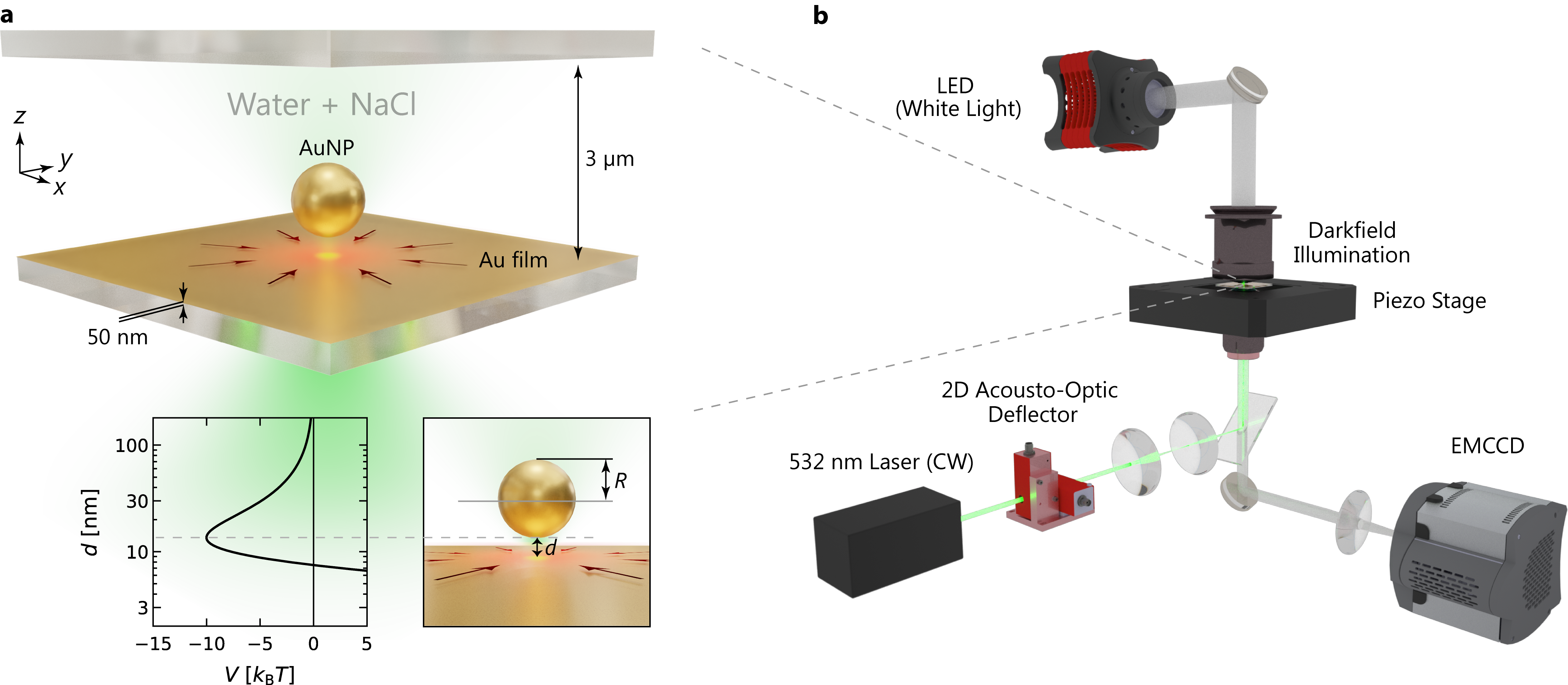
II.1 Experimental Configuration and Working Principle
Our experiments rely on a simple sample geometry with a gold film (50 nm) that is deposited on a microscopy glass coverslip (Fig. 1a). The sample chamber contains a suspension of gold nanoparticles (AuNPs) or other nano-objects (polystyrene NPs and ellipsoids) with a controlled amount of salt (NaCl), surfactants (SDS, …) or polymers (PEG). The gold film is heated locally in an inverted microscopy setup by a highly focused laser (532 nm) using beam steering optics (Acusto-Optic-Deflector, AOD). The nano-objects are observed using darkfield illumination with an oil-immersion darkfield condenser (NA 1.2) and a 100 oil-immersion objective set to NA 0.6 (Fig. 1b). Additional details of the experimental setup and sample preparation are provided in the Methods section.
The trapping of nano-objects as detailed in the following is comprising two effects. i) The vertical confinement of the suspended objects as achieved by an attractive interaction of the suspended nano-objects with the gold surface, which is found to be the vdW interaction for gold nanoparticles and can be replaced by depletion forces for other materials. ii) The generation of thermo-osmotic boundary flows that are induced by the local heating and the corresponding perturbation of the liquid-solid interactions. This boundary flow is directed radially inwards to the heated spot and provides a confining hydrodynamic force on suspended objects at the heating spot.
II.2 Dynamics of AuNPs Close to a Au Film
Consider a single AuNP with a radius of that is suspended in an aqueous solution of NaCl at a 10 mM concentration and diffusing in a thin liquid film of about thickness over a 50 nm Au film (Fig. 1a). Exploring the diffusion of the particle we observe a restriction of the -positions to a thin layer close to the gold film. The gold particle never defocuses under these conditions while it does in deionized (DI) water (Supplementary Video 1). This restricted out-of-plane motion is the result of interactions comprising an attractive vdW contribution and a repulsion of the electrostatic double layers of the particle and surface [32] as described by the DLVO theory and the gravitational potential (see Supplementary Information for details).
| (1) |
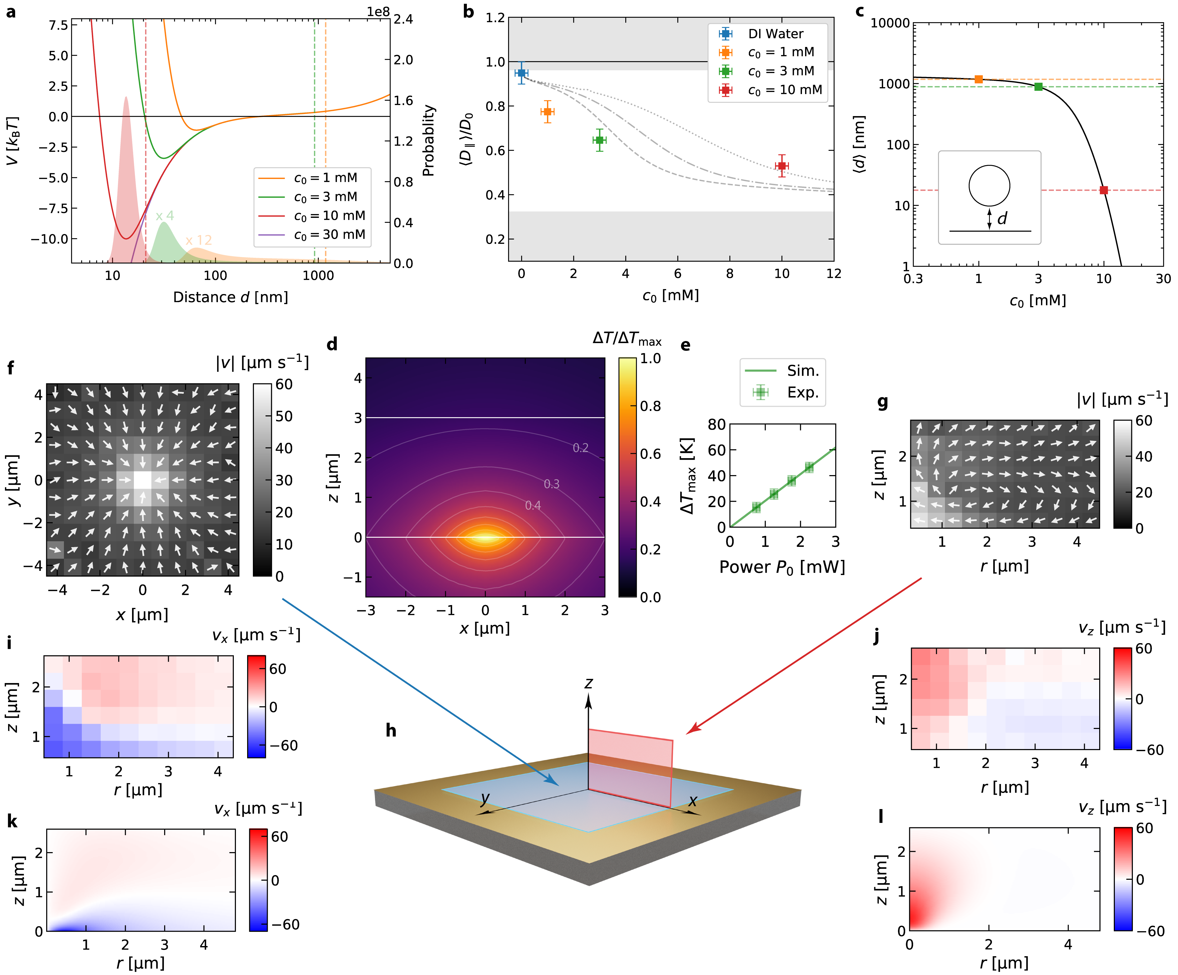
The total potential is depicted in Figure 2a for different salt concentrations (see SI for parameters) as a function of the surface-to-surface distance . The stronger screening of the surface charges at the gold film and the AuNP at higher salt concentration increase the importance of attractive vdW interactions to create this secondary minimum in the DLVO part of the potential. This potential also influences the observed dynamics as the particle couples with its hydrodynamic flow field to the solid boundary [33]. The in-plane (equation 2) and out-of-plane diffusion coefficient (see Supplementary Information for details) are modulated with the distance of the particle from the wall.
| (2) |
Over the course of a diffusion trajectory, the particle samples different diffusion coefficients according to its probability density to be at a distance from the surface (filled regions in Fig. 2a). The observed in-plane diffusion coefficient is thus a weighted average of the diffusion coefficient over the different vertical positions . Using we can calculate the corresponding salt concentration dependence of the diffusion coefficient and compare that to the experimental results. Figure 2b shows that the experimentally observed is decreasing with increasing salt concentration due to the hydrodynamic coupling in fair agreement with the theoretical predictions (see Supplementary Information for details). Calculating in addition the mean distance of the particle from the surface reveals that only in the case of mM the particle is confined in the DLVO potential well, while at lower salt concentrations an enhanced probability of finding the particle near the surface is the cause of the observed diffusion coefficient. These calculations help us to estimate the mean distance of the particle from the surface, which is about and for the lowest NaCl concentrations (Fig. 2c). At a concentration of mM the particle is hovering at a distance of nm surface. Note that this corresponds to values of , which is far below the commonly explored region of the hydrodynamic coupling of colloids to walls [33] allowing to experimentally explore new terrains also in the field of hydrodynamic wall coupling of colloids.
II.3 Hydrodynamic Particle Confinement
When tightly focusing the light of 532 nm wavelength to the gold film, a part of the incident energy (about 30 %) is absorbed and converted into heat that perturbs the liquid-solid interactions. The temperature rise at the gold surface can be determined using a thin nematic liquid crystal (5CB) film and substantiated by finite element simulations with the complete three-dimensional temperature profile in the solution (see Fig. 2d, e and Supplementary Information for details).
These local temperature perturbations of the solid-liquid interactions at the interface induce a thermo-osmotic flow [34, 35]. Taking a liquid volume element close to the solid from the cold side and exchanging that with one at the hot side would not only transport heat since the liquid volumes have different temperatures, but also additional free energy as the liquid has a different interaction with the solid in these regions. The flow is induced in an ultrathin boundary layer corresponding in thickness to the length scale of liquid-solid interactions. Since the interaction range of liquid-solid interactions is only a few nanometers (the characteristic length of interfacial interactions), the boundary flow is due to its dimension collapsed into a quasi-slip hydrodynamic boundary condition:
| (3) |
where is the excess enthalpy, the temperature and is the temperature gradient parallel to the surface. The integral can be summarized to a thermo-osmotic coefficient . The thermo-osmotic coefficient , therefore, contains all information about the interfacial interaction between the liquid and the solid. If the liquid is driven to the cold, whereas for , the liquid is driven to the hot. These boundary flows are present at all liquid-solid interfaces with tangential temperature gradients, though, they are commonly overlooked. They become particularly important for plasmonic and thermo-plasmonic trapping [16], as those techniques rely on the dynamics of molecules and particles in the direct vicinity of plasmonic nanostructures.
The boundary flow drives the flow field inside the fluid film. The resulting volumetric flow field can be tracked experimentally by single AuNPs in DI water, where the particles are not confined to a surface layer as reported above. We analyze the in-plane () position of the particle and its -position, where the latter is estimated from the radius of the defocussed particle images (see Supplementary Video 2 and Supplementary Information for details). The measured velocity distributions in the -plane near the gold layer and in the -plane are shown in Fig. 2f and g, respectively. The - and -component of the measured flow velocities are depicted in Fig. 2i, j and compare well to simulation results in Fig. 2k, l. From these measurements, we extract a thermo-osmotic coefficient on the order of (see Supplementary Information for details). We can break down the contributions to this value with equations (4) and (5) to estimate the double layer and vdW contributions using the experimental parameters. Note that AuNP do not show thermophoresis due to their high thermal conductivity and thus isothermal surface.
| (4) |
For the electrostatic contribution we used [36] and (see Methods section for details). An estimate of the vdW contribution can be given by
| (5) |
with being the thermal expansion coefficient of water and for the cut-off parameter [34] (see Supplementary Information for details). The sum of both contributions matches well the experimental result and suggests that thermo-osmosis at gold-water interfaces is governed by vdW interactions. The obtained quasi slip velocities are ranging up to 80 and provide, due to their omnipresence, a unique tool for nanofluidics. These thermo-osmotic flows are induced without any external pressure difference. They can be controlled by the light intensity heating laser and are quickly switched due to the extremely fast heat conduction at these length scales. Moreover the finding of the vdW dominated thermo-osmotic flows suggest that such contributions must be present in any plasmonic trapping experiment with extended gold structures [16, 17, 27, 12].
Using and , where and are the correction factors for the friction coefficient of a sphere close to a surface we are able to extract the hydrodynamic forces that are exerted on the AuNP tracers (see equation (2) and Supplementary Information for details). The lateral forces allow to confine objects at the heating spot, yet the hydrodynamic force normal to the surface (-direction) is repulsive without any additional interaction. Finally, such boundary flows with substantial vertical velocity gradients also exhibit a vorticity (see Supplementary Information for details) that generates a torque on suspended objects causing them to rotate [37].
II.4 Single Particle Trapping and Flow Field Multiplexing
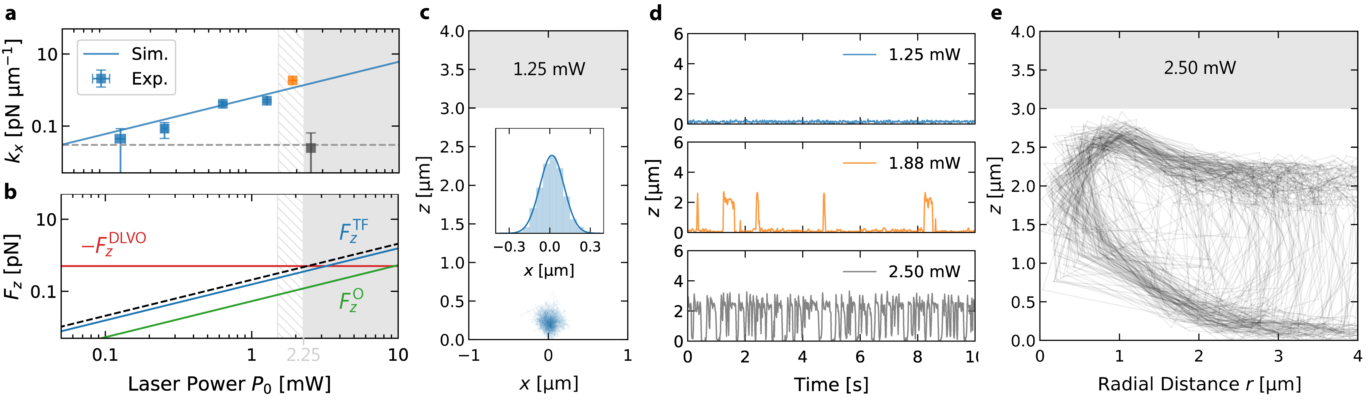
The repulsive normal component caused by the hydrodynamic drag is now superimposed with an attractive force due to the DLVO potential when increasing the NaCl concentration. The surface-to-surface distance between AuNP and gold film and the depth of the appearing secondary DLVO potential minimum can be controlled by the NaCl concentration. At a NaCl concentration of about mM, the attractive potential has a depth of about (see Fig. 2a) and is strong enough to compete with the vertical drag force and additional optical forces on the AuNP to trap the particle above the heating spot.
Supplementary Video 3 demonstrates this trapping of an AuNP above the hot spot on the Au surface. This is purely the result of the hydrodynamic drag forces generated by the thermo-osmotic flow and the attractive vdW interaction between the AuNP and the Au film. This observation is substantiated by a quantitative evaluation of the lateral trap stiffness and vertical forces, as depicted in Fig. 3a, b. The fluctuations of the particle in the hydrodynamic flow arise from a balance of the restoring hydrodynamic currents and the diffusive currents. Analysis of the lateral position histograms (inset in Fig. 3c for 1.25 mW) yields an effective stiffness of the trap (Fig. 3a) that well matches the predictions based on the thermo-osmotic flow (Fig. 2i, j). The hydrodynamic trapping stiffness increases linearly up to a heating power of about mW. At this power, the vertical forces become strong enough to let the particle escape the secondary DLVO minimum, which is visible from the -position time traces displayed in Fig. 3d. The AuNPs are then observed to move vertically out of the DLVO potential to follow the flow inside the sample and to eventually return to the boundary flow via sedimentation (Fig. 3e, Supplementary Video 4). The forces which eject the particle from the potential comprise the hydrodynamic and optical forces due to the radiation pressure from the heating laser leaked through the film. We have evaluated the individual contributions in simulations. They are shown together with the hydrodynamic force and the total vertical force as compared to the attractive force of the DLVO potential (Fig. 3b) and provide quantitative agreement (threshold heating power of mW) with our experimental results. Note that while the stationary distribution of particles in the vertical direction is not influenced by the diffusive dynamics, the escape rate from the potential well is heavily altered by the fact that the vertical diffusion coefficient of the particle is decreasing to zero when approaching the gold film. This is enhancing the trapping times considerably (see Supplementary Information for details) but also increases the time required for the particle to enter the DLVO minimum by diffusion.
The observed trapping is, hence, a vdW assisted thermo-hydrodynamic process. Vertical confinement is achieved by vdW attraction and double layer repulsion, while lateral confinement is the result of thermo-osmotic flows induced in an ultra-thin sheet of liquid at the interface. No additional contributions, for example, due to convective flows with similar flow patterns (see Supplementary Information for details) or thermo-electric effects are required for a quantitative description [38, 39, 25, 31].
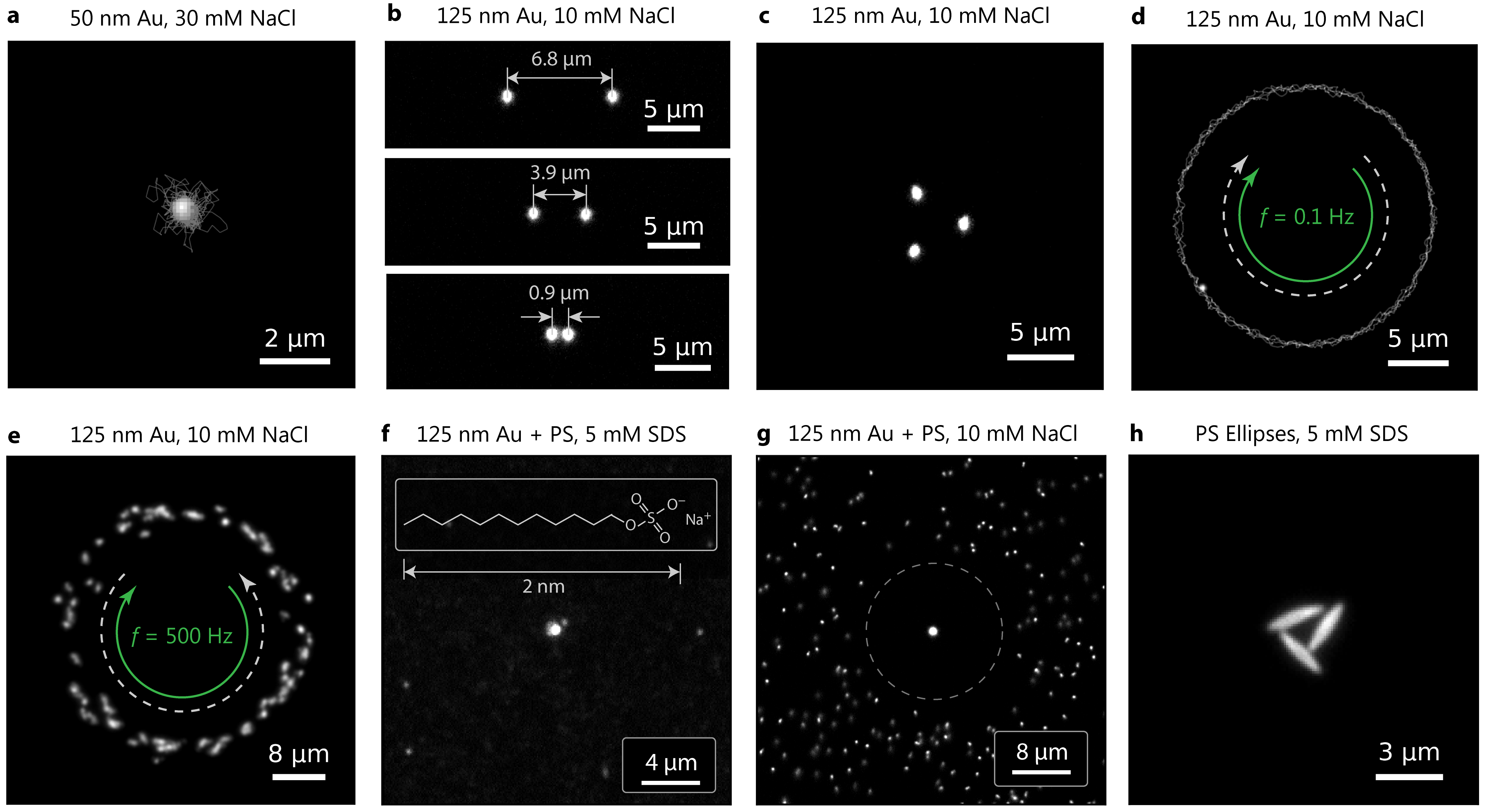
Precise tuning of the DLVO potential enables the trapping of even smaller Au NPs (Fig. 4a, Supplementary Video 5). The speed of heat diffusion, which is about 4 orders of magnitude faster than the particle diffusion [40] allows us to introduce a flow field multiplexing. Therefore, we switch the heating location between different locations inducing flow thermo-osmotic flow fields for time periods of about 100 . With the help of this multiplexing, we are able to hold multiple AuNPs (Fig. 4b, c) at distances of less than 1 , which would not be possible with continuous heating of close-by locations (Supplementary Videos 6 and 7). A trapped AuNP can also be guided along the predefined path over the Au film as fast as (Fig. 4d, Supplementary Video 8). At larger manipulation speeds ( Hz) and higher heating power ( mW) the thermo-osmotic attraction to the heating spot is combined with thermo-viscous flows [41, 42]. These flows originate from the temperature dependent viscosity of the liquid and are directed opposite to the scanning direction of the laser [42]. The result of this combination of thermo-osmosis and thermo-viscous flows is a rotating ring-like particle structure (Fig. 4e and Supplementary Video 9). These different effects that can be exploited in a simple planar geometry give rise to numerous applications including for example a freely configurable nanoparticle on mirror geometry for plasmonic sensing [43, 5].
II.5 Beyond Thermo-Osmotic van der Waals Trapping
So far, the presented manipulation is based on thermo-osmotic flows that drive the lateral motion of suspended colloids and a vertical confinement due to the secondary minimum of the DLVO potential between AuNP and Au film. While the thermo-osmotic flows are characteristic for all systems containing a heated gold/water interface including all previous studies on thermo-plasmonic trapping, the DLVO potential minimum is much weaker for other materials like polymer colloids or macromolecules due to their smaller vdW attraction. Often, those system even show a repulsion from the heat source due to thermophoresis, which is not present for AuNP. A more generalized strategy therefore needs additional attractive contributions, which confine suspended colloids or molecules to regions close to the gold surface to take advantage of the thermo-osmotic flow.
Such attractive contributions can arise from depletion interactions [44, 21]. Thereby a temperature gradient repels dissolved molecules from the heated regions generating a concentration gradient that drives suspended nano-objects to the heating spot. To demonstrate this effect we use the surfactant sodium dodecyl sulfate (SDS) at a concentration of 5 mM well below the critical micelle concentration (8.2 mM) to avoid complications of micelle formation. We suspend additional polystyrene particles(PS) and AuNPs of the same size () in the solution and compare their dynamics to a solution with AuNPs and PS particles without SDS but 10 mM NaCl. Remarkably, the heated spot is attractive for both AuNPs and for PS NPs (Fig. 4f, Supplementary Video 10) in the SDS solution showing even PS colloidal crystal growth, while only the AuNP is trapped in the NaCl solution and the PS particles are repelled by thermophoresis (Fig. 4g, Supplementary Video 11).
The observations in NaCl are readily explained by the fact that the AuNP is confined in the DLVO minimum as demonstrated above but the PS particle is not due to a 10 times lower Hamaker constant (see Supplementary Information for details). The PS particle samples the whole liquid film thickness equally and not preferentially the region close to the Au film and experiences an additional thermophoretic drift velocity given by
| (6) |
where is the thermophoretic mobility [34] and the temperature gradient (Fig. 4g, Supplementary Video 11). For the particle is driven to the cold. From equation (4) we find and , where we have used a measured zeta potential of . The vdW contribution, to either the thermophoretic drift or the attraction to the gold surface can be neglected due to the smaller Hamaker constant of PS. From the stationary probability distribution of the PS NP we find a Soret coefficient of (see Supplementary Information for details) in agreement with our theoretical prediction .
In the SDS solution, the additional surfactant molecules now undergo thermophoresis to yield a concentration gradient in which suspended colloidal particles drift. The lower concentration in the heated regions promotes an effective attractive interaction of suspended colloids with the gold surface due to depletion forces. The drift velocity is described by an additional term to the thermodiffusion coefficient , that is, the second term in brackets in equation (7) [21, 34, 44].
| (7) |
Here is the size of the SDS molecule, the concentration in units of and the Soret coefficient of SDS. For [45], and [46] we find for the additional depletion contribution, which exceeds the thermophoretic mobility, , rendering the overall mobility negative. The PS NPs and the AuNPs are thus driven to the the heated Au film surface(Fig. 4f, Supplementary Video 10) which allows for further transport in the thermo-osmotic boundary flow. Additional contributions, as for example thermo-electric fields may even enhance the attractive components. Overall, this concept is readily transferred to other objects as shown in Figure 4h and Supplementary Video 12, where we have trapped ellipsoidal PS particles in a 5 mM solution of SDS.
III Conclusion
In conclusion, we have demonstrated that thermo-hydrodynamic boundary flows can manipulate nano-objects with unprecedented flexibility in a very simple sample geometry. These flows are the key for future thermo-optofluidic implementations with an extensive range of applications in the fields of i) nanoparticle sorting and separation [7]; ii) assembly of nanophotonic circuits [47] and plasmonic quantum sensors [5, 4]; iii) biotechnology on-chip laboratories [48] and iv) manufacturing of nanomaterials [6, 4] and functional nanosurfaces [49, 50]. We have substantiated our experimental findings of thermo-osmotic flow assisted trapping with a quantitative theoretical description. A flow field multiplexing scheme has been further developed to allow for the simultaneous manipulation of many individual nano-objects. Our concept can be combined with other thermally induced effects such as thermophoresis, depletion forces and thermoviscous flows to form a fully-featured nanofluidic system-on-a-chip. Besides direct consequences for the field of plasmonic nano-tweezers and other thermoplasmonic trapping schemes, the use of thermo-hydrodynamic flows as a tool for nanofluidic applications will extend the limits at the forefront of nanotechnology.
References
- Maragò et al. [2013] O. M. Maragò, P. H. Jones, P. G. Gucciardi, G. Volpe, and A. C. Ferrari, Optical trapping and manipulation of nanostructures, Nat. Nanotechnol. 8, 807 (2013).
- Gao et al. [2017] D. Gao, W. Ding, M. Nieto-Vesperinas, X. Ding, M. Rahman, T. Zhang, C. Lim, and C.-W. Qiu, Optical manipulation from the microscale to the nanoscale: Fundamentals, advances and prospects, Light Sci. Appl. 6, 17039 (2017).
- Bradac [2018] C. Bradac, Nanoscale Optical Trapping: A Review, Adv. Opt. Mat. 6, 1800005 (2018).
- Xavier et al. [2018] J. Xavier, S. Vincent, F. Meder, and F. Vollmer, Advances in optoplasmonic sensors – combining optical nano/microcavities and photonic crystals with plasmonic nanostructures and nanoparticles, Nanophotonics 7, 1 (2018).
- Li et al. [2018] G.-C. Li, Q. Zhang, S. A. Maier, and D. Lei, Plasmonic particle-on-film nanocavities: A versatile platform for plasmon-enhanced spectroscopy and photochemistry, Nanophotonics 7, 1865 (2018).
- Lin et al. [2017a] L. Lin, X. Peng, and Y. Zheng, Reconfigurable opto-thermoelectric printing of colloidal particles, Chem. Commun. 53, 7357 (2017a).
- Xie et al. [2020] Y. Xie, J. Rufo, R. Zhong, J. Rich, P. Li, K. W. Leong, and T. J. Huang, Microfluidic Isolation and Enrichment of Nanoparticles, ACS Nano 14, 16220 (2020).
- Favre-Bulle et al. [2019] I. A. Favre-Bulle, A. B. Stilgoe, E. K. Scott, and H. Rubinsztein-Dunlop, Optical trapping in vivo: Theory, practice, and applications, Nanophotonics 8, 1023 (2019).
- Choudhary et al. [2019] D. Choudhary, A. Mossa, M. Jadhav, and C. Cecconi, Bio-Molecular Applications of Recent Developments in Optical Tweezers, Biomolecules 9, 23 (2019).
- Kayci et al. [2014] M. Kayci, H.-C. Chang, and A. Radenovic, Electron Spin Resonance of Nitrogen-Vacancy Defects Embedded in Single Nanodiamonds in an ABEL Trap, Nano Lett. 14, 5335 (2014).
- Juan et al. [2011] M. L. Juan, M. Righini, and R. Quidant, Plasmon nano-optical tweezers, Nat. Photonics 5, 349 (2011).
- Zhang et al. [2021] Y. Zhang, C. Min, X. Dou, X. Wang, H. P. Urbach, M. G. Somekh, and X. Yuan, Plasmonic tweezers: For nanoscale optical trapping and beyond, Light Sci. Appl. 10, 59 (2021).
- De Vlaminck and Dekker [2012] I. De Vlaminck and C. Dekker, Recent Advances in Magnetic Tweezers, Annu. Rev. Biophys. 41, 453 (2012).
- Wang and Moerner [2011] Q. Wang and W. E. Moerner, An Adaptive Anti-Brownian Electrokinetic Trap with Real-Time Information on Single-Molecule Diffusivity and Mobility, ACS Nano 5, 5792 (2011).
- Wu [2011] M. C. Wu, Optoelectronic tweezers, Nat. Photonics 5, 322 (2011).
- Kotnala and Gordon [2014] A. Kotnala and R. Gordon, Quantification of High-Efficiency Trapping of Nanoparticles in a Double Nanohole Optical Tweezer, Nano Lett. 14, 853 (2014).
- Jiang et al. [2020] Q. Jiang, B. Rogez, J.-B. Claude, G. Baffou, and J. Wenger, Quantifying the Role of the Surfactant and the Thermophoretic Force in Plasmonic Nano-optical Trapping, Nano Lett. 20, 8811 (2020).
- Braun and Cichos [2013] M. Braun and F. Cichos, Optically Controlled Thermophoretic Trapping of Single Nano-Objects, ACS Nano 7, 11200 (2013).
- Braun et al. [2015] M. Braun, A. P. Bregulla, K. Günther, M. Mertig, and F. Cichos, Single Molecules Trapped by Dynamic Inhomogeneous Temperature Fields, Nano Lett. 15, 5499 (2015).
- Fränzl et al. [2019] M. Fränzl, T. Thalheim, J. Adler, D. Huster, J. Posseckardt, M. Mertig, and F. Cichos, Thermophoretic trap for single amyloid fibril and protein aggregation studies, Nat. Methods 16, 611 (2019).
- Jiang et al. [2009] H.-R. Jiang, H. Wada, N. Yoshinaga, and M. Sano, Manipulation of Colloids by a Nonequilibrium Depletion Force in a Temperature Gradient, Phys. Rev. Lett. 102, 208301 (2009).
- Bregulla et al. [2014] A. P. Bregulla, H. Yang, and F. Cichos, Stochastic Localization of Microswimmers by Photon Nudging, ACS Nano 8, 6542 (2014).
- Khadka et al. [2018] U. Khadka, V. Holubec, H. Yang, and F. Cichos, Active particles bound by information flows, Nat. Commun. 9, 3864 (2018).
- Fränzl et al. [2021] M. Fränzl, S. Muiños-Landin, V. Holubec, and F. Cichos, Fully Steerable Symmetric Thermoplasmonic Microswimmers, ACS Nano 15, 3434 (2021).
- Lin et al. [2018] L. Lin, M. Wang, X. Peng, E. N. Lissek, Z. Mao, L. Scarabelli, E. Adkins, S. Coskun, H. E. Unalan, B. A. Korgel, L. M. Liz-Marzán, E.-L. Florin, and Y. Zheng, Opto-thermoelectric nanotweezers, Nat. Photonics 12, 195 (2018).
- Ndukaife et al. [2016] J. C. Ndukaife, A. V. Kildishev, A. G. A. Nnanna, V. M. Shalaev, S. T. Wereley, and A. Boltasseva, Long-range and rapid transport of individual nano-objects by a hybrid electrothermoplasmonic nanotweezer, Nat. Nanotechnol. 11, 53 (2016).
- Hong et al. [2020] C. Hong, S. Yang, and J. C. Ndukaife, Stand-off trapping and manipulation of sub-10 nm objects and biomolecules using opto-thermo-electrohydrodynamic tweezers, Nat. Nanotechnol. 15, 908 (2020).
- Donner et al. [2011] J. S. Donner, G. Baffou, D. McCloskey, and R. Quidant, Plasmon-Assisted Optofluidics, ACS Nano 5, 5457 (2011).
- Roxworthy et al. [2014] B. J. Roxworthy, A. M. Bhuiya, S. P. Vanka, and K. C. Toussaint, Understanding and controlling plasmon-induced convection, Nat. Commun. 5, 3173 (2014).
- Chen et al. [2020] J. Chen, J. F.-C. Loo, D. Wang, Y. Zhang, S.-K. Kong, and H.-P. Ho, Thermal Optofluidics: Principles and Applications, Adv. Opt. Mater. 8, 1900829 (2020).
- Ciraulo et al. [2021] B. Ciraulo, J. Garcia-Guirado, I. de Miguel, J. Ortega Arroyo, and R. Quidant, Long-range optofluidic control with plasmon heating, Nat. Commun. 12, 2001 (2021).
- Israelachvili [2011] J. N. Israelachvili, Intermolecular and Surface Forces, 3rd ed. (Elsevier, Academic Press, Amsterdam, 2011).
- Lin et al. [2000] B. Lin, J. Yu, and S. A. Rice, Direct measurements of constrained Brownian motion of an isolated sphere between two walls, Phys. Rev. E 62, 3909 (2000).
- Würger [2010] A. Würger, Thermal non-equilibrium transport in colloids, Rep. Prog. Phys. 73, 126601 (2010).
- Bregulla et al. [2016] A. P. Bregulla, A. Würger, K. Günther, M. Mertig, and F. Cichos, Thermo-Osmotic Flow in Thin Films, Phys. Rev. Lett. 116, 188303 (2016).
- Giesbers et al. [2002] M. Giesbers, J. Kleijn, and M. A. Cohen Stuart, The Electrical Double Layer on Gold Probed by Electrokinetic and Surface Force Measurements, J. Colloid Interface Sci. 248, 88 (2002).
- Bluemink et al. [2008] J. J. Bluemink, D. Lohse, A. Prosperetti, and L. Van Wijngaarden, A sphere in a uniformly rotating or shearing flow, J. Fluid Mech. 600, 201 (2008).
- Lin et al. [2016] L. Lin, X. Peng, M. Wang, L. Scarabelli, Z. Mao, L. M. Liz-Marzán, M. F. Becker, and Y. Zheng, Light-Directed Reversible Assembly of Plasmonic Nanoparticles Using Plasmon-Enhanced Thermophoresis, ACS Nano 10, 9659 (2016).
- Lin et al. [2017b] L. Lin, J. Zhang, X. Peng, Z. Wu, A. C. H. Coughlan, Z. Mao, M. A. Bevan, and Y. Zheng, Opto-thermophoretic assembly of colloidal matter, Sci. Adv. 3, 1700458 (2017b).
- Baffou et al. [2020] G. Baffou, F. Cichos, and R. Quidant, Applications and challenges of thermoplasmonics, Nat. Mater. 19, 946 (2020).
- Weinert et al. [2008] F. M. Weinert, J. A. Kraus, T. Franosch, and D. Braun, Microscale Fluid Flow Induced by Thermoviscous Expansion Along a Traveling Wave, Phys. Rev. Lett. 100, 164501 (2008).
- Weinert et al. [2011] F. M. Weinert, C. B. Mast, and D. Braun, Optical fluid and biomolecule transport with thermal fields, Phys. Chem. Chem. Phys. 13, 9918 (2011).
- Chikkaraddy et al. [2016] R. Chikkaraddy, B. de Nijs, F. Benz, S. J. Barrow, O. A. Scherman, E. Rosta, A. Demetriadou, P. Fox, O. Hess, and J. J. Baumberg, Single-molecule strong coupling at room temperature in plasmonic nanocavities, Nature 535, 127 (2016).
- Maeda et al. [2011] Y. T. Maeda, A. Buguin, and A. Libchaber, Thermal Separation: Interplay between the Soret Effect and Entropic Force Gradient, Phys. Rev. Lett. 107, 038301 (2011).
- Syshchyk et al. [2016] O. Syshchyk, D. Afanasenkau, Z. Wang, H. Kriegs, J. Buitenhuis, and S. Wiegand, Influence of temperature and charge effects on thermophoresis of polystyrene beads, Eur. Phys. J. E 39, 129 (2016).
- Vigolo et al. [2010] D. Vigolo, S. Buzzaccaro, and R. Piazza, Thermophoresis and Thermoelectricity in Surfactant Solutions, Langmuir 26, 7792 (2010).
- Zhang et al. [2019] Q. Zhang, H. Yu, M. Barbiero, B. Wang, and M. Gu, Artificial neural networks enabled by nanophotonics, Light Sci Appl 8, 42 (2019).
- Xavier et al. [2021] J. Xavier, D. Yu, C. Jones, E. Zossimova, and F. Vollmer, Quantum nanophotonic and nanoplasmonic sensing: Towards quantum optical bioscience laboratories on chip, Nanophotonics 10, 1387 (2021).
- Peng et al. [2019] J. Peng, H.-H. Jeong, Q. Lin, S. Cormier, H.-L. Liang, M. F. L. De Volder, S. Vignolini, and J. J. Baumberg, Scalable electrochromic nanopixels using plasmonics, Sci. Adv. 5, 10.1126/sciadv.aaw2205 (2019).
- Mohammadi et al. [2021] E. Mohammadi, A. Tittl, K. L. Tsakmakidis, T. V. Raziman, and A. G. Curto, Dual Nanoresonators for Ultrasensitive Chiral Detection, ACS Photonics 8, 1754 (2021).