Nonclassical Exciton Diffusion in Monolayer WSe2
Abstract
We experimentally demonstrate time-resolved exciton propagation in a monolayer semiconductor at cryogenic temperatures. Monitoring phonon-assisted recombination of dark states, we find a highly unusual case of exciton diffusion. While at 5 K the diffusivity is intrinsically limited by acoustic phonon scattering, we observe a pronounced decrease of the diffusion coefficient with increasing temperature, far below the activation threshold of higher-energy phonon modes. This behavior corresponds neither to well-known regimes of semiclassical free-particle transport nor to the thermally activated hopping in systems with strong localization. Its origin is discussed in the framework of both microscopic numerical and semi-phenomenological analytical models illustrating the observed characteristics of nonclassical propagation. Challenging the established description of mobile excitons in monolayer semiconductors, these results open up avenues to study quantum transport phenomena for excitonic quasiparticles in atomically-thin van der Waals materials and their heterostructures.
Correlated motion of Coulomb-bound electron-hole pairs, commonly known as excitons Frenkel (1931); Gross and Karrjew (1952), represents a vibrant field of research. From their electronic constituents excitons naturally inherit the ability to propagate through the crystal Ivchenko (2005); Klingshirn (2007). Moreover, optically active excitons can directly visualize transport phenomena and possess emerging properties associated with interacting, composite bosons Haug and Koch (2009), including discussions of superfluidity Mysyrowicz et al. (1996); Benson et al. (1997), condensation Moskalenko and Snoke (2005), phonon wind Tikhodeev et al. (1998), and ring formation Butov et al. (2004). Particularly interesting in this context are layered two-dimensional (2D) materials Ginsberg and Tisdale (2020) such as semiconducting transition-metal dichalcogenides (TMDCs) Wilson and Yoffe (1969); Novoselov et al. (2005); Mak et al. (2010); Splendiani et al. (2010). As monolayers they host robust exciton states with high binding energies Xiao et al. (2017); Wang et al. (2018) and the possibility to carry spin-valley information quanta Xu et al. (2014); Yu et al. (2015); Glazov and Golub (2020). The excitons in TMDCs have been shown to be mobile Kumar et al. (2014); Kato and Kaneko (2016); Yuan et al. (2017); Cadiz et al. (2018); Fu et al. (2019); Raja et al. (2019); Uddin et al. (2020); Zipfel et al. (2020), guided by gradients Cordovilla Leon et al. (2018); Shahnazaryan et al. (2019); Hao et al. (2020), exhibit non-linear diffusion Mouri et al. (2014); Kulig et al. (2018); Wang et al. (2019); Glazov (2019); Perea-Causín et al. (2019); Rosati et al. (2020a), strain-dependence Rosati et al. (2021), as well as intriguing propagation in heterostructures Rivera et al. (2016); Calman et al. (2018); Unuchek et al. (2018); Yuan et al. (2020).
In general, systems hosting mobile excitons such as TMDCs fall into two main categories, exhibiting either semiclassical free-particle transport Smith et al. (1988); Erland et al. (1993); Grosse et al. (1997) or hopping between localization sites Anderson (1958); Mikhnenko et al. (2015). These mechanisms are typical for Wannier-Mott excitons with spatially extended wavefunctions in inorganic semiconductors and tightly bound Frenkel-like states in molecular crystals, respectively. Excitons in TMDC monolayers, however, present a particularly interesting, intermediate case. They uniquely combine the characteristics of the two descriptions, with wavefunctions being delocalized across many unit cells but also exhibiting high binding energies Wang et al. (2018). This duality is expected to manifest itself prominently in the exciton transport behavior, including potential emergence of quantum interference phenomena Ivchenko et al. (1977); Altshuler and Aronov (1985); Arseev and Dzyubenko (1998); Evers and Mirlin (2008); Glazov (2020). Despite much progress, however, only little is known regarding the appropriate picture for the exciton propagation in monolayer semiconductors, currently based on the assumption of a purely semiclassical framework Mouri et al. (2014); Kato and Kaneko (2016); Cadiz et al. (2018); Kulig et al. (2018); Shahnazaryan et al. (2019); Perea-Causín et al. (2019); Zipfel et al. (2020); Rosati et al. (2020a); Harats et al. (2020).
Here, we address the question of the fundamental description of mobile excitons in 2D TMDCs, demonstrating the unusual nature of the exciton diffusion in these systems. In the experiments, we take advantage of dark excitons in hBN-encapsulated WSe2 monolayers with suppressed long-range disorder Raja et al. (2019). In contrast to bright excitons with picosecond lifetimes Robert et al. (2016), dark states live up to 100’s of ps Zhang et al. (2015); Robert et al. (2017) and allow us to study them under thermal equilibrium conditions. Dark exciton emission is monitored through phonon-assisted recombination channels Lindlau et al. (2017); Christiansen et al. (2017); Liu et al. (2019); Li et al. (2019); Brem et al. (2020); Rosati et al. (2020b); He et al. (2020) via temporally and spatially-resolved microscopy at cryogenic temperatures after weak, strictly resonant excitation of the bright state. Importantly, the characteristic spectral shape of the phonon sidebands (PSBs) Klingshirn (2007); Christiansen et al. (2017); Brem et al. (2020) allows for an independent evaluation of the exciton temperature and scattering rates, making it possible to extract key parameters governing exciton propagation from experiments.
At the lowest studied temperature of 5 K, we detect linear diffusion with a coefficient of cm2/s, essentially limited by the exciton-phonon coupling. As the temperature increases, we observe an unusually strong decrease already in the low-temperature range of 4 to 30 K, far below activation threshold of high-energy phonons. Under these conditions, the observations agree neither with the semiclassical free-particle description nor with thermally activated hopping. These conclusions are further supported by quantitative analysis involving experimentally determined scattering rates as well as many-body calculations of the spatio-temporal exciton dynamics. In view of the recently predicted quantum interference phenomena for TMDC monolayers Glazov (2020), it allows us to experimentally demonstrate an interesting scenario that requires nonclassical effects in the exciton transport.
The studied hBN-encapsulated WSe2 monolayers were obtained by mechanical exfoliation and stamping Castellanos-Gomez et al. (2014) of bulk crystals (WSe2 from “HQgraphene”, hBN from NIMS) onto SiO2/Si substrates (see Supplemental Material). For the measurements the samples were placed in a microscopy cryostat. We used a 80 MHz, 140 fs-pulsed Ti:sapphire as excitation source, tuned into resonance with the bright exciton at 1.726 eV to minimize excess energy and avoid contributions of unbound electron-hole pairs. The incident light was focused to a spot with about 1 m diameter. The emission was collected from a lateral cross-section and guided through an imaging spectrometer equipped with a mirror and a grating to provide spatial and spectral resolutions, respectively. We employed a streak camera for time-resolved detection and a CCD-sensor for time-integrated signals, also see Refs. Kulig et al. (2018); Zipfel et al. (2020).
The optical fingerprints used to monitor dark excitons in WSe2 monolayer are schematically illustrated in Fig. 1 (a). The short-lived, bright exciton transition () is denoted by the respective valence and conduction band valleys of the empty and filled electron states that form the exciton. The schematic and the notation are chosen from the valley perspective and apply equally for . After optical injection, bright excitons rapidly redistribute toward lower-lying states Zhang et al. (2015); Selig et al. (2016); Rosati et al. (2020b). These are commonly labeled as dark excitons Zhang et al. (2015) due to strongly suppressed light-matter coupling from either nonzero spin (intravalley triplets) or large center-of-mass momentum (intervalley singlets).
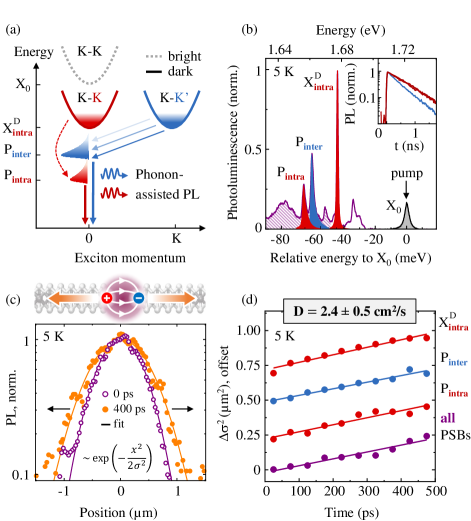
While being essentially dark in absorption, these states can be detected in photoluminescence (PL) at sufficiently low temperatures Wang et al. (2018). Intra-valley triplets couple weakly to light via out-of-plane-polarization Wang et al. (2017); Robert et al. (2017) and are observed with large collection apertures. They give rise to the prominent transition in the PL spectrum at K, presented in Fig. 1 (b). In addition, they couple to the in-plane polarization through a phonon-assisted process Liu et al. (2019); Li et al. (2019); He et al. (2020) leading to the emergence of the sideband, indicated in Figs. 1 (a) and (b). Similarly, intervalley singlets recombine under emission of zone-edge phonons Brem et al. (2020); Rosati et al. (2020b); He et al. (2020) and exhibit PL labeled as , while their direct recombination is forbidden due to momentum conservation. Additional, weak signatures stem largely from the higher order PSBs below , a feature attributed to the sideband at about meV Brem et al. (2020); Rosati et al. (2020b), nearly negligible PL from negative trions ( and meV) as well as a peak at meV, consistent with Refs. Liu et al. (2019); Li et al. (2019); He et al. (2020).
Importantly, all dark states are long-lived with population lifetimes of 500 and 800 ps for and (), respectively, as illustrated in the inset of Fig. 1 (b) (also see Supplemental Material). Following resonant excitation of the bright state, relaxation and cooling of dark excitons occur on much shorter timescales, during the first 10’s of ps Rosati et al. (2020b). During their lifetimes of many 100’s of ps, these excitons should be thus thermally equilibrated within their respective intra- and intervalley subsets.
Representative profiles of the spatially-resolved dark state emission are presented in Fig. 1 (c) for 0 and 400 ps after the excitation at K. They illustrate broadening of the spatial exciton distribution over time. The employed excitation density of 50 nJ cm-2 corresponds to the linear regime with the estimated electron-hole pair density of several 1010 cm-2 per pulse. For the quantitative analysis we fit the PL profiles with a Gauss function, , where is the coordinate along the detected cross-section. From this procedure we extract time-dependent change of the variance , commonly labeled as the mean squared displacement. Corresponding values, obtained from the individual, spectrally filtered emission features of the dark states, are presented in Fig. 1 (d). All of them exhibit very similar behavior with the linear increase of over time being a clear hallmark of diffusive propagation Ginsberg and Tisdale (2020). From we extract an average diffusion coefficient cm2/s, corresponding to diffusion lengths on the order of 0.5 m.
According to the semiclassical description Erland et al. (1993), the diffusion coefficient is determined by the total mass (that is about 0.75 of the free electron mass for dark excitons in WSe2 Kormányos et al. (2015)), temperature , and the scattering rate :
| (1) |
where denotes the Boltzmann constant. Ideally, the primary mechanism limiting the diffusion at low temperatures is the quasielastic exciton scattering with long-wavelength acoustic phonons. The corresponding rate scales linearly with the temperature and is expected to yield a temperature-independent, constant diffusivity for the purely semiclassical behavior Glazov (2020). To elucidate the nature of the exciton transport it is thus necessary to gain independent access to both diffusion coefficient and scattering rate as functions of the exciton temperature. As we demonstrate in the following, the rates and the temperatures are directly obtained from spectrally-resolved, characteristic PSB profiles. That also allows us to confirm the equilibrated state of the photoexcited exciton system.
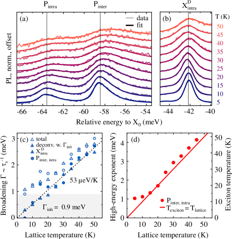
PL spectra in the temperature range between 5 and 50 K are presented in Figs. 2 (a) and (b) for the PSBs and the peak, respectively. In order to fulfill both momentum and energy conservation, the direct radiative transition is only allowed for vanishing momenta and near-zero kinetic energies. It results in a fully symmetric peak that motivates the use of a Voigt profile with a temperature-dependent linewidth for analysis. In contrast to that, recombination via phonon-assisted emission can involve excitons with arbitrary large center-of-mass momenta. This yields the typically asymmetric shape of the sidebands, directly reflecting the exciton distribution in momentum space Hägele et al. (1999); Kozhevnikov et al. (1997); Klingshirn (2007), as illustrated in Fig. 1 (a). To fit the observed PSBs we thus convolute a Lorentzian peak with an exponential high-energy flank (see Supplemental Material). The symmetric broadening then accounts for the scattering rate and the asymmetric flank represents the exciton distribution in kinetic energy, corresponding to an effective temperature .
As shown in Figs. 2 (a) and (b), this choice of the fit functions describes the data very well, allowing for a meaningful extraction of the parameters. Temperature-dependent broadening is presented in Fig. 2 (c) as both total and deconvoluted linewidths. For the latter we assume an additional, constant broadening of 0.9 meV to account for residual, spatially-extended inhomogeneities. The observed linear increase of the linewidth with a coefficient of eV/K is characteristic for quasielastic scattering with phonons from the linear acoustic branch Rudin et al. (1990); Moody et al. (2015); Brem et al. (2019) (see Supplemental Material for additional discussion). In this case, each phonon-scattering event randomly changes the propagation direction of the exciton wavepacket. The optical phase scattering rate determining the spectral broadening is then equal to that for momentum scattering governing the diffusion in Eq. (1). Moreover, the broadening obtained from the symmetric component of the PSBs is nearly identical to that of the peak, supporting the consistency of the applied model. Finally, the extracted exciton temperature presented in Fig. 2 (d) corresponds to that of the lattice with only small deviations.
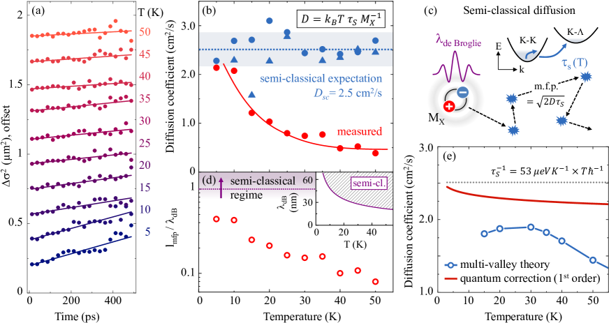
To illustrate temperature-dependent diffusion, time-dependent expansion of the spatially-resolved PL profiles is presented in Fig. 3 (a) for a series of temperatures up to 50 K. Here, we detect the accumulated signal of all sidebands taking advantage of an enhanced signal-to-noise ratio and spectrally-independent diffusion coefficients (c.f. Fig. 1). The extracted diffusivity is shown in Fig. 3 (b) as a function of temperature. At lowest temperatures the measured values are close to the semi-classical expectation cm2/s using the measured broadening coefficient of eV/K and Eq. (1). As the temperature increases, we do not observe any thermally activated increase of diffusion that would otherwise point to hopping Mikhnenko et al. (2015) or defect-assisted scattering Hillmer et al. (1988); Oberhauser et al. (1993). Instead, we find a pronounced decrease of the diffusivity already during the first 10’s of K.
This peculiar observation strongly contrasts the expectation of a constant diffusivity from the semi-classical model, Eq.(1), using independently determined scattering rates from Fig. 2 (c), shown for direct comparison. Importantly, the general description via equation (1) does not depend on a specific origin of the scattering. We further emphasize the absence of non-equilibrium effects due to the long time-scales in our observations, far beyond the initial relaxation during the first 10’s of ps Rosati et al. (2020b). Finally, our findings are robust, confirmed using a sample in the neutral-doping regime, and do not depend on the excitation density (see Supplemental Material). The latter allows us to exclude non-linearities, such as bimolecular processes Warren et al. (2000); Kulig et al. (2018) or phonon-wind effects Smith et al. (1989); Glazov (2019).
The observed inadequacy of the semi-classical description can be rationalized in view of the formal applicability limit, known as the Mott-Ioffe-Regel criterion Landau and Lifshitz (1981). In the semi-classical picture, schematically illustrated in Fig. 3 (c), the exciton diffusion is dictated by temperature and scattering rate . The model is expected to break down when the mean-free-path of the particle becomes similar or smaller than the wavepacket size characterized by the de Broglie wavelength . From the scattering rates in Fig. 2 (c) we indeed obtain for all studied temperatures. We also present the ratio between , extracted from measured diffusion coefficients under the assumption of Eq.(1), and in Fig. 3 (d). This ratio decreases far below unity at elevated temperatures, further illustrating the inconsistency of the semi-classical description. Moreover, due to the similarity of the key exciton parameters and scattering rates, the above considerations should apply for other TMDC monolayers as well.
It is thus instructive to consider current theoretical understanding of the exciton transport in 2D TMDCs both in a semi-classical framework with a more comprehensive description of the exciton band structure and from the perspective of quantum corrections. For this purpose, the results of the calculations for the exciton diffusivity in thermal equilibrium are presented in Fig. 3 (e). In the multi-valley approach we use a model Hamiltonian in the excitonic basis, including carrier–light, carrier–phonon, and carrier–carrier interactions to set up equations of motion for the excitons Perea-Causín et al. (2019); Rosati et al. (2020b, 2021). The required input parameters for monolayer WSe2 are taken from first-principle studies Kormányos et al. (2015); Jin et al. (2014). The diffusion coefficient is extracted from the spatio-temporal evolution of the excitons in the semi-classical approximation neglecting exciton-exciton mechanisms Perea-Causín et al. (2019); Rosati et al. (2020a, 2021). The model takes explicitly into account both a realistic multi-valley band structure of WSe2 and the exciton-phonon coupling beyond the long-wavelength acoustic branches. In particular, we include thermal activation of higher-energy phonon absorption that leads to additional intervalley scattering for excitons, schematically illustrated in Fig. 3 (c). At low temperatures we find an essentially constant value of the diffusion coefficient, close to the experimental result at 5 K and the semi-classical estimation via Eq. (1). Only above about 30 K the model predicts a small decrease of the diffusivity. This onset depends on the energy of the phonons involved in the intervalley scattering and, most importantly, is always accompanied by a non-linear increase of the linewidth (cf. Supplemental Material and Ref. Brem et al. (2019)).
First-order quantum corrections to a simplified semi-classical picture of constant diffusivity are illustrated in Fig. 3 (e). Recently developed for 2D TMDCs Glazov (2020), the calculations are adapted for the WSe2 monolayer by using the exciton mass and the sound velocity of 0.75 and 3.3 km/s, respectively. The model accounts perturbatively for quantum interference effects in the exciton transport. Constructive interference can arise between clock- and counterclockwise propagation of exciton wave packets through closed loops, leading to an effective localization of excitons (also see Supplementary Material). For quasielastic exciton-phonon interaction Glazov (2020), the specific interplay between the loss of the relative phase in the loop and momentum scattering results in an initial decrease of the effective diffusivity with increasing temperature. Interestingly, the functional form of the quantum corrections and their temperature dependence indeed resemble experimental observations. However, the magnitude of the measured effect is almost an order of magnitude higher. It follows that while nonclassical contributions to the exciton diffusion are clearly necessary, further development of the theory beyond the commonly studied first order in quantum corrections is required.
In conclusion, we have explored the nature of the exciton transport in monolayer semiconductors via transient microscopy at cryogenic temperatures. The excitons are found to exhibit neither the characteristic behavior of diffusive free particle propagation nor that of thermally activated hopping between localized states. Measured diffusion coefficients strongly deviate from the semi-classical expectation of a temperature-independent diffusivity based on independently obtained momentum scattering rates. Instead, we find evidence for nonclassical effects playing a key role, consistent with comparable scales of the free propagation and de Broglie lengths. The obtained results should be relevant for optoelectronic devices based on mobile optical excitations in 2D materials and provide a solid platform to understand exciton propagation in more complex heterostructures that involve monolayers as building blocks. The observed unusual behavior in the exciton diffusion highlights van der Waals monolayer semiconductors as a particularly promising platform to merge the rich field of quantum transport phenomena with the physics of composite excitonic quasiparticles.
I Acknowledgments
We thank Alexander Högele and Victor Funk for fruitful discussions as well as Christian Bäuml and Nicola Paradiso for their assistance with pre-patterned substrate preparation. Financial support by the DFG via Emmy Noether Initiative (CH 1672/1), SFB 1277 (project B05), as well as the Würzburg-Dresden Cluster of Excellence on Complexity and Topology in Quantum Matter ct.qmat (EXC 2147) and SFB 1083 (project B09) is gratefully acknowledged. The theoretical part of the work by R.R., S.B., R. P.-C., and E.M. was further supported by the European Union’s Horizon 2020 research and innovation program under grant agreement no. 881603. The computations were enabled by resources provided by the Swedish National Infrastructure for Computing (SNIC) at C3SE and HPC2N. We acknowledge the funding provided by 2D TECH VINNOVA competence Center (Ref. 2019-00068). K. Watanabe and T.T. acknowledge support from the Elemental Strategy Initiative, conducted by the MEXT, Japan, Grant Number JPMXP0112101001, JSPS KAKENHI Grant Numbers JP20H00354 and the CREST (JPMJCR15F3), JST. The development of the analytical theory by M.M.G. has been supported by RSF Project No. 19-12-00051.
References
- Frenkel (1931) J. Frenkel, Phys. Rev. 37, 17 (1931).
- Gross and Karrjew (1952) E. F. Gross and N. A. Karrjew, Dokl. Akad. Nauk SSSR 84, 471 (1952).
- Ivchenko (2005) E. L. Ivchenko, Optical spectroscopy of semiconductor nanostructures (Alpha Science, Harrow England, 2005).
- Klingshirn (2007) C. Klingshirn, Semiconductor Optics, 3rd ed. (Springer, Berlin Heidelberg New York, 2007).
- Haug and Koch (2009) H. Haug and S. W. Koch, Quantum theory of the optical and electronic properties of semiconductors, 5th ed. (World Scientific, Singapore, 2009).
- Mysyrowicz et al. (1996) A. Mysyrowicz, E. Benson, and E. Fortin, Phys. Rev. Lett. 77, 896 (1996).
- Benson et al. (1997) E. Benson, E. Fortin, and A. Mysyrowicz, Solid State Commun. 101, 313 (1997).
- Moskalenko and Snoke (2005) S. A. Moskalenko and D. W. Snoke, Bose-Einstein condensation of excitons and biexcitons (Cambridge University Press, 2005).
- Tikhodeev et al. (1998) S. Tikhodeev, G. Kopelevich, and N. Gippius, Phys. status solidi 206, 45 (1998).
- Butov et al. (2004) L. V. Butov, L. S. Levitov, A. V. Mintsev, B. D. Simons, A. C. Gossard, and D. S. Chemla, Phys. Rev. Lett. 92, 117404 (2004).
- Ginsberg and Tisdale (2020) N. S. Ginsberg and W. A. Tisdale, Annu. Rev. Phys. Chem. 71, 1 (2020).
- Wilson and Yoffe (1969) J. Wilson and A. Yoffe, Adv. Phys. 18, 193 (1969).
- Novoselov et al. (2005) K. S. Novoselov, D. Jiang, F. Schedin, T. J. Booth, V. V. Khotkevich, S. V. Morozov, and A. K. Geim, Proc. Natl. Acad. Sci. USA 102, 10451 (2005).
- Mak et al. (2010) K. F. Mak, C. Lee, J. Hone, J. Shan, and T. F. Heinz, Phys. Rev. Lett. 105, 136805 (2010).
- Splendiani et al. (2010) A. Splendiani, L. Sun, Y. Zhang, T. Li, J. Kim, C.-Y. Chim, G. Galli, and F. Wang, Nano Lett. 10, 1271 (2010).
- Xiao et al. (2017) J. Xiao, M. Zhao, Y. Wang, and X. Zhang, Nanophotonics 6, 1309 (2017).
- Wang et al. (2018) G. Wang, A. Chernikov, M. M. Glazov, T. F. Heinz, X. Marie, T. Amand, and B. Urbaszek, Rev. Mod. Phys. 90, 021001 (2018).
- Xu et al. (2014) X. Xu, W. Yao, D. Xiao, and T. F. Heinz, Nat. Phys. 10, 343 (2014).
- Yu et al. (2015) H. Yu, X. Cui, X. Xu, and W. Yao, Natl. Sci. Rev. 2, 57 (2015).
- Glazov and Golub (2020) M. M. Glazov and L. E. Golub, Phys. Rev. Lett. 125, 157403 (2020).
- Kumar et al. (2014) N. Kumar, Q. Cui, F. Ceballos, D. He, Y. Wang, and H. Zhao, Phys. Rev. B 89, 125427 (2014).
- Kato and Kaneko (2016) T. Kato and T. Kaneko, ACS Nano 10, 9687 (2016).
- Yuan et al. (2017) L. Yuan, T. Wang, T. Zhu, M. Zhou, and L. Huang, J. Phys. Chem. Lett. 8, 3371 (2017).
- Cadiz et al. (2018) F. Cadiz, C. Robert, E. Courtade, M. Manca, L. Martinelli, T. Taniguchi, K. Watanabe, T. Amand, A. C. H. Rowe, D. Paget, B. Urbaszek, and X. Marie, Appl. Phys. Lett. 112, 152106 (2018).
- Fu et al. (2019) Y. Fu, D. He, J. He, A. Bian, L. Zhang, S. Liu, Y. Wang, and H. Zhao, Adv. Mater. Interfaces 6, 1901307 (2019).
- Raja et al. (2019) A. Raja, L. Waldecker, J. Zipfel, Y. Cho, S. Brem, J. D. Ziegler, M. Kulig, T. Taniguchi, K. Watanabe, E. Malic, T. F. Heinz, T. C. Berkelbach, and A. Chernikov, Nat. Nanotechnol. 14, 832 (2019).
- Uddin et al. (2020) S. Z. Uddin, H. Kim, M. Lorenzon, M. Yeh, D.-H. Lien, E. S. Barnard, H. Htoon, A. Weber-Bargioni, and A. Javey, ACS Nano 14, 13433 (2020).
- Zipfel et al. (2020) J. Zipfel, M. Kulig, R. Perea-Causín, S. Brem, J. D. Ziegler, R. Rosati, T. Taniguchi, K. Watanabe, M. M. Glazov, E. Malic, and A. Chernikov, Phys. Rev. B 101, 115430 (2020).
- Cordovilla Leon et al. (2018) D. F. Cordovilla Leon, Z. Li, S. W. Jang, C.-H. Cheng, and P. B. Deotare, Appl. Phys. Lett. 113, 252101 (2018).
- Shahnazaryan et al. (2019) V. Shahnazaryan, O. Kyriienko, and H. Rostami, Phys. Rev. B 100, 165303 (2019).
- Hao et al. (2020) S. Hao, M. Z. Bellus, D. He, Y. Wang, and H. Zhao, Nanoscale Horizons 5, 139 (2020).
- Mouri et al. (2014) S. Mouri, Y. Miyauchi, M. Toh, W. Zhao, G. Eda, and K. Matsuda, Phys. Rev. B 90, 155449 (2014).
- Kulig et al. (2018) M. Kulig, J. Zipfel, P. Nagler, S. Blanter, C. Schüller, T. Korn, N. Paradiso, M. M. Glazov, and A. Chernikov, Phys. Rev. Lett. 120, 207401 (2018).
- Wang et al. (2019) J. Wang, Y. Guo, Y. Huang, H. Luo, X. Zhou, C. Gu, and B. Liu, Appl. Phys. Lett. 115, 131902 (2019).
- Glazov (2019) M. M. Glazov, Phys. Rev. B 100, 045426 (2019).
- Perea-Causín et al. (2019) R. Perea-Causín, S. Brem, R. Rosati, R. Jago, M. Kulig, J. D. Ziegler, J. Zipfel, A. Chernikov, and E. Malic, Nano Lett. 19, 7317 (2019).
- Rosati et al. (2020a) R. Rosati, R. Perea-Causín, S. Brem, and E. Malic, Nanoscale 12, 356 (2020a).
- Rosati et al. (2021) R. Rosati, S. Brem, R. Perea-Causín, R. Schmidt, I. Niehues, S. Michaelis de Vasconcellos, R. Bratschitsch, and E. Malic, 2D Mater. 8, 015030 (2021).
- Rivera et al. (2016) P. Rivera, K. L. Seyler, H. Yu, J. R. Schaibley, J. Yan, D. G. Mandrus, W. Yao, and X. Xu, Science (80-. ). 351, 688 (2016).
- Calman et al. (2018) E. V. Calman, M. M. Fogler, L. V. Butov, S. Hu, A. Mishchenko, and A. K. Geim, Nat. Commun. 9, 1895 (2018).
- Unuchek et al. (2018) D. Unuchek, A. Ciarrocchi, A. Avsar, K. Watanabe, T. Taniguchi, and A. Kis, Nature 560, 340 (2018).
- Yuan et al. (2020) L. Yuan, B. Zheng, J. Kunstmann, T. Brumme, A. B. Kuc, C. Ma, S. Deng, D. Blach, A. Pan, and L. Huang, Nat. Mater. 19, 617 (2020).
- Smith et al. (1988) L. M. Smith, D. R. Wake, J. P. Wolfe, D. Levi, M. V. Klein, J. Klem, T. Henderson, and H. Morkoç, Phys. Rev. B 38, 5788 (1988).
- Erland et al. (1993) J. Erland, B. S. Razbirin, K.-H. Pantke, V. G. Lyssenko, and J. M. Hvam, Phys. Rev. B 47, 3582 (1993).
- Grosse et al. (1997) S. Grosse, R. Arnold, G. von Plessen, M. Koch, J. Feldmann, V. M. Axt, T. Kuhn, R. Rettig, and W. Stolz, Phys. status solidi 204, 147 (1997).
- Anderson (1958) P. W. Anderson, Phys. Rev. 109, 1492 (1958).
- Mikhnenko et al. (2015) O. V. Mikhnenko, P. W. M. Blom, and T.-Q. Nguyen, Energy Environ. Sci. 8, 1867 (2015).
- Ivchenko et al. (1977) E. L. Ivchenko, G. E. Pikus, B. S. Razbirin, and A. I. Starukhin, JETP 45, 1172 (1977).
- Altshuler and Aronov (1985) B. Altshuler and A. Aronov, Electron-Electron Interactions in Disordered Systems, edited by A. E. and M. and Pollak (North Holland, Amsterdam, 1985).
- Arseev and Dzyubenko (1998) P. I. Arseev and A. B. Dzyubenko, J. Exp. Theor. Phys. 87, 200 (1998).
- Evers and Mirlin (2008) F. Evers and A. D. Mirlin, Rev. Mod. Phys. 80, 1355 (2008).
- Glazov (2020) M. M. Glazov, Phys. Rev. Lett. 124, 166802 (2020).
- Harats et al. (2020) M. G. Harats, J. N. Kirchhof, M. Qiao, K. Greben, and K. I. Bolotin, Nat. Photonics 14, 324 (2020).
- Robert et al. (2016) C. Robert, R. Picard, D. Lagarde, G. Wang, J. P. Echeverry, F. Cadiz, P. Renucci, A. Högele, T. Amand, X. Marie, I. C. Gerber, and B. Urbaszek, Phys. Rev. B 94, 155425 (2016).
- Zhang et al. (2015) X.-X. Zhang, Y. You, S. Y. F. Zhao, and T. F. Heinz, Phys. Rev. Lett. 115, 257403 (2015).
- Robert et al. (2017) C. Robert, T. Amand, F. Cadiz, D. Lagarde, E. Courtade, M. Manca, T. Taniguchi, K. Watanabe, B. Urbaszek, and X. Marie, Phys. Rev. B 96, 155423 (2017).
- Lindlau et al. (2017) J. Lindlau, C. Robert, V. Funk, J. Förste, M. Förg, L. Colombier, A. Neumann, E. Courtade, S. Shree, T. Taniguchi, K. Watanabe, M. M. Glazov, X. Marie, B. Urbaszek, and A. Högele, (2017), arXiv:1710.00988 .
- Christiansen et al. (2017) D. Christiansen, M. Selig, G. Berghäuser, R. Schmidt, I. Niehues, R. Schneider, A. Arora, S. M. de Vasconcellos, R. Bratschitsch, E. Malic, and A. Knorr, Phys. Rev. Lett. 119, 187402 (2017).
- Liu et al. (2019) C. Liu, H. Hong, Q. Wang, P. Liu, Y. Zuo, J. Liang, Y. Cheng, X. Zhou, J. Wang, Y. Zhao, J. Xiong, B. Xiang, J. Zhang, and K. Liu, Nanoscale 11, 17195 (2019).
- Li et al. (2019) Z. Li, T. Wang, C. Jin, Z. Lu, Z. Lian, Y. Meng, M. Blei, S. Gao, T. Taniguchi, K. Watanabe, T. Ren, S. Tongay, L. Yang, D. Smirnov, T. Cao, and S.-F. Shi, Nat. Commun. 10, 2469 (2019).
- Brem et al. (2020) S. Brem, A. Ekman, D. Christiansen, F. Katsch, M. Selig, C. Robert, X. Marie, B. Urbaszek, A. Knorr, and E. Malic, Nano Lett. 20, 2849 (2020).
- Rosati et al. (2020b) R. Rosati, K. Wagner, S. Brem, R. Perea-Causín, E. Wietek, J. Zipfel, J. D. Ziegler, M. Selig, T. Taniguchi, K. Watanabe, A. Knorr, A. Chernikov, and E. Malic, ACS Photonics 7, 2756 (2020b).
- He et al. (2020) M. He, P. Rivera, D. Van Tuan, N. P. Wilson, M. Yang, T. Taniguchi, K. Watanabe, J. Yan, D. G. Mandrus, H. Yu, H. Dery, W. Yao, and X. Xu, Nat. Commun. 11, 618 (2020).
- Castellanos-Gomez et al. (2014) A. Castellanos-Gomez, M. Buscema, R. Molenaar, V. Singh, L. Janssen, H. S. J. van der Zant, and G. A. Steele, 2D Mater. 1, 011002 (2014).
- Jin et al. (2014) Z. Jin, X. Li, J. T. Mullen, and K. W. Kim, Phys. Rev. B 90, 045422 (2014).
- Selig et al. (2016) M. Selig, G. Berghäuser, A. Raja, P. Nagler, C. Schüller, T. F. Heinz, T. Korn, A. Chernikov, E. Malic, and A. Knorr, Nat. Commun. 7, 13279 (2016).
- Wang et al. (2017) G. Wang, C. Robert, M. M. Glazov, F. Cadiz, E. Courtade, T. Amand, D. Lagarde, T. Taniguchi, K. Watanabe, B. Urbaszek, and X. Marie, Phys. Rev. Lett. 119, 047401 (2017).
- Kormányos et al. (2015) A. Kormányos, G. Burkard, M. Gmitra, J. Fabian, V. Zólyomi, N. D. Drummond, and V. Fal’ko, 2D Mater. 2, 022001 (2015).
- Hägele et al. (1999) D. Hägele, R. Zimmermann, M. Oestreich, M. R. Hofmann, W. W. Rühle, B. K. Meyer, H. Amano, and I. Akasaki, Phys. Rev. B 59, R7797 (1999).
- Kozhevnikov et al. (1997) M. Kozhevnikov, B. Ashkinadze, E. Cohen, and A. Ron, J. Lumin. 72-74, 312 (1997).
- Rudin et al. (1990) S. Rudin, T. Reinecke, and B. Segall, Phys. Rev. B 42, 11218 (1990).
- Moody et al. (2015) G. Moody, C. Kavir Dass, K. Hao, C.-H. Chen, L.-J. Li, A. Singh, K. Tran, G. Clark, X. Xu, G. Berghäuser, E. Malic, A. Knorr, and X. Li, Nat. Commun. 6, 8315 (2015).
- Brem et al. (2019) S. Brem, J. Zipfel, M. Selig, A. Raja, L. Waldecker, J. D. Ziegler, T. Taniguchi, K. Watanabe, A. Chernikov, and E. Malic, Nanoscale 11, 12381 (2019).
- Hillmer et al. (1988) H. Hillmer, S. Hansmann, A. Forchel, M. Morohashi, E. Lopez, H. P. Meier, and K. Ploog, Appl. Phys. Lett. 53, 1937 (1988).
- Oberhauser et al. (1993) D. Oberhauser, K.-H. Pantke, J. M. Hvam, G. Weimann, and C. Klingshirn, Phys. Rev. B 47, 6827 (1993).
- Warren et al. (2000) J. T. Warren, K. E. O’Hara, and J. P. Wolfe, Phys. Rev. B 61, 8215 (2000).
- Smith et al. (1989) L. M. Smith, J. S. Preston, J. P. Wolfe, D. R. Wake, J. Klem, T. Henderson, and H. Morkoç, Phys. Rev. B 39, 1862 (1989).
- Landau and Lifshitz (1981) L. Landau and E. Lifshitz, Physical Kinetics (Butterworth-Heinemann, Oxford, 1981).