Spectral diffusion of phosphorus donors in silicon at high magnetic field
Abstract
We characterize the phase memory time of phosphorus donor electron spins in lightly-doped natural silicon at high magnetic field (8.58 T) in the dark and under low-power optical excitation. The spin echo decays are dominated by spectral diffusion due to the presence of the 4.7 % abundant spin-1/2 silicon-29 nuclei. At 4.2 K, the spectral diffusion time (T) measured in the dark is s, a factor of 2 smaller than that measured at low magnetic fields (0.35 T). Using a tunable laser we also measured the echo decay as the wavelength of the optical excitation is swept across the band edge from 1050 nm to 1090 nm. Above-bandgap optical excitation is seen to increase the spectral diffusion time of the donor electron spin to s. The physical mechanism underlying both the decrease of T at high field and the subsequent increase under optical excitation remains unclear.
Donor and defect electronic spins in solids are promising platforms for quantum technologies Awschalom et al. (2013, 2018). Understanding how systems such as donors in silicon or defects in diamond and silicon carbide decohere under different experimental conditions is key to enabling improved materials design and to identifying optimal operating conditions.
The donor electrons in phosphorus-doped silicon (Si:P) have some of the longest coherence times observed in solid-state spin systems Tyryshkin et al. (2006, 2011). Natural silicon consists of 3 isotopes: 28Si, 29Si and 30Si, whose relative abundances are 92.23%, 4.67% and 3.1% respectively. While 29Si is a spin-1/2 nucleus, 28Si and 30Si are spin-0 nuclei. Spectral diffusion due to the 29Si spins is the dominant source of spin echo decay in lightly-doped natural Si:P samples at and below 4 K in low magnetic fields de Sousa and Das Sarma (2003); Tyryshkin et al. (2011); Ma et al. (2014, 2015); Witzel and Das Sarma (2006); Witzel et al. (2007). The spin dynamics of the isolated donor spin are determined by its hyperfine interactions with surrounding nuclear spins – an instance of the classic central spin problem Yang et al. (2017). Many-body magnetic dipolar interactions between the nuclear spins induce a fluctuating nuclear magnetic field at the site of the donor electron spin causing the echo decay Schweiger and Jeschke (2001). The elimination of the spin-1/2 29Si nuclei was seen to dramatically suppress spectral diffusion of the phosphorus donor electron resonance in low-field experiments Tyryshkin et al. (2011), enabling the development of coherent single and two-qubit devices Morello et al. (2010); Pla et al. (2012); Muhonen et al. (2014); Veldhorst et al. (2015); He et al. (2019).
Here we measure the phase memory time of the donor electron in a lightly-doped (N cm-3) natural abundance Si:P sample at liquid helium temperatures at 8.58 T, both in the dark and with low power optical excitation. The experiments were performed on the 240 GHz electron magnetic resonance setup at the National High Field Magnet Laboratory (NHFML) van Tol et al. (2005).
At low donor concentrations, the Hamiltonian of the isolated phosphorus donor at high magnetic field is
| (1) |
which results in two electron spin resonance (ESR) transitions at frequencies . Figure 1(a) shows the continuous wave ESR spectrum of the sample with the two hyperfine-split transitions separated by 4.2 mT (117.5 MHz). The small central peak is due to contributions from coupled donor pairs Slichter (1955); Dementyev et al. (2011). The intensity of the two ESR transitions are nearly equal, indicating a low polarization of the 31P nuclear spins. As these two transitions are well separated in energy, it is possible to address and measure their coherence times individually.

Figure 1(b) shows the measured echo decay in the dark (red) and with 1051 nm (E = 1.18 eV) optical excitation (blue). The optical excitation is clearly seen to extend the timescale of the signal decays. This is a surprising effect as optical excitation – though typically at higher intensities – has been shown to dramatically reduce both T1 and T2 relaxation times for donors in silicon Morley et al. (2008); McCamey et al. (2009, 2010). Each data point is an average over 16 scans. The signal intensity is calculated from the total area of the echo using both in-phase and quadrature channels.
The inset to the figure shows a schematic of the two-pulse spin echo experiment. The durations of the and pulses were 500 ns and 980 ns respectively. The echo delay was incremented from 2 s to 100 s in 2 s steps. The repetition time of the experiment was set to 1 s, which is much longer than the electron spin T1 ( 20 ms McCamey et al. (2012)), but still short compared to any nuclear spin T1 time (typically several hours Verhulst et al. (2005)). Laser excitation (when used) was applied continuously to the sample throughout the experiment. The echo decay measured in the dark was acquired immediately after the conclusion of the experiment with optical illumination. The size of the Si:P sample was about mm. The orientation of the crystal in the magnetic field is unknown in our experiments.
In general, there are different phenomena that contribute to the decay of a spin echo signal in systems such as Si:P. The echo intensity at time
| (2) |
includes contributions from instantaneous diffusion (ID), spectral diffusion (SD) and the T2 coherence time of the spins Schweiger and Jeschke (2001). In silicon the parameter has been observed to be in the range from 2–3 Tyryshkin et al. (2006). Instantaneous diffusion describes the decay observed due to dipolar-induced spin-flips with surrounding electron spins that are also excited by the microwave pulse. For electron spins/cm3 the characteristic time T for instantaneous diffusion is about 1.2 ms Schweiger and Jeschke (2001). At high fields the T1 relaxation time is known to shorten considerably and has been measured to be on the order of 20 ms in the dark in a similarly doped sample McCamey et al. (2012). In the absence of coupling to other spins – which is accounted for by the instantaneous diffusion and spectral diffusion terms – we expect T T 20 ms. T2 and T are thus both significantly longer than the timescales on which the decays are observed and do not play a significant role in these experiments. Fitting the echo decays to the spectral diffusion component of Equation [2] using as measured both theoretically Witzel and Das Sarma (2006) and experimentally Tyryshkin et al. (2006) for Si:P, we obtain T s in the dark and T s under 1051 nm excitation. At low field T was found to range from 270 s to 620 s depending on the orientation of crystal in the magnetic field Tyryshkin et al. (2006). The T observed in the dark is thus more than a factor 2 smaller than that measured at lower magnetic fields. Under optical excitation T is seen to increase and is closer to the value of T measured in the dark at low magnetic fields. Figure 1(b) also shows the simulated echo decay calculated using the pair correlation approach of Witzel et al. which was found to be in excellent agreement with low-field experiments Witzel and Das Sarma (2006); Witzel et al. (2007).
In an effort to better understand these surprising experimental results, it is useful to examine the microscopic origins of the T variations. Following Witzel and Das Sarma (2006); Witzel et al. (2007); Yang and Liu (2008); Witzel et al. (2010); Yang et al. (2017) we can cast this problem in terms of a qubit-bath Hamiltonian with the donor electron as the spin qubit and the nuclear spins as the bath. Since we are exploring the coherence properties of a single ESR manifold, we can treat this as a single spin and ignore the donor nucleus. The Hamiltonian describing the interaction of this electron spin with the surrounding 29Si spins can be expressed as
| (3) |
consisting of the electron Zeeman, electron-nuclear hyperfine, nuclear Zeeman and nuclear dipolar interactions respectively. The pairwise nuclear dipolar interaction is given by , where .
The hyperfine interactions contain both contact and dipolar contributions. The electron and nuclear Zeeman interaction terms can be dropped as they independently commute with all other terms in the Hamiltonian and play no role in the decay of the electron spin coherence in a Hahn-echo experiment. The qubit-bath Hamiltonian can be re-written as
| (4) |
where are different bath Hamiltonians that depend on the qubit state ( or ) Yang et al. (2017). If the bath nuclear spins are initially in a state – a random thermal equilibrium nuclear spin configuration, then an initial coherent superposition state of the qubit
| (5) |
evolves into a state
| (6) |
at time , entangling the spin and bath. Evaluating the state of the spins at time in a Hahn echo experiment and tracing over the nuclear spins gives us the magnitude of the electron spin echo
| (7) |
For a mixed state of the nuclear spin bath , we average equation [7] over the different bath states. The magnetic field dependence of this expression for the spectral diffusion only arises from the probabilities for the different bath configurations. At T = 4 K and B = 8.58 T we have a thermal equilibrium 29Si nuclear spin polarization of which indicates that the nuclear spins are effectively at infinite temperature and all bath configurations are equally likely. The resulting echo amplitude
| (8) |
is independent of magnetic field in contrast to our experimental result, as long as the Hamiltonian parameters of the system remain unchanged.
Different approaches have been used to estimate the decay due to this many-body dynamic Yang et al. (2017). For a 2-pulse spin echo experiment the pair correlation function approach has been shown to give very good agreement to experimental results at low magnetic fields Witzel and Das Sarma (2006); Witzel et al. (2007). The resulting echo intensity is
| (9) |
where . Figure 1(b) compares numerical simulations of this model to the experimental data acquired at high field. The simulated curve has been multiplied by an decaying exponential to account for instantaneous diffusion effects (T= 1.2 ms). Details of the simulation are provided in the Supplementary Information.
Ultimately the observed changes in T indicate changes in the local magnetic field fluctuations seen by the donor electron. The decrease in T in the dark suggests that either the magnitude of the magnetic noise has increased or that the fluctuations have become more rapid leading to imperfect refocusing by the spin echo. One possibility is that the Kohn-Luttinger wavefunctions Kohn and Luttinger (1955a, b) are perturbed by the high-field conditions used here so that the magnitudes of the 29Si hyperfine interactions become larger, thus increasing the strength of the magnetic noise seen by the electron spin. While the ESR conditions are a sensitive probe of the phosphorus donor hyperfine interaction strength and were not observed to change under optical excitation, they do not tell us about the 29Si hyperfine coupling strengths, which would require electron-nuclear double resonance (ENDOR) experiments under the same conditions.

In order to further understand the enhancement of spectral diffusion time under light excitation, we repeated the experiment with a tunable laser (O/E Land OETLS-300-1060 electrically-tunable external-cavity fiber laser) that could be tuned across the band edge from 1050 nm to 1090 nm (corresponding to the energy window 1.1375 – 1.18 eV). Silicon is an indirect band-gap material with a band-gap of 1.17 eV at 4.2 K Green (1990). The first optical absorption edge at E = 1.174 eV (1056 nm) is mediated by the emission of 18.2 meV TA phonons and the creation of a free exciton Green (2013). The power of the tunable laser varies between 250 W and 2 mW - depending on the wavelength as shown in the Supplementary Materials. The optical linewidth is specified to be below 0.04 nm. We also ran the experiment with 650 nm (E = 1.91 eV) and 980 nm (E = 1.27 eV) excitation, using free space lasers coupled into the fiber.
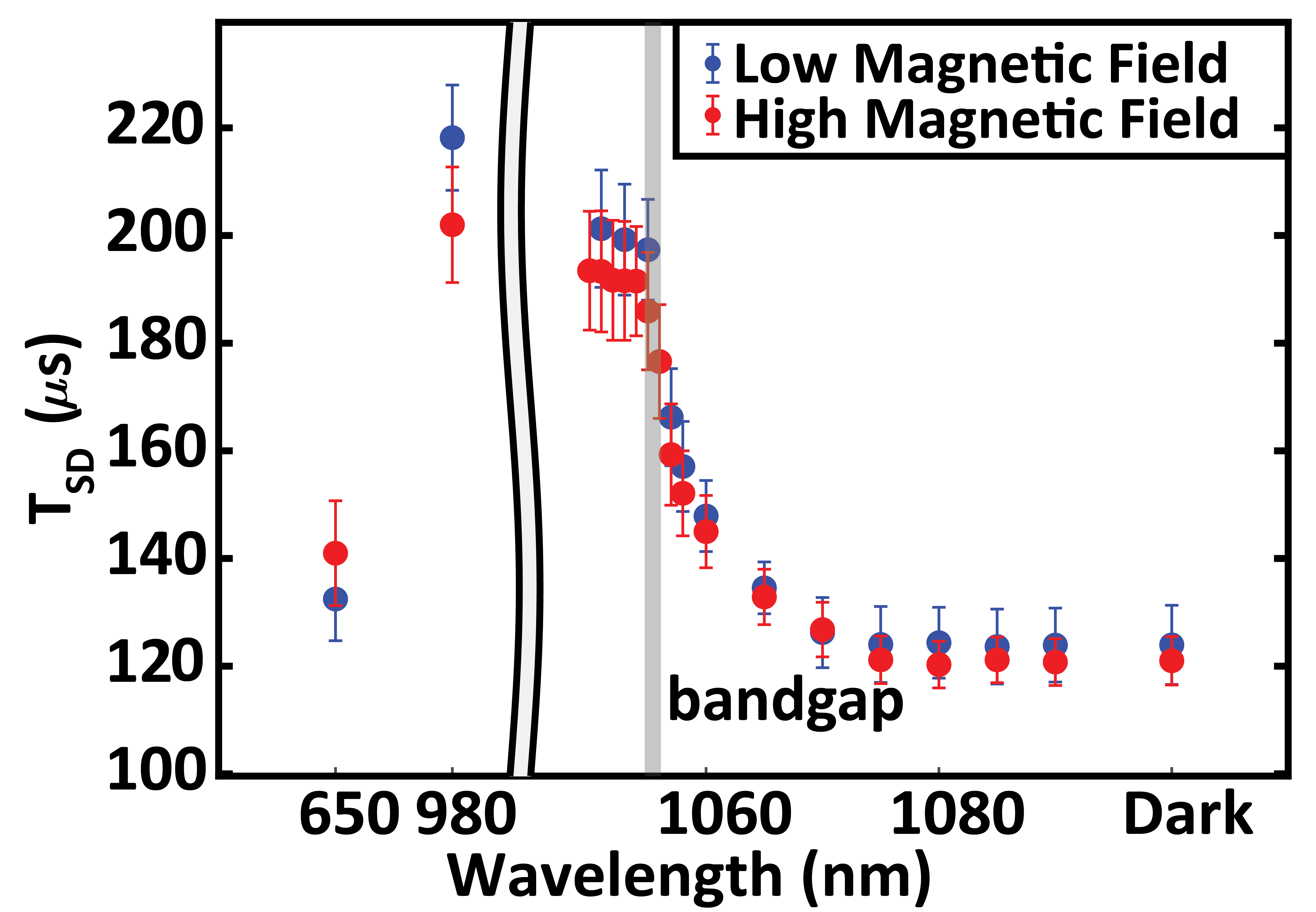
Figure 2 shows the measured spectral diffusion time at different laser wavelength for both the high-field and the low-field phosphorus peaks of Figure 1(b). The signal decays are fit to just the T term using . The error bars are calculated by the 95 percent confidence interval of the T. The data show that T increases as the energy of the optical excitation is increased above the silicon band-gap, and that the change in spectral diffusion time is the same for both the high-field and low-field phosphorus peaks. The longer T is also observed with the 980 nm laser, but falls off again at 650 nm, likely due to the limited penetration depth into the sample. It should be noted that the signal measured in these inductively detected pulsed ESR experiments arises from the entire sample. At cryogenic temperatures the optical penetration depth of light into silicon is known to be strongly wavelength dependent Macfarlane et al. (1958). For wavelengths of 1050 nm and longer the optical penetration depth is several centimeters, while it reduces to about 100 m at 980 nm and to 2-3 m at 650 nm. Thus the 3 mm thick sample is expected to be uniformly illuminated at the longer wavelengths, fairly inhomogeneously illuminated at 980 nm and only the very surface of the sample is optically excited by the 650 nm wavelength. The photo-excited carriers can travel further into the sample, with free excitons traveling up to a millimeter in silicon under these conditions Tamor and Wolfe (1980).
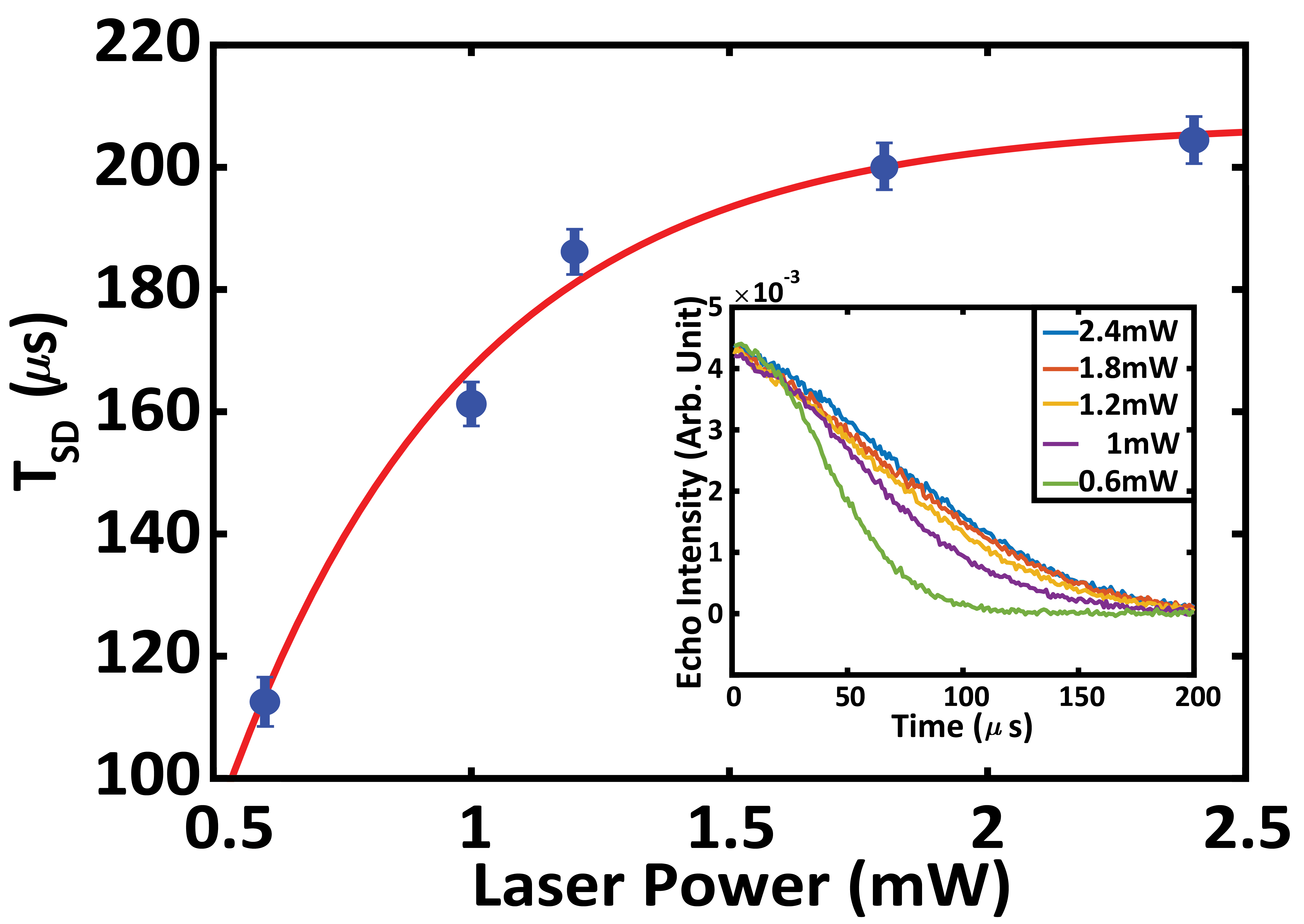
Figure 3 shows the change in T as a function of nominal laser power (measured at the output of the optical fiber) with the 980 nm laser for the high magnetic field transition. The fit indicates that T saturates to a maximum of 208 s with increasing laser power. This is close to the value measured at low magnetic fields.
The increase in T indicates either that the magnitude of the noise reduces, or the spectral features change – either slowing down and enabling better refocusing by the spin echo or fluctuating rapidly enough to enable a ”motional narrowing” of the noise Klauder and Anderson (1962). For NV centers in diamond, polarizing the spin bath (electronic P1 centers) has been shown to suppress spectral diffusion and increase TTakahashi et al. (2008). While above bandgap illumination has been shown to hyperpolarize the phosphorus donor spins at high field and low temperature McCamey et al. (2009); Gumann et al. (2014, 2018), only weak optical hyperpolarization of the silicon spins has been observed to date at high magnetic fields Verhulst et al. (2005). Verhulst et al. observed a 29Si spin polarization of 0.25% at 500 mW after 30 minutes of laser irradiation at 7 T and 4.2 K. With 1024.8 nm excitation, they initially observed a reduction in T1 of the silicon spins at low optical power ( 1 mW) and a growth of 29Si hyperpolarization at higher optical power ( 1 mW). The 1-2 mW optical power coming out of the fiber in our experiments is just on the threshold of when 29Si hyperpolarization was observed, so it is unlikely that the 29Si nuclear spins polarization is sufficiently high to change T significantly. More importantly, given the long T1 of the silicon nuclear spins, the enhanced T should persist for several hours. However, as shown in Figure 1(b), the enhanced spectra diffusion times were not observed when the experiment was repeated in the dark 15 minutes later.
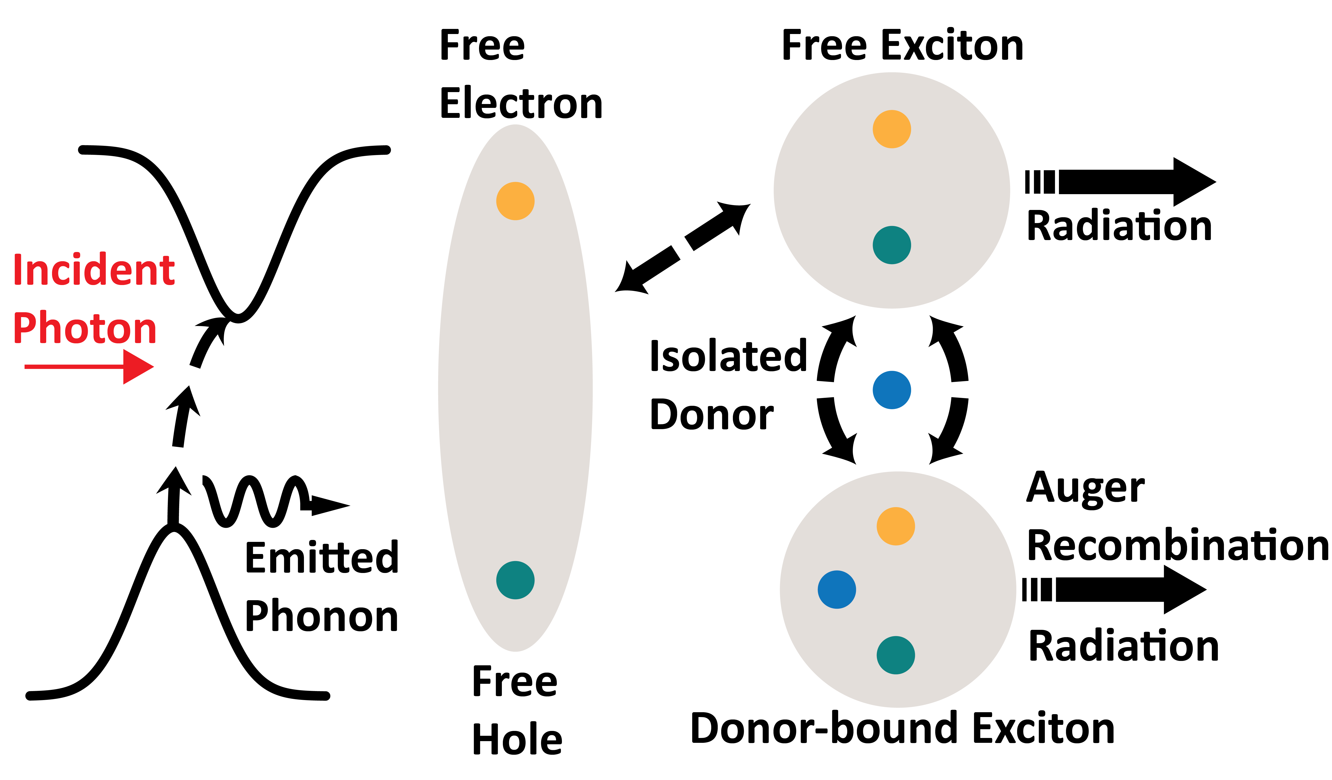
Alternatively, spectral diffusion effects can also be reduced if the effective strength of either the inter-nuclear dipolar coupling or the hyperfine coupling are reduced or acquire rapid random rapid fluctuations that lead to motional narrowing of the local field fluctuations. One possibility is that modulation of the donor electron wavefunction by mobile carriers modulates and reduces the silicon hyperfine couplings. While noise spectroscopy Álvarez and Suter (2011); Bylander et al. (2011) could be used in principle to directly measure the spectrum of magnetic noise seen by the donors, the lack of phase control on the 240 GHz instruments does not permit robust multiple-pulse dynamical decoupling experiments. Disentangling the different potential sources of magnetic field fluctuations in the presence of light is a non-trivial task. Optical excitation creates a dynamically rich environment for the spins as can be seen from the schematic in Figure 4.

At the absorption edge, the electrons and holes are primarily in the form of free excitons (the exciton binding energy is meV Green (2013)), while at higher optical excitation energies above 1.19 eV, it is possible to generate free electrons and holes - which in turn form free excitons as they lose energy. The optical generation of free excitons is followed by continuous interconversion between free and bound excitons, and both radiative recombination of free excitons and non-radiative Auger recombination of bound excitons Hammond and Silver (1980). A 1 mW excitation with 1 eV photons would result in photoelectrons/excitons generated per second if all the power is deposited in the sample. Given the penetration depth of 1 eV photons in silicon only about 10% of the photons are absorbed. Since the free exciton lifetime at 4.2 K is on the order of 1 s Hammond and Silver (1980), this results in an estimated exciton density of about cm-3 in the sample - significantly lower than the donor concentration. However, the excitonic Bohr radius is about 5 nm McLean and Loudon (1960) - larger than that of the donor electron - and they are known to have extremely high mobilities Tamor and Wolfe (1980). It is possible that that exciton dynamics contributes to the observed increase in T as the electrons and holes comprising these excitons can have fairly strong hyperfine couplings to the silicon nuclei in the lattice Philippopoulos et al. (2020).

In conclusion, the spectral diffusion time measured in the dark at high field (8.59 T) was observed to be more than a factor of 2 shorter than the values measured at 0.35 T, though the simple theory outlined here would suggest that the values should be identical. In the presence of weak above band-gap optical excitation, T was observed to increase by almost a factor of 2 suggesting a motional narrowing of the spectrum of magnetic field fluctuations seen by the donor.
I Acknowledgements
We thank William Coish and Susumu Takahashi for helpful discussions. This work was supported in part by the National Science Foundation under grants OIA-1921199. Part of the work was done at the National High Magnetic Field Laboratory, which is supported by the NSF under grant DMR-1644779 and the State of Florida.
References
- Awschalom et al. (2013) D. D. Awschalom, L. C. Bassett, A. S. Dzurak, E. L. Hu, and J. R. Petta, Science 339, 1174 (2013).
- Awschalom et al. (2018) D. D. Awschalom, R. Hanson, J. Wrachtrup, and B. B. Zhou, Nature Photonics 12, 516 (2018).
- Tyryshkin et al. (2006) A. M. Tyryshkin, J. J. L. Morton, S. C. Benjamin, A. Ardavan, G. A. D. Briggs, J. W. Ager, and S. A. Lyon, J Phys-Condens Mat 18, S783 (2006).
- Tyryshkin et al. (2011) A. M. Tyryshkin, S. Tojo, J. J. L. Morton, H. Riemann, N. V. Abrosimov, P. Becker, H.-J. Pohl, T. Schenkel, M. L. W. Thewalt, K. M. Itoh, et al., Nature Materials 11, 143 (2011).
- de Sousa and Das Sarma (2003) R. de Sousa and S. Das Sarma, Phys. Rev. B 68, 115322 (2003).
- Ma et al. (2014) W.-L. Ma, G. Wolfowicz, N. Zhao, S.-S. Li, J. J. Morton, and R.-B. Liu, Nat Commun 5, 4822 (2014).
- Ma et al. (2015) W.-L. Ma, G. Wolfowicz, S.-S. Li, J. J. L. Morton, and R.-B. Liu, Phys. Rev. B 92, 161403 (2015).
- Witzel and Das Sarma (2006) W. M. Witzel and S. Das Sarma, Phys. Rev. B 74, 035322 (2006).
- Witzel et al. (2007) W. M. Witzel, X. Hu, and S. Das Sarma, Phys. Rev. B 76, 737 (2007).
- Yang et al. (2017) W. Yang, W.-L. Ma, and R.-B. Liu, Rep. Prog. Phys. 80, 016001 (2017).
- Schweiger and Jeschke (2001) A. Schweiger and G. Jeschke, Principles of pulse electron paramagnetic resonance (Oxford, 2001).
- Morello et al. (2010) A. Morello, J. J. Pla, F. A. Zwanenburg, K. W. Chan, K. Y. Tan, H. Huebl, M. Mottonen, C. D. Nugroho, C. Yang, J. A. van Donkelaar, et al., Nature 467, 687 (2010).
- Pla et al. (2012) J. J. Pla, K. Y. Tan, J. P. Dehollain, J. J. L. Lim, W. H.and Morton, D. N. Jamieson, A. S. Dzurak, and A. A. Morello, Nature 489, 541 (2012).
- Muhonen et al. (2014) J. T. Muhonen, J. P. Dehollain, A. Laucht, F. E. Hudson, R. Kalra, T. Sekiguchi, K. M. Itoh, D. N. Jamieson, J. C. McCallum, A. S. Dzurak, et al., Nature nanotechnology 9, 986 (2014).
- Veldhorst et al. (2015) M. Veldhorst, C. Yang, J. Hwang, W. Huang, J. Dehollain, J. Muhonen, S. Simmons, A. Laucht, F. Hudson, K. M. Itoh, et al., Nature 526, 410 (2015).
- He et al. (2019) Y. He, S. K. Gorman, D. Keith, L. Kranz, J. G. Keizer, and M. Y. Simmons, Nature 571, 371 (2019).
- van Tol et al. (2005) J. van Tol, L.-C. Brunel, and R. J. Wylde, Review of Scientific Instruments 76, 074101 (2005).
- Slichter (1955) C. P. Slichter, Phys. Rev. 99, 479 (1955).
- Dementyev et al. (2011) A. E. Dementyev, D. G. Cory, and C. Ramanathan, J. Chem. Phys. 134 (2011).
- Morley et al. (2008) G. Morley, D. McCamey, and H. Seipel, Phys. Rev. Lett. 101, 207602 (2008).
- McCamey et al. (2009) D. R. McCamey, J. van Tol, G. W. Morley, and C. Boehme, Phys. Rev. Lett. 102, 027601 (2009).
- McCamey et al. (2010) D. R. McCamey, J. van Tol, G. W. Morley, and C. Boehme, Science 330, 1652 (2010).
- McCamey et al. (2012) D. R. McCamey, C. Boehme, G. W. Morley, and J. van Tol, Phys. Rev. B 85, 073201 (2012).
- Verhulst et al. (2005) A. S. Verhulst, I. G. Rau, Y. Yamamoto, and K. M. Itoh, Phys. Rev. B 71, 235206 (2005).
- Yang and Liu (2008) W. Yang and R.-B. Liu, Phys. Rev. B 78, 935 (2008).
- Witzel et al. (2010) W. M. Witzel, M. S. Carroll, A. Morello, L. Cywinski, and S. Das Sarma, Phys. Rev. Lett. 105, 187602 (2010).
- Kohn and Luttinger (1955a) W. Kohn and J. M. Luttinger, Phys. Rev. 97, 883 (1955a).
- Kohn and Luttinger (1955b) W. Kohn and J. M. Luttinger, Phys. Rev. 98, 915 (1955b).
- Green (1990) M. A. Green, Journal of Applied Physics 67, 2944 (1990).
- Green (2013) M. A. Green, AIP Advances 3, 112104 (2013).
- Macfarlane et al. (1958) G. G. Macfarlane, T. P. McLean, J. E. Quarrington, and V. Roberts, Physical Review 111, 1245 (1958).
- Tamor and Wolfe (1980) M. A. Tamor and J. P. Wolfe, Phys. Rev. Lett. 44, 1703 (1980).
- Klauder and Anderson (1962) J. R. Klauder and P. W. Anderson, Phys Rev 125, 912 (1962).
- Takahashi et al. (2008) S. Takahashi, R. Hanson, J. van Tol, M. S. Sherwin, and D. D. Awschalom, Phys. Rev. Lett. 101, 047601 (2008).
- Gumann et al. (2014) P. Gumann, O. Patange, C. Ramanathan, H. Haas, O. Moussa, M. L. W. Thewalt, H. Riemann, N. V. Abrosimov, P. Becker, H. J. Pohl, et al., Phys. Rev. Lett. 113, 267604 (2014).
- Gumann et al. (2018) P. Gumann, H. Haas, S. Sheldon, L. Zhu, R. Deshpande, T. Alexander, M. L. W. Thewalt, D. G. Cory, and C. Ramanathan, Phys. Rev. B 98, 180405 (2018).
- Álvarez and Suter (2011) G. A. Álvarez and D. Suter, Phys. Rev. Lett. 107, 230501 (2011).
- Bylander et al. (2011) J. Bylander, S. Gustavsson, F. Yan, F. Yoshihara, K. Harrabi, G. Fitch, D. G. Cory, Y. Nakamura, J.-S. Tsai, and W. D. Oliver, Nature Phys 7, 565 (2011).
- Hammond and Silver (1980) R. B. Hammond and R. N. Silver, Appl. Phys. Lett. 36, 68 (1980).
- McLean and Loudon (1960) T. McLean and R. Loudon, Journal of Physics and Chemistry of Solids 13, 1 (1960).
- Philippopoulos et al. (2020) P. Philippopoulos, S. Chesi, and W. A. Coish, Phys. Rev. B 101, 115302 (2020).