MIDLMedical Imaging with Deep Learning
\jmlryear2021
\midlauthor
\NameBarhoumi Yassine \Emailbarhou29@students.rowan.edu
\NameGhulam Rasool \Emailrasool@rowan.edu
\addrRowan University, New Jersey, USA
Scopeformer: n-CNN-ViT Hybrid Model for Intracranial Hemorrhage Classification
Abstract
We propose a feature generator backbone composed of an ensemble of convolutional neural networks (CNNs) to improve the recently emerging Vision Transformer (ViT) models. We tackled the RSNA intracranial hemorrhage classification problem, i.e., identifying various hemorrhage types from computed tomography (CT) slices. We show that by gradually stacking several feature maps extracted using multiple Xception CNNs, we can develop a feature-rich input for the ViT model. Our approach allowed the ViT model to pay attention to relevant features at multiple levels. Moreover, pretraining the ”n” CNNs using various paradigms leads to a diverse feature set and further improves the performance of the proposed n-CNN-ViT. We achieved a test accuracy of 98.04% with a weighted logarithmic loss value of 0.0708. The proposed architecture is modular and scalable in both the number of CNNs used for feature extraction and the size of the ViT.
keywords:
Computed Tomography slices, Intracranial hemorrhage, CNN, ViT.1 Introduction
Motivated by the recently emerging vision transformer model [Dosovitskiy et al.(2020)Dosovitskiy, Beyer, Kolesnikov, Weissenborn, Zhai, Unterthiner, Dehghani, Minderer, Heigold, Gelly, Uszkoreit, and Houlsby], we propose a hybrid architecture composed of multiple CNNs for feature extraction and a vision transformer designated for intracranial hemorrhage classification. In our work, we hypothesize that utilizing feature maps extracted from highly crafted CNNs can improve the information content the ViT is processing and the resolution input it attends to. Furthermore, we hypothesize that generating features from the same input image using multiple CNNs leads to a richer feature content with higher resolution. To this end, we used multiple Xception CNN feature extractors, pretrained on separate paradigms using two distinguished datasets. The first CNN model used the ImageNet dataset for pretraining, which was then fine-tuned on the RSNA dataset. The second CNN model was priorly pretrained on the ImageNet dataset and then further pretrained on data generated from ImageNet using a Generative adversarial network (GAN) [Goodfellow et al.(2014)Goodfellow, Pouget-Abadie, Mirza, Xu, Warde-Farley, Ozair, Courville, and Bengio] applied on several brain computed tomography images. The idea behind generating the dataset using GAN was motivated by the dissimilarities of ImageNet natural images and the 2D medical images. We reduce these dissimilarities by further pretraining on the generated GAN images to approach a better inductive bias for our target computed tomography dataset.
2 Methodology
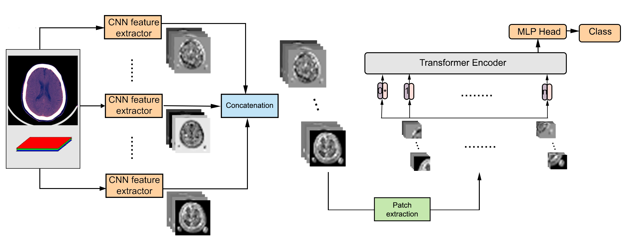
The Scopeformer model, presented in figure 1, is an extension of the vision transformer (ViT) architecture. It is applied either directly to raw images or to a given ”n” number of feature maps extracted from the latest Xception Add layers and concatenated to a single feature map. We adopted the base ViT variant with 12 encoder layers and a latent vector dimension of 1456. In our experiments, we used the RSNA intracranial hemorrhage dataset [Flanders AE(2019)] by generating 2242243 images from the DICOM files [Burduja M(2020)]. The input image to each feature extractor is 2242243, and the output dimension is 771024. For multiple CNNs, the size of the input vector will be 77(n1024). A smaller version of n-CNN-ViT models was introduced to reduce the computational complexity of the ViT input, where we use a 11 CNN filter after the Xception Add layer to reduce the dimension from 1024 to 128.
3 Results and discusion
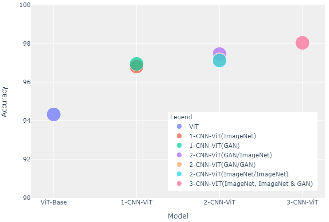
The performances of our models are evaluated by the multi-label weighted mean logarithmic loss [Goodfellow et al.(2014)Goodfellow, Pouget-Abadie, Mirza, Xu, Warde-Farley, Ozair, Courville, and Bengio]. Figure 2 shows the classification accuracy of our models against the number of CNNs in the feature extractor backbone for different pretraining paradigm settings. We observe that the classification accuracy is directly proportional to the number of Xception models. Moreover, using the same pretrained weights for the 2-CNN-ViT model results in lower accuracies compared to using diverse backbones. Table 1, summarizes best models within each variant. Applying the ViT encoder directly on the raw intracranial hemorrhage images shows that the proposed ViT model by [Dosovitskiy et al.(2020)Dosovitskiy, Beyer, Kolesnikov, Weissenborn, Zhai, Unterthiner, Dehghani, Minderer, Heigold, Gelly, Uszkoreit, and Houlsby] cannot overcome models without including CNNs as claimed. In fact, The more we add features with a more diverse CNN pretraining paradigms, the richer the feature content will be and the better the ViT attends to the input to extract global attention among patches. The n-CNN-ViT model uses a base ViT variant and relatively small Xception CNN feature maps. Furthermore, we show comparable results for the smaller version of the 2-CNN-ViT compared to larger ViT inputs. This implies the degree of modularity and scalability of the proposed model.
| Model | ViT input dimension | Validation accuracy | Loss |
|---|---|---|---|
| ViT | 2562563 | 94.33% | 0.1822 |
| 1-CNN-ViT (GAN) | 771024 | 96.95% | 0.08272 |
| 2-CNN-ViT (ImageNet/GAN) | 772048 | 97.46% | 0.07754 |
| 3-CNN-ViT (ImageNet/ImageNet/GAN) | 773072 | 98.04% | 0.07050 |
| 2-CNN-ViT (ImageNet/GAN) | 7x7x256 | 97.58% | 0.07903 |
References
- [Burduja M(2020)] Verga N. Burduja M, Ionescu RT. Accurate and efficient intracranial hemorrhage detection and subtype classification in 3d ct scans with convolutional and long short-term memory neural networks. sensors., 2020.
- [Dosovitskiy et al.(2020)Dosovitskiy, Beyer, Kolesnikov, Weissenborn, Zhai, Unterthiner, Dehghani, Minderer, Heigold, Gelly, Uszkoreit, and Houlsby] Alexey Dosovitskiy, Lucas Beyer, Alexander Kolesnikov, Dirk Weissenborn, Xiaohua Zhai, Thomas Unterthiner, Mostafa Dehghani, Matthias Minderer, Georg Heigold, Sylvain Gelly, Jakob Uszkoreit, and Neil Houlsby. An image is worth 16x16 words: Transformers for image recognition at scale, 2020.
- [Flanders AE(2019)] Shih G et al (2020) Flanders AE, Prevedello LM. Construction of a machine learning dataset through collaboration: the rsna 2019 brain ct hemorrhage challenge. radiology, 2019.
- [Goodfellow et al.(2014)Goodfellow, Pouget-Abadie, Mirza, Xu, Warde-Farley, Ozair, Courville, and Bengio] Ian J. Goodfellow, Jean Pouget-Abadie, Mehdi Mirza, Bing Xu, David Warde-Farley, Sherjil Ozair, Aaron Courville, and Yoshua Bengio. Generative adversarial networks, 2014.