High Resolution Spectroscopy of Neutral Yb Atoms in a Solid Ne Matrix
Abstract
We present an experimental and theoretical study of the absorption and emission spectra of Yb atoms in a solid Ne matrix at a resolution of 0.025 nm. Five absorption bands were identified as due to transitions from the ground state configuration to and configurations. The two lowest energy bands were assigned to outer-shell transitions to and atomic states and displayed the structure of a broad doublet and an asymmetric triplet, respectively. The remaining three higher-frequency bands were assigned to inner-shell transitions to distinct states arising from the configuration and were highly structured with narrow linewidths. A classical simulation was performed to identify the stability and symmetry of possible trapping sites in the Ne crystal. It showed that the overarching 1+2 structure of the high frequency bands could be predominantly ascribed to crystal field splitting in the axial field of a 10-atom vacancy of symmetry. Their prominent substructures were shown to be manifestations of phonon sidebands associated with the zero-phonon lines on each crystal field state. Unprecedented for a metal-rare gas system, resolution of individual phonon states on an allowed electronic transition was possible under excitation spectroscopy which reflects the semi-quantum nature of solid Ne. In contrast to the absorption spectra, emission spectra produced by steady-state excitation into the absorption band consisted of simple, unstructured fluorescence bands.
I Introduction

Rare gas (RG) solids, formed at cryogenic temperatures, are a promising host medium for the capture, detection and quantum state manipulation of guest atoms and molecules. They provide stable and chemically inert isolation and confinement for a wide variety of guest species at a tunable density—from a single isolated atom to a number possibly exceeding atoms/cm3. Because RG solids are transparent at optical wavelengths, the guest species can be probed using lasers and the induced fluorescence efficiently detected outside the solid. Spin coherence times of the guest species, which are ultimately dominated by long-range dipolar couplings vv48 ; ka53 , could be made as long as s for nuclear spins and s for electronic spins by minimizing spin impurities within the host matrix. Applications of this “matrix isolation” technique include tests of fundamental symmetries kd06 ; edm3 , magnetometry kanagin13 ; upadhyay20 and quantum information science wolf21 .
Examples of systems studied specifically for these applications include alkali atoms in both solid RGs wp65 and parahydrogen upadhyay19a , Yb atoms in solid Ne xu11 ; xu14 and Tm atoms in solid Ar and Kr gaire19 . For these species, medium effects generally broaden linewidths and shift transition frequencies by several hundred wavenumbers wp65 . Despite substantial matrix-induced perturbations to their -lines, however, alkali atoms have been successfully optically pumped in matrix isolation kanagin13 ; upadhyay16 . Furthermore, spin coherence times as long as 0.1 s have been observed for Rb atoms in solid parahydrogen upadhyay20 .
Ytterbium (Yb) is a heavy divalent atom with a electronic ground state, several optical transitions and a naturally abundant isotope, 171Yb (14%), whose nuclear spin is 1/2. These features combine to make matrix-isolated 171Yb a promising candidate for a solid state search for a permanent electric dipole moment rmp19 or for a nuclear spin-based qubit. Such applications depend on the ability to both optically prepare and readout the 171Yb nuclear spin state. This in turn depends crucially on the strength of the hyperfine interaction in the excited state compared to the size of the matrix-induced perturbations. The need to characterize the latter motivates the high resolution spectroscopic study of Yb atoms in solid Ne (sNe) presented here.
Recent work at relatively low resolution ( 0.5 nm) has detailed some of the complexities of Yb/RG systems. When solid Xe was used as the matrix host, absorption and emission bands were found to have a two-fold structure due to Yb occupation in tetravacancies and single substitutional sites kleshhina19Yb . Earlier work found that when solid Ar was the matrix host, the same bands had a three-fold structure, due to Yb occupation in hexavacancies, tetravacancies and single substitutional sites Tao2015 . The symmetry of these trapping sites generally induces crystal field splitting (CFS) of degenerate atomic states upon excitation CrepinTramer . Low symmetries further multiply the number of bands, while high symmetries create secondary structures due to Jahn-Teller electron-phonon coupling Bersuker . The phonon structure itself is usually not visible—except on certain forbidden transitions of the Mn/Kr by10 and Eu/Ar br11 systems—and predominantly contributes to the width of strongly non-Frank-Condon bright absorptions.
The above hierarchy of matrix-induced spectral perturbations is typical of the heavy “classical” RG solids made of Ar, Kr or Xe. In “quantum” matrices, such as those of He and H2, for which the effects of nuclear motion are expected to be much stronger, the identity of distinct trapping sites is eroded and large-amplitude motions magnify electron-nuclear couplings. Solid Ne is often regarded as a “semi-quantum” crystal in which one may expect incipient nuclear quantum effects Cazorla2017 . At the same time, the Ne crystal is a less perturbing environment than the heavier RGs. Isolated in it, the Yb electronic structure is still qualitatively the same as in vacuum, while the lifetimes of excited states are not significantly different xu11 ; xu14 . Based on these observations, single atom detection of Yb in sNe would appear feasible, which could then be extended for use in detecting rare nuclear reactions loseth19 . This would be in close analogy with the recent demonstration of single atom imaging of Ba in solid Xe, performed with a view to searching for neutrinoless double beta decaychambers19 .
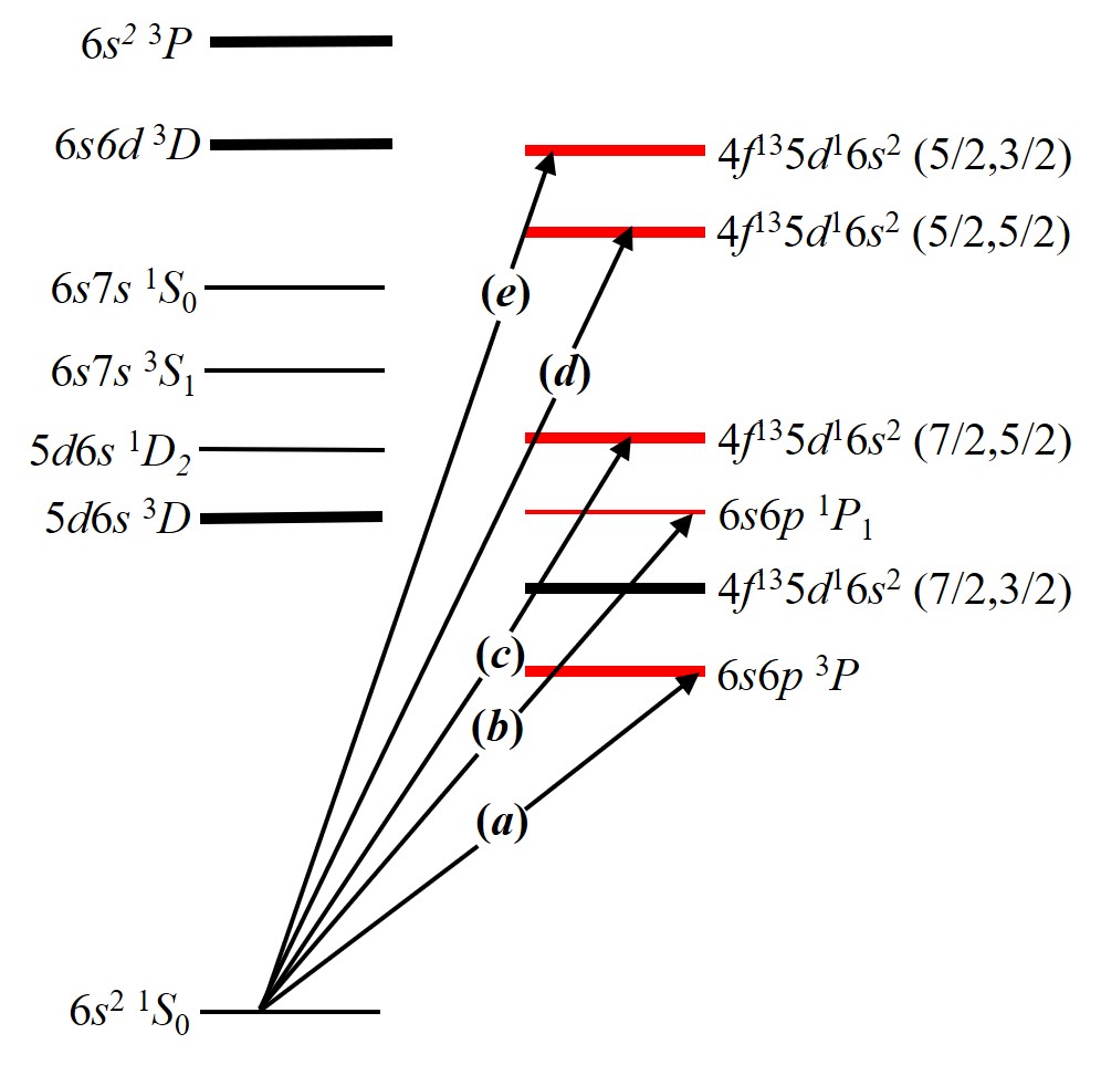
In order to investigate all these prospects, we have performed the first high resolution (0.025 nm) broadband spectroscopy of the low-lying states of the Yb/Ne system (Fig. 1). Five absorption bands were identified as due to Yb atomic transitions from the ground state of the configuration to states of the or configuration. These appear as either broad, largely featureless bands or narrow, highly structured ones depending on their susceptibility to the matrix-induced perturbations described above. To assist in their interpretation, we performed classical simulations that gave the structure and symmetry of the Yb/Ne trapping sites and whose corresponding theoretical spectra adequately explained the observed lineshapes. The spectrum resulting from steady state excitation was also recorded at the same high resolution. Knowledge of the decay paths originating from the excited singlet state will be important for single atom spectroscopy.
II Experimental Setup
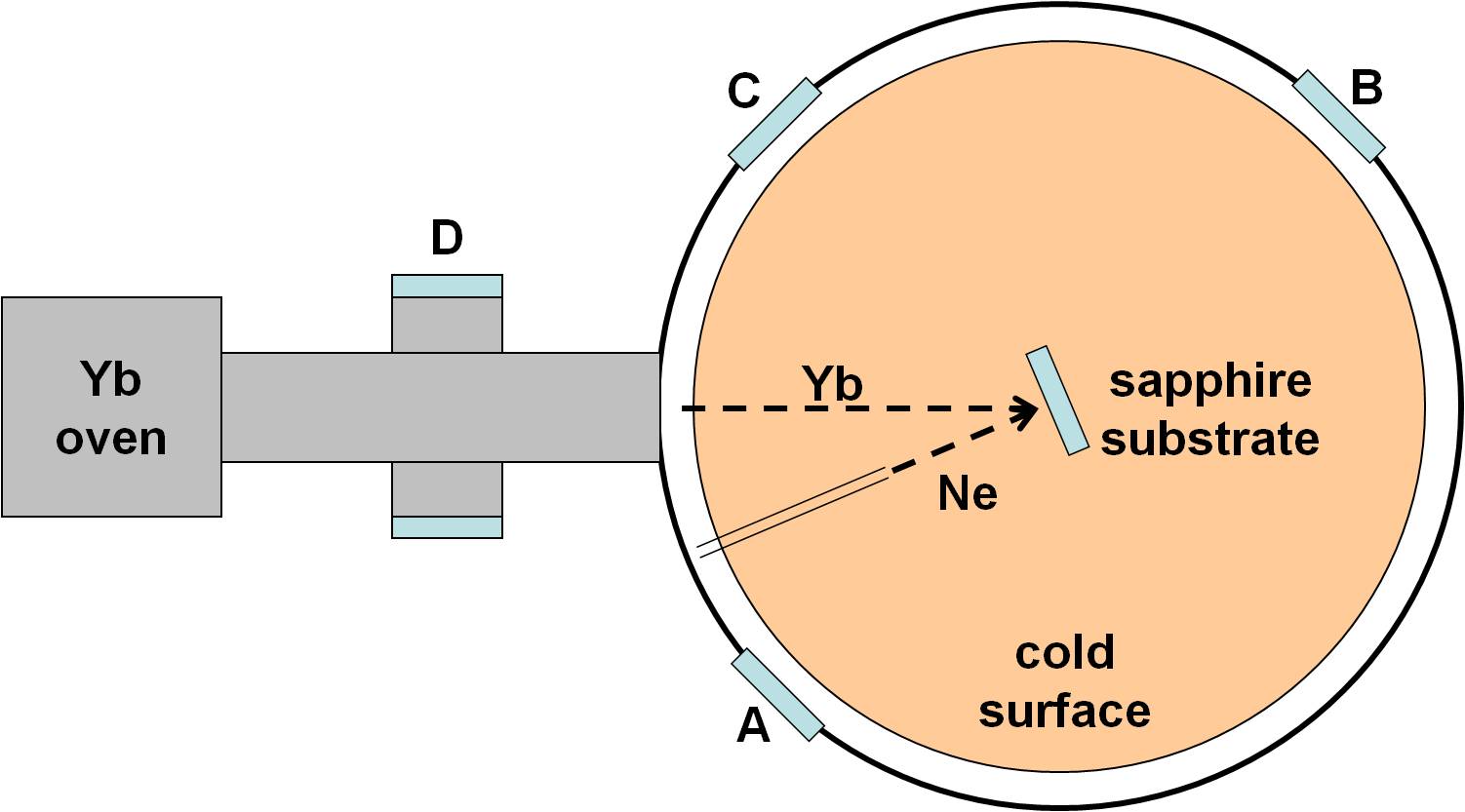
All the experiments were conducted with the liquid helium cryostat illustrated in Fig. 2. Its cold surface has an area of 300 cm2 that is cooled by the liquid helium bath to 4.2 K, at which temperature the pressure of the vacuum falls below Torr. The Yb/Ne samples were grown on a 2.54 cm diameter c-plane sapphire substrate installed vertically on the cold surface using a copper mount. Indium wires and low vapor pressure grease (Apiezon N) were used to achieve good thermal contact between the substrate and the cold surface. The temperature of the substrate was measured by a resistance temperature detector (Lakeshore CX-1050-AA) and was typically at 4.2-5.0 K depending on the thermal contact.
There were four 2.54 cm diameter ports on the perimeter of the cryostat. One port was connected to an effusion oven whose crucible was loaded with metallic Yb of natural isotope abundance. The other three ports (A, B and C in Fig. 2) were closed with fused silica windows and used as viewports. Viewports A and B, which were each at an angle of 22.5∘ to the normal direction of the substrate surface, were used during measurements of the sample thickness and the absorption spectroscopy. Viewport C, which was at an angle of 67.5∘ to the normal direction of the substrate surface, was used in the emission study to collect fluorescence from the sample upon excitation.
The sample was prepared as follows: the Ne gas first flowed through a purifier (LDetek LDP1000) and a 77 K charcoal trap and then leaked into the cryostat via a capillary tube that ended 5 cm away from the substrate. The implantation of Yb atoms was achieved by directing the beam from the effision oven at the substrate while the sNe crystal grew. The sample thickness was determined by recording the transmitted light of a He-Ne laser and counting the fringes due to the thin-film interference effect. Meanwhile, the Yb intensity was measured using the cross-beam-fluorescence method at the six-way cross (viewport D in Fig. 2). A 398.9 nm laser beam resonant with the Yb transition in vacuum was applied perpendicularly to the Yb beam and the fluorescence measured using a photomultiplier tube along an axis perpendicular to both beams.

We found empirically that individual Yb atoms (as opposed to higher aggregates) were isolated in sNe only when the Yb:Ne ratio was kept below 5 ppm. In practice, we held that ratio at 1 ppm for which a Yb beam intensity of atoms/cmhr and an oven temperature of 620 K were required. The steady state Ne growth rate was typically 50 m/hr and the Yb areal and volumetric densities were estimated to be atoms/cm2 and atoms/cm3 respectively. Under these conditions, the heat load due to the deposition of Ne and the blackbody radiation of the oven increased the substrate temperature by only 50 mK. A ramp-up time of two hours was required to preserve the transparency of the sample and, once this rate was reached, it could be grown for five hours before it cracked under its own internal strain.
In the absorption study, a weak broadband light source (Ocean Optics DH2000-DUV) illuminated the sample through viewport A, and the transmitted light was collected through viewport B and fiber-coupled to a spectrometer. In the emission study, a light-emitting diode (LED) centered at 385 nm illuminated the sample through viewport A, and the induced fluorescence was collected at viewport C and also fiber-coupled to a spectrometer. Two spectrometers were available to us to analyze the absorption and the emission signals, both of which used line CCD cameras as the detectors. The Ocean Optics USB4000-UV-VIS spectrometer covered a broad range from 200 nm to 1100 nm and had a resolution of 1.5 nm. The McPherson 225 spectrometer had a resolution of 0.025 nm and covered a range from 0 nm to 600 nm but with a camera frame that was only 40 nm wide.
To obtain the highest possible spectral resolution, we also used an optical parametric oscillator (OPO) (Continuum Sunlite EX) as the probe light. The OPO was pumped by a third harmonic pulsed Nd:YAG laser (Continuum Powerlite DLS) before passing through a frequency doubler (Inrad Autotracker II) to provide UV light. Each pulse was about 10 ns long and contained about 1 mJ of energy. The light was attenuated by four orders of magnitude to avoid melting the sample and damaging the detector. The output beam was split into a probe beam that went through the sample and a reference beam that traveled outside the cryostat. The linewidth of the OPO was about 1 GHz and the frequency was scanned in 2 GHz steps to map out the lineshape of the whole absorption band.
III Results
III.1 Probe with Broadband Light
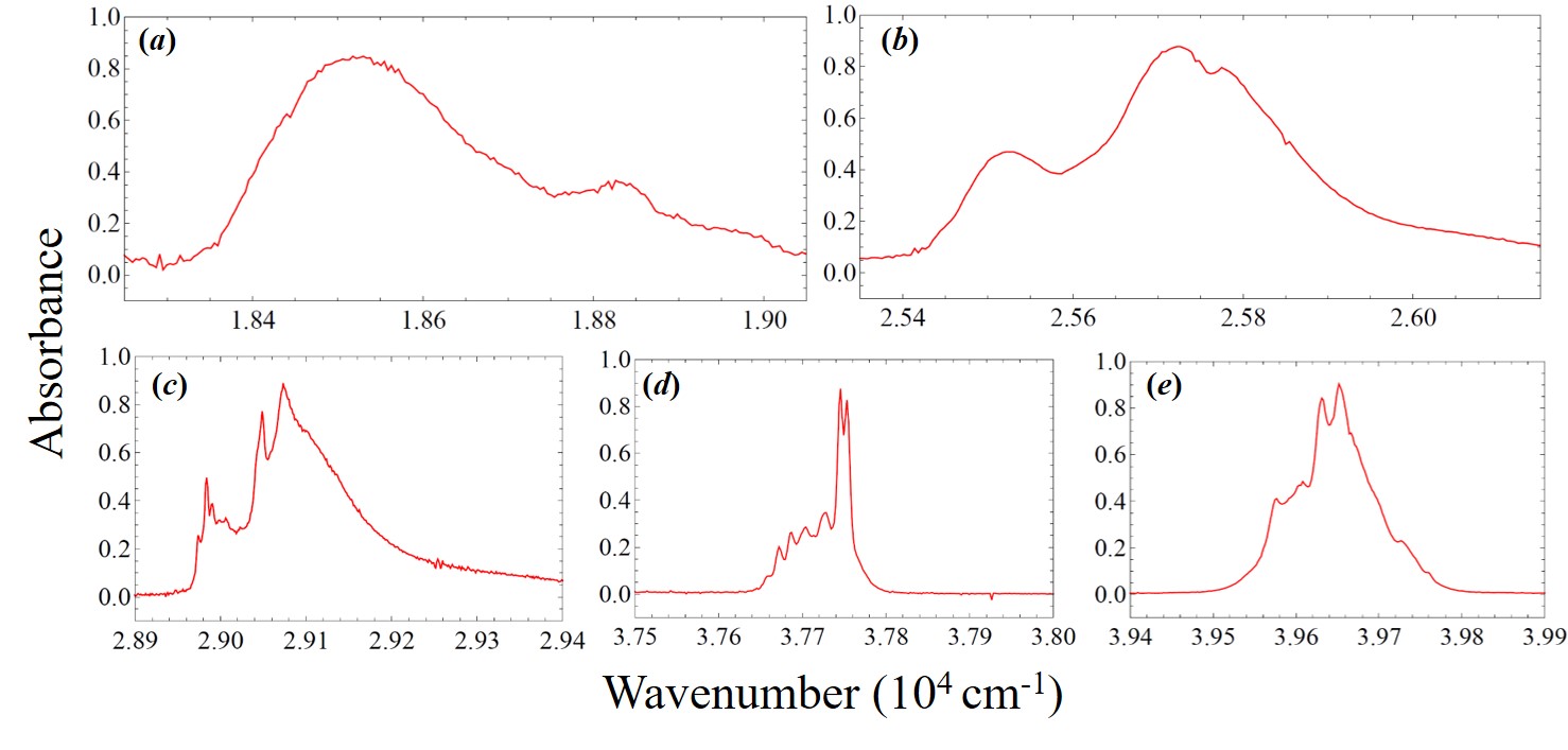
Figures 3 and 4 show the white-light absorption spectra of a typical sample taken, respectively, with the 1.5 nm resolution Ocean Optics and 0.025 nm McPherson spectrometers. Sample deposition leads to a decrease in the transparency of the subtrate which manifests itself as frequency-dependent distortions in the baselines. Nonetheless, five absorption bands, represented in terms of absorbance, can be clearly seen and are identified as due to electronic transitions originating from ground state Yb atoms. Given that the configuration of the ground state is , low-lying excitations can either be outer-shell transitions or inner-shell transitions, the latter of which break the full shell and leave the shell intact. In the spectra, bands (a) and (b) correspond to outer-shell transitions whose final levels are and respectively. In contrast, bands (c), (d) and (e) correspond to inner-shell transitions and their final states are in the coupling scheme with equal to , and , respectively. A comparison between the wavenumbers of transitions in vacuum and those in sNe is summarized in Table 1.

| Band | Transition | |||
|---|---|---|---|---|
| (a) | 17,992 | 18,510 | +518 | |
| (b) | 25,068 | 25,760 | +692 | |
| (c) | 28,857 | 29,140 | +283 | |
| (d) | 37,415 | 37,730 | +315 | |
| (e) | 38,422 | 39,680 | +1,260 |

Band (a), assigned to the intercombination transition, was not observed in our earlier work but becomes visible here due to an increase in the Yb areal density by an order of magnitude xu11 . Another difference is in our reassignment of band (d). Recent spectroscopic data and calculations of free Yb atoms wc79 ; zdwh02 ; ko10 indicate that it should be associated the state rather than, as previously thought, the one. In the case of band (e), there was also some ambiguity in its assignment originally. In earlier work it has been assigned to to the excited level, the energy of which is 40,564 cm-1 above the ground state in vacuum sa88 . This implies a red-shift in its transition frequency in sNe that is peculiar given that the other four transitions are all blue-shifted and makes the narrowness of its peaks inconsistent with the typical effects of the matrix on an transition. Reassigning it to the level removes these inconsistencies, though at the cost of a larger-than-expected absolute shift.
In matrix isolation, outer-shell and inner-shell transitions have different lineshapes due to the screening of medium effects by the outer-shell electrons sa88 . For Yb in sNe, the difference can be readily seen by comparing bands (a) and (b) () with bands (c), (d) and (e) () at the higher spectral resolution of Figure 4. Band (a) has two principal structures with maxima at 18,530 cm-1 and 18,826 cm-1, while band (b) has three principal structures with maxima at 25,525 cm-1, 25,724 cm-1 and 25,774 cm-1. Although both bands (a) and (b) display some splitting, the linewidth of each peak remains a few hundred wavenumbers. By contrast, bands (c), (d) and (e) have more complex structures in which the narrowest linewidth is comparable to the instrument resolution. The three principal structures in band (c) have their peaks at 28,988 cm-1, 29,049 cm-1 and 29,073 cm-1. In band (e) there are two clear peaks at 39,631 cm-1 and 39,652 cm-1 and an unresolved shoulder extending from 39,580 to 39,610 cm-1. In band (d) we have a distinct doublet with peaks at 37,741 cm-1 and 37,751 cm-1 and a series of smaller peaks constituting a substructure that extends to lower frequencies.
In the subsequent sections, we propose that the principal peaks of these latter bands are connected to ZPLs on individual crystal field levels. We also propose that substructures on these bands are the manifestations of phonon excitations that produce the phonon sidebands typically associated with ZPLs, of which band (d) offers a prime example.
III.2 Probe with Laser Light
In order to investigate further the vibronic progression seen in band (d), the transition was probed at 2 GHz resolution using frequency doubled light from the OPO. Figure 5 shows a plot of the absorbed light intensity (normalized against the incident light intensity) versus wavenumber. As with the original white-light spectra, the two peaks centered at 37,741 cm-1 and 37,751 cm-1 were assigned to the state. Extending to lower frequencies, between 37,631 cm-1 and 37,733 cm-1, one can see a series of 9 smaller, consecutive peaks. Possible explanations for this series include multiple site occupancy and/or CFS. However, both of these seem unlikely in this case. First, there is no analogous structure in bands (c) and (e), which indicates that this is a phenomenon peculiar to the transition. Second, attributing this series to multiple site occupancy would require the existence of too many additional trapping sites: at least 3 of axial symmetry, given that CFS gives rise to 3 features for each state; and at most 9, if we assume a one-to-one correspondence between the peaks and trapping sites. This is unsupported by the theoretical calculations of Section IV.1, while the regularity of the peaks—a spacing of 14 cm-1 for the first four—has never been observed in other cases of multiple site occupation.
Instead, we interpret the harmonic series of Fig. 5 as evidence of coupling between the electronic transition and a small number of local phonon modes. This type of structure is usually not visible in RG matrices as electronic transitions tend to couple strongly to a large number of lattice modes to produce broad unstructured bands in the spectra. In ionic crystals, the emergence of strong vibronic sidebands in intra 4fN electronic transitions is associated with strong configuration interaction and coupling to charge transfer states gu14 . These mechanisms likely also operate here since crystal field splitting induced by van der Waals forces in the neutral Mn/Kr system is attributed to the dominance of: (i) inter-electronic repulsion in the metal atom in the case of weak splitting; or (ii) ligand-metal electron repulsion in the case of strong splitting by10 .
In the most common treatment, the strength of the electron-phonon coupling is measured by the Huang-Rhys parameter, , which is related to the displacement in the lattice equilibrium position near a defect site, , accompanying an electron transition according to:
| (1) |
for which and are the frequencies of the vibrational mode in the initial and final electronic states, respectively, and is the effective mass of the defect site. At very low temperature (i.e. cm-1) vibronic transitions can be approximated as arising from the zero-phonon () state and , the probability that the th vibrational state is populated, can be straightforwardly shown under the Franck-Condon approximation to obey which is also the form of the overall envelope of the vibronic band zh19 . Within that band, on the assumption that coupling involves a single vibrational mode and that the frequencies of the oscillators follow a Gaussian distribution around the mode’s average frequency, , the intensity function of the phonon line is wa64 :
| (2) |
for which is its linewidth and is the energy difference between the initial and final electronic states.
We therefore expect to see a harmonic progression in the absorption spectrum in intervals of and, in Fig. 5, the three phonon lines corresponding to can be clearly distinguished, centered at cm-1, cm-1 and cm-1, respectively. Per Equation (2), the order corresponding to the ZPL should be a -function. In reality, because of electronic dispersion and inhomogeneous broadening due to trapping site inhomogeneity it has a finite width, . Higher order () phonon lines are also visible in the range of 37,690 cm-1 to 37,733 cm-1, though they are not so easily resolved, possibly because the single phonon approximation no longer holds and interference between multiple modes should be considered. For the sake of simplicity, we only fit the orders using a function for the absorption intensity, , that superimposes the ZPL on the single frequency mode harmonic progressing band gu13 :
| (3) |
The small fixed offset cm accounts for the non-zero background in Fig. 5 produced by light scattered from the matrix. The same figure gives the phonon and ZPL frequencies as cm-1 and cm-1, respectively. The black trace shows that the fit was performed in the range of 37,630 to 37,685 cm-1 and gives the following values for the parameters: , , cm-1 and cm-1. The lattice displacement, , can be calculated using Equation (1) under the approximations that (i.e. ) and is equal to the sum of the masses of the Yb atom and its nearest neighbor Ne atoms. We thus obtain Å, which is approximately 25% of the ground-state Yb–Ne bond length. This displacement is quite realistic and supports our value for . It can be easily seen that the fit is least accurate for the feature. The ZPL at cm-1 is only minimally visible because its intensity is reduced in favor of the phonon sideband. It should be more prominent at lower temperatures. The maxima of the peaks at 37,741 cm-1 and 37,751 cm-1 cannot be completely discerned due to saturation of the detector. However, their sharpness in Fig. 3 (d) strongly suggests that they are also ZPLs.

III.3 Emission Spectroscopy
In earlier work, we showed that strong fluorescence of Yb atoms trapped in sNe can be induced by driving the transition Lambo_2012 ; xu14 . Both a 1 MHz linewidth laser centered at 388 nm (25,770 cm-1) and a 10 nm linewidth LED centered at 385 nm (25,970 cm-1) were able to resonantly excite this transition. This results in a significant population transfer to the metastable level via intersystem crossing, the branching ratio of which is enhanced by level mixing induced by the crystal field of the matrix. Due to the long lifetime of the state in sNe, excited Yb atoms continue absorbing photons to make transitions from this state to even higher excited ones. A strong absorption peak centered at 26,740 cm-1, corresponding to the excitation, has been previously reported xu11 . Given the broad linewidths of absorption peaks in the solid state, the 385 nm LED can also excite this transition and emission from states at higher energies than the exciting photons are thus observed.
| Transition | ||||
| 17,288 | 17,730 | |||
| 17,992 | 18,320 | |||
| 18,737 | 18,900 | |||
| 19,710 | 20,080 | |||
| 23,189 | 23,070 | |||
| 24,489 | 24,490 | |||
| 25,068 | 25,320 | |||
| 27,678 | 27,860 | |||
| 28,857 | 29,030 | |||
| 35,197 | 35,590 | |||
| 37,415 | 37,730 | |||
| 38,422 | 39,610 |
Figure 6 shows the full steady state emission spectrum of Yb atoms in sNe excited by the 385 nm LED and recorded at a resolution of 0.025 nm. We divide the emission lines into the three wavenumber regions given below and summarize those we were able to identify in Table 2.

Region 1 ( cm-1): The prominent emission lines in this region are responsible for a green glow from the sample when its is pumped by the LED. They include the decays of the fine-structure triplet group whose spacing in sNe is approximately the same as in the free atom. Although metastable in vacuum, the levels decay radiatively in matrix isolation due to a Stark-like mixing induced by the crystal field. The assignments of the states have been validated by lifetime measurements in earlier work xu14 . It should be noted that the strong emission line at 18,900 cm-1 does not belong to this triplet group but corresponds to the decay following the excitation mentioned above xu11 . The emission line at 20,710 cm-1 is the most prominent one that remains unidentified in this region.
Region 2 ( cm-1): Aside from the decay at 25,320 cm-1, two strong emission lines are seen in this region. We tentatively propose that the line at 23,070 cm-1 corresponds to the decay of the state. This excited level has the same electronic configuration as those of absorption peaks (c), (d) and (e), but the coupling of and does not give and its decay is thus forbidden in vacuum. This assignment results in the only negative matrix shift in Table 2. However, attempts to assign it to other nearby states, such as , result in unnaturally large matrix shifts. On similar grounds, the parity forbidden transition is proposed to be responsible for the 24,490 cm-1 line.
Region 3 ( cm-1): Emission lines in this region have photon energies higher than the exciting photon. We again assign the decay of various excited states according to the proximity of their wavenumbers to those of excited states obtained in vacuum. Thus, the 28,857 cm-1 emission corresponds to decay; the 37,730 cm-1 emission corresponds to decay; and the 39,610 cm-1 emission corresponds to decay. The profile of the absorption energies that populate these excited states are, respectively, peaks (c), (d) and (e) of Fig. 4. Although the matrix shift of the transition appears rather large, it is comparable to the size of the matrix shift that occurs in the absorption spectrum.
IV Discussion
In order to interpret the structure of the absorption bands, we discuss here the consistency of all experimental data invoking also the results of limited theoretical simulations possible for the Yb/Ne system.
IV.1 Stable trapping sites
The thermodynamically stable trapping sites of ground-state atomic Yb in a perfect face-centered cubic (fcc) Ne crystals were modeled following the approach suggested in our earlier works Tao2015 ; kleshhina19Yb ; kleshchina19Ba . The full details of the simulations are summarized in the Supplemental Materials to this paper. In brief, we used ab initio Yb()–Ne Lambo_2012 and slightly modified Aziz-Slaman Ne–Ne potentials Aziz1989 to represent the pairwise force field. For a large fragment of the Ne crystal, composed of more than 3,000 atoms, the Yb accommodation energy, , was obtained as a function of , the number of Ne atoms removed from the crystal. For each , the lowest energy was found by minimization of the trapping site structure in configuration space and corrected by the energy required to remove crystal atoms.
The meaningful part of the resulting diagram is shown in Fig. 7. It identifies thermodynamically stable sites as those whose energies lie on its convex hull, in close analogy to the analysis of the discrete variable composition phase diagrams Zhu2013 . Four such sites are evident: the ground one, having the lowest accommodation energy, with Yb in an vacancy and three others lying higher in energy with Yb in and 13 vacancies (referred to in what follows as V for “-atom vacancy”). Thus, the Ne crystal tends to accommodate the Yb atom in quite spacious multiple-atom vacancies. By contrast, and sites compete with each other in solid Ar, an site dominates in solid Kr, and a single substitutional site is the most stable one in solid Xe kleshhina19Yb . Moreover, the energies of the perfect octahedral site, 6V, and the cuboctahedral site, 13V, are quite large, while the more energetically stable 10V and 8V sites have only axial coordination symmetries, and , respectively (see Supplemental Materials).
These findings have important implications for the CFS of the absorption bands related to level excitations. In the rigid crystal environment of symmetry, they should appear as two degenerate ZPLs and a single ZPL (with associated substructures if multiple phonons are excited) according to the projection of onto the site axis and 0, falling into and irreducible representations, respectively. The electron-phonon Jahn-Teller interaction should lift the remaining degeneracy, producing a 2+1 or 1+2 generic band structure, as was observed for the axial trapping site of the Ba atom dm18 ; dgm18 ; kleshhina19YbBa . For the site of symmetry, CFS produces three individual ZPLs.


IV.2 Absorption Spectra
The absorption bands due to transitions in Figs. 4 (a) and (b), appear as broad, weakly structured features, typical of allowed or weakly forbidden outer-electron transitions by10 ; dm16 ; dm18 . Upon electron promotion, the atom-matrix interaction changes dramatically and becomes strongly anisotropic, so that multiple phonon excitations superimpose relatively large crystal field splittings. At a glance, the and transition profiles of Figs. 4 (a) and 4 (b), respectively, can be interpreted as 2+1 and 1+2 split bands originating from the ground 10V site.

To verify this interpretation, we simulated the absorption spectra using the site geometries found for the ground state and the diatomics-in-molecule model Ryan2010 ; Boatz1994 ; Visticot1994 ; Kryloval1996 parametrized by the Yb(,)–Ne ab initio potentials available in Ref. 32. For the multiplet, we assumed a weak crystal field limit noting that its spin-orbit coupling constant is on the order of 800 cm-1, while the relative matrix-induced shifts in different types of sites do not exceed 100 cm-1 (Fig. 8). Further details are presented in the Supplemental Materials to this paper. Figure 8(a) shows the spin-orbit-coupled interaction potentials for the and atomic states. The latter state exhibits much stronger interaction anisotropy—i.e. the difference between the potentials for and 1 components —than the former state. Moreover, the sign of the anisotropy for each state is the opposite of the other state’s. Nevertheless, for both states, strong repulsion of one component overpowers weak attraction of the other component and leads to an overall positive matrix shift in their absorption frequencies.
Panels (b) and (c) of Fig. 8 present the vertical transition frequency shifts relative to the corresponding atomic transition in vacuum for the lowest energy sites at each . Those corresponding to the stable 6V, 8V, 10V and 13V sites are marked by vertical lines. Three adiabatic potentials, which correlate to atomic and diatomic terms , split out differently depending on the crystal field symmetry of the site: the polyhedral 6V and 13V sites maintain threefold degeneracy; the 10V site of symmetry provides a 2+1 degeneracy pattern; and the 7V site of symmetry, which is unstable for Yb in solid Ne but stable for Ba in solid Ar, Kr and Xe kleshhina19YbBa , produces 1+2 splitting. In the sites of lower symmetries, degeneracy is completely lifted. Despite anisotropies of opposite signs, the CFS patterns of the singlet and triplet states are qualitatively similar, though the larger splittings in the case of the singlet state reflect its larger interaction anisotropy.
Molecular dynamics simulations were performed for the absorption bandshapes, as described in the Supplemental Materials. Figure 9 compares the bands simulated for each stable trapping site with bands (a) and (b) of Fig. 4. The simulations systematically overestimate the matrix shift of the absorption and, to a lesser extent, underestimate the shift of the absorption. These errors are attributed to uncertainties in the short-range repulsive branches of the diatomic potentials, which strongly affect the transition frequencies. In agreement with the above analysis of the vertical transition frequencies, the simulated bands are very broad and exhibit a remarkable dependence on the symmetry of the trapping site. The structures seen in their shapes fully agree with the CFS of the vertical transitions: 2+1 for the 10V site with the two-fold degeneracy broken by the Jahn-Teller effect; a symmetric Jahn-Teller triplet for the triply degenerate 6V site; and an asymmetric triplet for the 8V site. An unstructured asymmetric lineshape is obtained for the polyhedral 13V site due to the large dynamical distortion of this spacious and labile structure. By contrast, the simulations of the absorption predict much narrower and strongly overlapping bands. The smaller anisotropy of this state leads to smaller CFS, which is partially washed out for the 8V site. The Jahn-Teller structure is not resolved at all and only contributes to the broadening of the bands.
The simulations confirm that the observed absorption band bears a 2+1 CFS structure typical of axially-symmetric sites. It may be due to occupation in a single site, most likely the 10V one, or involve contributions from the other trapping sites, which all produce bands of very similar shape, as can be seen in Fig. 9(a). Interpretation of the absorption is less obvious. Uncertainty in the excited-state Yb()–Ne potentials is unlikely to be so large as to cause an inverted CFS structure for the 10V site, and we therefore explain the observed 1+2 structure as a result of multiple site absorptions, which, for this particular transition, produce the distinct band shapes seen in Fig. 9(b). Attempts to fit the observed envelopes of the absorption spectrum to the weighted sum of the simulated ones gave ambiguous results. Excitation spectroscopy, which we reserve for future work, is expected be more informative for the discrimination of the distinct trapping sites.

IV.3 Absorption Spectra
The high-resolution absorption spectra of the Yb/Ne system shown in Fig. 4 exhibit a number of features that have not been previously detected in heavier RG matrices. In the latter, at low resolution, the spectra are structured in a way that reflects occupation in multiple trapping sites. Here, however, we observe much sharper structures that have qualitative similarities across all three absorption bands of Figs. 4 (c)-(e). As described in Sec. III.1: band (c) has a low frequency peak at 28,988 cm-1 and a high-frequency doublet with peaks at 29,049 cm-1 and 29,073 cm-1; band (e) also has a doublet, with peaks at 39,631 cm-1 and 39,652 cm-1, and a less resolved shoulder to its red from 39,580 to 39,610 cm-1; and in band (d) the doublet has its peaks at 37,741 cm-1 and 37,751 cm-1. The latter band has a long harmonic progression in its red wing, which Fig. 5 shows to be especially well-resolved under laser excitation. We attributed this progression to phonon sidebands and the fit presented in Sec. III.2 fully confirms this assignment. A more erratic secondary structure seen in band (c) may have the same origin, though the spacings between the first four peaks vary from 6 to 10 cm-1.
It is extremely difficult to address the states of Yb by means of ab initio methods and thus perform a theoretical analysis similar to that presented above for the absorption spectra. Nonetheless, it is possible to make plausible inferences. First, the structure of the atomic states originating from the configuration is dominated by -coupling and shows large spin-orbit splitting wc79 ; zdwh02 ; ko10 . The weak-field approximation used for the multiplet should be equally valid. It is thus reasonable to attribute the overarching band structure to the 1+2 CFS of the level in the axial crystal environment. As discussed above, simulations indicate that the 10V and 8V the trapping sites predominate.
Second, it is well-known from experiments in atomic isolation and cooling in magnetic traps and their theoretical interpretation Hancox2004 ; Krems2005 that interaction anisotropy of the open-shell lanthanide atoms with He is strongly suppressed. As both and shells of the Yb atom are beneath the outer spherical shell, the same suppression should be expected for the states arising from the configuration. For the same reason, the interaction potentials of these states with RG atoms should be similar to those for the ground state. This accounts for why the peaks in Figs. 4 (c) to (e) are essentially diagonal Frank-Condon envelopes accompanied by phonon overtones. This also explains why the 1+2 crystal field splittings of the bands do not exceed 100 cm-1, whereas they are 200 and 300 cm-1 for the outer-shell and absorptions, respectively.
Smaller state anisotropy should also diminish Jahn-Teller splitting of the doublet peaks. Observed splittings for bands (c), (d) and (e) are 25, 9 and 21 cm-1, respectively. Significantly larger values of 80 and 140 cm-1 for bands (a) and (b), respectively, are predicted by the absorption simulations. Similarity of the pairs of excited- and ground-state interaction potentials in transitions to those in transitions implies similar frequencies for the phonons involved. Calculations for the 10V site simulated in Sec.IV.2 predict 16 and 19 cm-1 for and symmetry phonons, respectively—close to the value of 14 cm-1 deduced from the sideband progression of symmetry phonons in Fig. 5.
IV.4 Emission Spectrum
The spectrum resulting from excitation has been discussed in detail in earlier reports xu11 ; Lambo_2012 . They found corroborating evidence for the presence of a crystal field in the observation of state decay. The quenching of this state is posited to occur through a Stark-like coupling to the and states mediated by a field the order of 20 MV/m xu14 . A common feature of these reports, though, is that they only considered the fluorescence products at energies below those of the excitation energy i.e., in the cm-1 range. The present work is the first one to consider the spectrum due to decay from higher energy levels, which are reached through the absorption of a photon by the excited Yb() atom. Thus, the emission counterparts to the absorption bands (c), (d) and (e) in Fig. 4 are observed at 29,030 cm-1, 37,730 cm-1 and 39,610 cm-1 in Fig. 6.
The above is important for the proposed single atom detection of Yb in sNe loseth19 . The imaging of a single s2 ground state atom in RG isolation has already been demonstrated for Ba in solid Xe through the observation of the 6s6p state decay following its resonant excitation chambers19 . But efficient detection of singlet state fluorescence is only possible when its branching ratio to other states is low. Thus, for the majority of M/RG systems, in which such fluorescence is heavily quenched by matrix-enhanced intersystem crossing, a proof-of-principle using the Yb/Ne system is highly relevant. The latter’s state fluorescence has the advantage of being spectroscopically distinct from the source’s exciting photons and its decay rate of 1.47 MHz is orders of magnitude greater than the dark count rate ( 25 Hz) of widely-available single photon counting modules (e.g. Perkin Elmer AQR-16 SPCM). A drawback, though, of leveraging its triplet manifold for single atom detection is that its relatively long-lived state acts as a population trap into which electrons will be transferred on a timescale that is the order of that state’s lifetime (17 s in sNe). Fortunately, the observation here of the transition, whose decay rate is at least an order of magnitude higher than that of the state, shows that repumping is a viable method for returning the dark state Yb atom back to the ground state.
For metal atoms isolated in the heavier RGs, structures observed in the absorption spectra due to matrix effects such as multiple site occupation, Jahn-Teller coupling and CFS, usually have counterparts in the emission spectra. In sNe, however, this is rarely the case and emission bands typically have a simple unstructured Gaussian profile Healy2012 . Some matrix effects can still be resolved by lifetime measurements, as each component of the emission band may have distinct decay probabilities. For instance, previous lifetime measurements of the Yb() state in sNe were fitted to a triple exponential and the various decay constants attributed to multiple site occupation and isotope composition xu11 . However, to identify such components in the lifetimes of all the excited state decays reported here would require a significant upgrade in our apparatus and so is left for future work.
Spin-polarization of 171Yb nuclei in a matrix by optical pumping would enable the kinds of precision experiments envisioned for optically addressable solid-state spin systems. If the hyperfine structure of a particular transition is resolved, then it is expected that the optical pumping efficiency (the fraction of angular momentum transferred from the photon to the atom) will be high. However, if the hyperfine structure is unresolved, the optical pumping efficiency will likely depend on the details of the line-broadening mechanism. The hyperfine structure for the naturally abundant 171Yb(,14%) and 173Yb(,16%) isotopes do not appear to be resolved for any of the transitions studied here. This implies that the optical pumping efficiency maybe low, which could be compensated for by a high number density. Because the transitions involving the inner shell electrons have unusually narrow features, a more detailed study of them using circularly polarized light has the potential to find evidence of optical pumping.
V Conclusion
High-resolution spectroscopy of Yb atoms in a cryogenic Ne matrix over a wide excitation range revealed five asymmetric absorption features. The two lowest are assigned to outer-shell transitions to and atomic states and appear, respectively, as a broad doublet and an asymmetric triplet. Three bands that correspond to inner-shell transitions to distinct states arising from the configuration are much narrower and have rich structural features.
Interpretation of the band structures relied on the modeling of the stable trapping sites of the Yb atom in a perfect Ne fcc crystal. This indicated that the most energetically stable site is a 10-atom vacancy of symmetry and that there are three other stable sites—6-, 8- and 13-atom vacancies—lying relatively close by in energy. In this environment, any atomic state should split into one-dimensional and two-dimensional representation. The dynamic Jahn-Teller effect will then lift the remaining degeneracy producing the generic 1+2 or 2+1 absorption bandshapes observed. The magnitude of the splittings depends on the interaction anisotropy which varies from strong for the state to medium for the state and is largely suppressed for the states.
Similarly, the degree of phonon excitation reflects the difference in the ground- and excited-state interaction potentials. It ranges from significant band broadening in the transitions to the perfectly resolved phonon sideband progression in the transition. While multiple site occupation complicates the detailed assignment of all features of the transitions, the evidence of resolved individual phonon lines for CFS components is very strong as their fits give plausible values for the Huang-Rhys parameter and the lattice displacement.
Following the Mn/Kr by10 and Eu/Ar systems br11 , the Yb/Ne system is only the third example of a metal-rare gas matrix whose crystal field states yield ZPLs with accompanying phonon sidebands. It is the first one for which this occurs on an allowed transition, for which sNe is the matrix host and for which the trapping site has non-polyhedral symmetry. These results also indicate the peculiarities of the semi-quantum Ne matrix. On the one hand, well-defined CFS reflects the classical nature of relatively rigid trapping sites. On the other, the existence of multiple spacious trapping sites of axial symmetry and discernible individual phonon excitations manifest the onset of quantum solid-state physics.
VI Acknowledgment
We would like to thank T. Oka and R. W. Dunford for helpful discussions and the use of their equipment. Experimental work and analysis is supported by Department of Energy, Office of Nuclear Physics, under Contract No. DEAC02-06CH11357, while the simulations are performed in the frame of Russian Science Foundation project No. 17-13-01466. H. X. and S. T. P. are supported by the U.S. Department of Energy, Office of Science, Office of Basic Energy Sciences, Division of Chemical Sciences, Geosciences, and Biosciences under contract No. DE-AC02-06CH11357. J. T. S. is supported by Argonne Director’s postdoctoral fellowship and R. L. is supported by the China Postdoctoral Science Foundation.
References
- (1) J. H.. Van Vleck. The Dipolar Broadening of Magnetic Resonance Lines in Crystals. Phys. Rev., 74:1168–1183, Nov 1948.
- (2) C. Kittel and Elihu Abrahams. Dipolar Broadening of Magnetic Resonance Lines in Magnetically Diluted Crystals. Phys. Rev., 90:238–239, Apr 1953.
- (3) M. G. Kozlov and Andrei Derevianko. Proposal for a Sensitive Search for the Electric Dipole Moment of the Electron with Matrix-Isolated Radicals. Phys. Rev. Lett., 97(6), aug 2006.
- (4) A. C. Vutha, M. Horbatsch, and E. A. Hessels. Orientation-dependent hyperfine structure of polar molecules in a rare-gas matrix: A scheme for measuring the electron electric dipole moment. Phys. Rev. A, 98:032513, Sep 2018.
- (5) Andrew N. Kanagin, Sameer K. Regmi, Pawan Pathak, and Jonathan D. Weinstein. Optical pumping of rubidium atoms frozen in solid argon. Phys. Rev. A, 88:063404, Dec 2013.
- (6) Sunil Upadhyay, Ugne Dargyte, David Patterson, and Jonathan D. Weinstein. Ultralong spin-coherence times for rubidium atoms in solid parahydrogen via dynamical decoupling. Phys. Rev. Lett., 125:043601, Jul 2020.
- (7) Gary Wolfowicz, F. Joseph Heremans, Christopher P. Anderson, Shun Kanai, Hosung Seo, Adam Gali, Giulia Galli, and David D. Awschalom. Quantum guidelines for solid-state spin defects. Nat. Rev. Mater., pages 1–20, 2021.
- (8) W. Weyhmann and F. M. Pipkin. Optical absorption spectra of alkali atoms in rare-gas matrices. Phys. Rev., 137:A490–A496, Jan 1965.
- (9) Sunil Upadhyay, Ugne Dargyte, Robert P. Prater, Vsevolod D. Dergachev, Sergey A. Varganov, Timur V. Tscherbul, David Patterson, and Jonathan D. Weinstein. Enhanced spin coherence of rubidium atoms in solid parahydrogen. Phys. Rev. B, 100:024106, Jul 2019.
- (10) C.-Y. Xu, S.-M. Hu, J. Singh, K. Bailey, Z.-T. Lu, P. Mueller, T. P. O’Connor, and U. Welp. Optical excitation and decay dynamics of ytterbium atoms embedded in a solid neon matrix. Phys. Rev. Letters, 107(9), aug 2011.
- (11) C.-Y. Xu, J. Singh, J. C. Zappala, K. G. Bailey, M. R. Dietrich, J. P. Greene, W. Jiang, N. D. Lemke, Z.-T. Lu, P. Mueller, and T. P. O’Connor. Measurement of the hyperfine quenching rate of the clock transition in Yb171. Phys. Rev. Letters, 113(3), jul 2014.
- (12) Vinod Gaire, Chandra S. Raman, and Colin V. Parker. Subnanometer optical linewidth of thulium atoms in rare-gas crystals. Phys. Rev. A, 99:022505, Feb 2019.
- (13) Sunil Upadhyay, Andrew N. Kanagin, Chase Hartzell, Tim Christy, W. Patrick Arnott, Takamasa Momose, David Patterson, and Jonathan D. Weinstein. Longitudinal Spin Relaxation of Optically Pumped Rubidium Atoms in Solid Parahydrogen. Phys. Rev. Lett., 117:175301, Oct 2016.
- (14) T. E. Chupp, P. Fierlinger, M. J. Ramsey-Musolf, and J. T. Singh. Electric dipole moments of atoms, molecules, nuclei, and particles. Rev. Mod. Phys., 91:015001, Jan 2019.
- (15) N. N. Kleshchina, I. S. Kalinina, R. Lambo, A. A. Buchachenko, D. S. Bezrukov, and S.-M. Hu. Triplet emission of atomic ytterbium isolated in a xenon matrix. Low Temp. Phys., 45(7):707–714, 2019.
- (16) L.-G. Tao, N. N. Kleshchina, R. Lambo, A. A. Buchachenko, X.-G. Zhou, D. S. Bezrukov, and S.-M. Hu. Heat- and light-induced transformations of Yb trapping sites in an Ar matrix. J. Chem. Phys., 143(17):174306, nov 2015.
- (17) C. Crepin-Gilbert and A. Tramer. Photophysics of metal atoms in rare-gas complexes, clusters and matrices. Int. Rev. Phys. Chem., 18(4):485–556, oct 1999.
- (18) I. B. Bersuker. The Jahn–Teller Effect. Cambridge University Press, Cambridge, 2006.
- (19) O. Byrne, M. A. Collier, M. C. Ryan, and J. G. McCaffrey. Crystal field splitting on DS transitions of atomic manganese isolated in solid krypton. Low Temp. Phys., 36(5):417–423, may 2010.
- (20) Owen Byrne and John G. McCaffrey. Eu/RG absorption and excitation spectroscopy in the solid rare gases: State dependence of crystal field splitting and jahn–teller coupling. J. Chem. Phys., 134(12):124501, mar 2011.
- (21) Claudio Cazorla and Jordi Boronat. Simulation and understanding of atomic and molecular quantum crystals. Rev. Mod. Phys., 89(3), aug 2017.
- (22) B. Loseth, R. Fang, D. Frisbie, K. Parzuchowski, C. Ugalde, J. Wenzl, and J. T. Singh. Detection of atomic nuclear reaction products via optical imaging. Phys. Rev. C, 99:065805, Jun 2019.
- (23) Imaging individual barium atoms in solid xenon for barium tagging in nEXO. Nature, 569(7755):203–207, apr 2019.
- (24) J-F Wyart and P Camus. Extended Analysis of the Emission Spectrum of Neutral Ytterbium (Yb I). Phys. Scr., 20(1):43–59, jul 1979.
- (25) R Zinkstok, E J van Duijn, S Witte, and W Hogervorst. Hyperfine structure and isotope shift of transitions in yb i using UV and deep-UV cw laser light and the angular distribution of fluorescence radiation. J. Phys. B, 35(12):2693–2701, jun 2002.
- (26) B. Karaçoban and L. Özdemır. Energies, Landé Factors, and Lifetimes for Some Excited Levels of Neutral Ytterbium (Z = 70). Acta Phys. Pol. A, 119(3):342–353, mar 2011.
- (27) Sefik Suzer and Lester Andrews. Optical spectra of yb atoms and dimers in rare gas matrices. J. Chem. Phys., 89(9):5514–5516, nov 1988.
- (28) Guokui Liu. Advances in the theoretical understanding of photon upconversion in rare-earth activated nanophosphors. Chem. Soc. Rev., 44(6):1635–1652, 2015.
- (29) Yong Zhang. Applications of Huang–Rhys theory in semiconductor optical spectroscopy. J. Semicond., 40(9):091102, sep 2019.
- (30) Max Wagner. Structural form of vibronic bands in crystals. J. Chem. Phys., 41(12):3939–3943, dec 1964.
- (31) Guokui Liu. A degenerate model of vibronic transitions for analyzing 4f–5d spectra. J. Lumin., 152:7–10, aug 2014.
- (32) R. Lambo, A. A. Buchachenko, L. Wu, Y. Tan, J. Wang, Y. R. Sun, A.-W. Liu, and S.-M. Hu. Electronic spectroscopy of ytterbium in a neon matrix. J. Chem. Phys., 137(20):204315, nov 2012.
- (33) Nadezhda N. Kleshchina, Inna S. Kalinina, Iosif V. Leibin, Dmitry S. Bezrukov, and Alexei A. Buchachenko. Stable axially symmetric atomic impurity in an fcc solid—ba in rare gases. J. Chem. Phys., 151(12):121104, sep 2019.
- (34) Ronald A. Aziz and M.J. Slaman. The ne-ne interatomic potential revisited. Chemical Physics, 130(1-3):187–194, feb 1989.
- (35) Qiang Zhu, Li Li, Artem R. Oganov, and Philip B. Allen. Evolutionary method for predicting surface reconstructions with variable stoichiometry. Phys. Rev. B, 87(19), may 2013.
- (36) Barry Davis and John G McCaffrey. Luminescence of Atomic Barium in Rare Gas Matrices: A Two-Dimensional Excitation/Emission Spectroscopy Study. J. Phys. Chem. A, 122(37):7339–7350, 2018.
- (37) Barry M. Davis, Benoit Gervais, and John G. McCaffrey. An investigation of the sites occupied by atomic barium in solid xenon—A 2D-EE luminescence spectroscopy and molecular dynamics study. J. Chem. Phys., 148(12):124308, 2018.
- (38) Nadezhda N. Kleshchina, Inna S. Kalinina, Iosif V. Leibin, Dmitry S. Bezrukov, and Alexei A. Buchachenko. Stable axially symmetric atomic impurity in an fcc solid—Ba in rare gases. J. Chem. Phys., 151(12):121104, sep 2019.
- (39) Barry M. Davis and John G. McCaffrey. Aborption spectroscopy of heavy alkaline earth metals Ba and Sr in rare gas matrices—CCSD(T) calculations and atomic site occupanciesancies. J. Chem. Phys., 144(4):044308, 2016.
- (40) Maryanne Ryan, Martin Collier, Patrick de Pujo, Claudine Crépin, and John G. McCaffrey. Investigations of the optical spectroscopy of atomic sodium isolated in solid argon and krypton: Experiments and simulations. J. Phys. Chem. A, 114(9):3011–3024, sep 2009.
- (41) Jerry A. Boatz and Mario E. Fajardo. Monte Carlo simulations of the structures and optical absorption spectra of Na atoms in Ar clusters, surfaces, and solids. J. Chem. Phys., 101(5):3472–3487, sep 1994.
- (42) J. P. Visticot, P. de Pujo, J. M. Mestdagh, A. Lallement, J. Berlande, O. Sublemontier, P. Meynadier, and J. Cuvellier. Experiment versus molecular dynamics simulation: Spectroscopy of Ba–(Ar)n clusters. J. Chem. Phys., 100(1):158–164, jan 1994.
- (43) A. I. Krylov, R. B. Gerber, and R. D. Coalson. Nonadiabatic dynamics and electronic energy relaxation of cl(2p) atoms in solid Ar. J. Chem. Phys., 105(11):4626–4635, sep 1996.
- (44) Cindy I. Hancox, S. Charles Doret, Matthew T. Hummon, Linjiao Luo, and John M. Doyle. Magnetic trapping of rare-earth atoms at millikelvin temperatures. Nature, 431(7006):281–284, sep 2004.
- (45) R. V. Krems and A. A. Buchachenko. Electronic interaction anisotropy between open-shell lanthanide atoms and helium from cold collision experiment. J. Chem. Phys., 123(10):101101, sep 2005.
- (46) Brendan Healy, Paul Kerins, and John G. McCaffrey. Metal atom (Zn, Cd and Mg) luminescence in solid neon. Low Temp. Phys., 38(8):679–687, aug 2012.