-
June 2021
Intense narrowband XUV pulses from a compact setup
Abstract
We report on a compact and spectrally intense extreme-ultraviolet (XUV) source, which is based on high-harmonic generation (HHG) driven by 395 nm pulses. In order to minimize the XUV virtual source size and to maximize the XUV flux, HHG is performed several Rayleigh lengths away from the driving laser focal plane in a high-density gas jet. As a result, a high focused XUV intensity of W/cm2 is achieved, using a beamline with a length of only two meters and a modest driving laser pulse energy of 3 mJ. The high XUV intensity is demonstrated by performing a nonlinear ionization experiment in argon, using an XUV spectrum that is dominated by a single harmonic at 22 eV. Ion charge states up to Ar3+ are observed, which requires the absorption of at least four XUV photons. The high XUV intensity and the narrow bandwidth are ideally suited for a variety of applications including photoelectron spectroscopy, the coherent control of resonant transitions and the imaging of nanoscale structures.
Ultrashort, intense extreme-ultraviolet (XUV) pulses are used in a growing number of applications, which include nonlinear multiphoton ionization of atoms [1, 2, 3, 4, 5, 6], molecules [7, 8] and clusters [9, 10, 11], second-harmonic generation in thin films [12] and the study of XUV strong-field physics [13, 14]. Intense XUV pulses are also a prerequisite for performing XUV-pump XUV-probe experiments where XUV pulse durations down to the attosecond regime have already been used [15, 16, 17]. While the study of electron dynamics on these extremely short timescales requires broadband XUV pulses, intense XUV pulses with a narrower bandwidth are advantageous for a range of applications including photoelectron spectroscopy [18, 19] and the study of resonant transitions, e.g. within four-wave mixing [20], superfluorescence [21] and the control of Rabi oscillations [22]. Furthermore, XUV pulses with a narrow bandwidth and a high spectral intensity are ideally suited for single-shot coherent diffractive imaging (CDI) of nanostructures and nanoscale targets [23, 24, 25].
Narrowband intense XUV pulses are available from free-electron laser (FEL) facilities [26, 27, 28], but the limited access and the large size of these facilities can make experiments very challenging. Alternatively, long high-harmonic generation (HHG) beamlines with lengths around 10 meters or even longer have been developed for the generation of intense XUV pulses [29, 15, 16, 11, 30, 5, 4, 6]. Intense XUV sources based on HHG are currently being developed, including the user facilities ELI beamlines in Prague [31] and ELI-ALPS in Szeged [32]. A disadvantage of these sources is that they require very powerful laser systems for driving the HHG process, often reaching the multi-terawatt range. At the same time, these large-scale setups lead to high demands regarding the laser stability. This can be challenging, especially when considering that the XUV pulses are typically focused to micrometer or even nanometer spot sizes [33, 34] and that some of these experiments require attosecond stability. A number of more compact XUV sources have been reported that were used to study two-photon ionization and absorption in He, resulting in the generation of singly-charged He ions [35, 36, 37].
In state-of-the-art setups devoted to the generation of intense XUV pulses based on HHG, the focus often lies on maximizing the XUV flux by using powerful driving lasers and by loosely focusing the driving laser pulses into the HHG medium [32, 4, 5, 38]. Recently, we have used a different approach, demonstrating that optimization of the XUV intensity on target requires a choice of parameters entirely different from the parameters needed to optimize the XUV pulse energy [6]. This approach was based on using a modest focal distance ( m) for the near-infrared (NIR) driving laser, followed by a long propagation distance of the generated XUV beam. This enables large demagnification of the XUV source size and resulted in a high XUV intensity of W/cm2. Using these pulses for multiphoton ionization, charge states up to Ar5+ were observed following the absorption of at least 10 XUV photons. At the same time, a moderate NIR pulse energy of 11 mJ was used [6]. However, this setup still required a lot of space, since overall an 18-m-long beamline was used.
An important consideration for the generation of intense XUV pulses is that optical elements should only be used where absolutely necessary, because these typically suffer from high reflection / transmission losses and / or aberrations. Furthermore, only modest demagnification of the XUV source size can be achieved even in complex optical arrangements, e.g. when three toroidal mirrors are used [39]. Instead, a promising path is to exploit the inherent properties of the generated XUV pulses. Recently, we demonstrated that generating high harmonics several Rayleigh lengths away from the driving laser focus can result in the generation of a large high-harmonic generation volume and a small virtual XUV source size of only a few micrometers. After refocusing the XUV pulses, a high XUV intensity of W/cm2 was achieved in a compact setup with a length of only 2 meters [40], enabling triple ionization of argon atoms. In these experiments the XUV pulses used for the ionization of argon atoms consisted of four different harmonic orders, which made it difficult to understand the ionization pathways in detail.
Here we demonstrate the generation of intense narrowband XUV pulses in a simple and compact setup, where high harmonics are generated several Rayleigh lengths away from the driving laser focus in a short, high-density gas jet. The HHG is driven by the second harmonic of the fundamental NIR pulses, resulting in a large spectral separation between the individual harmonic orders. Using an Al filter to block the driving laser and a normal-incidence XUV focusing mirror, an XUV pulse dominated by a single harmonic order at 22 eV is obtained. In the ionization of Ar atoms, Ar2+ and Ar3+ ions are observed, which requires the absorption of at least two and four XUV photons, respectively.
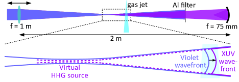
The concept for generating intense XUV pulses in a compact setup is presented in Fig. 1. First, NIR pulses centered at 790 nm with an energy of 13 mJ and a duration of 40 fs [41] were frequency-doubled in a 150-m-thick beta-barium borate (BBO) crystal by second-harmonic generation (SHG), resulting in a pulse energy of 3 mJ at 395 nm. Two dichroic mirrors were used to separate the generated violet beam from the fundamental beam. Next, as shown in Fig. 1, these pulses were focused using a spherical lens with a focal length of 1 m, resulting in an estimated peak intensity of W/cm2 in the focal plane when no gas was present. High harmonics were generated in a high-density gas jet produced by a cantilever piezoelectric valve with a nozzle diameter of 0.5 mm [42] that was placed either before or behind the focal plane of the violet laser. By generating high harmonics several Rayleigh lengths away from the focal plane of the violet laser, curved wavefronts are transferred from the driving laser to the generated XUV beam (see lower graph in Fig. 1). As a consequence, the violet and the XUV beams are expected to have similar divergences. Due to the shorter wavelength, however, the XUV virtual source size is significantly smaller than the focus size of the driving laser (see also Ref. [40]). By further demagnifying the virtual XUV source size using a spherical mirror (coated with B4C) with a short focal length of 75 mm, a high XUV intensity can be achieved using a setup that has a length of only 2 meters. An important advantage of generating harmonics in this scheme is the large generation volume leading to a comparably high XUV photon flux.
A 100-nm-thick Al filter was used to block the driving laser, while transmitting XUV photons with energies higher than about 16 eV. The XUV beam profile was recorded using a microchannel plate (MCP) / phosphor screen assembly that was placed at a distance of about 50 cm from the gas jet. For the nonlinear ionization of argon, a pulsed atomic jet of argon atoms was generated by a second cantilever piezoelectric valve with a nozzle diameter of 0.5 mm. The central part of the atomic jet was selected by a skimmer with an orifice diameter of 0.5 mm. Following interaction of the focused XUV beam with the atomic beam, the generated ions were recorded using a velocity-map imaging spectrometer (VMIS) [43] that was operated in spatial-map imaging mode [44]. Individual ion traces were separately recorded by gating the detector and changing the delay of the gating window.
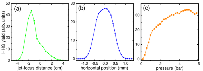
Fig. 2(a) shows the dependence of the obtained HHG yield in Kr using a backing pressure of 4 bar on the distance between the focal plane of the driving laser and the gas jet. This curve has a maximum at cm, which means that the focus is located approximately 5 Rayleigh lengths before the gas jet. Under these conditions, both the XUV beam size at the jet and its divergence are expected to be similar to the size and the divergence of the driving laser (see Fig. 1 and Ref. [40]). To obtain information about the width of the gas jet, the dependence of the HHG yield on the transverse position of the gas jet with respect to the driving laser beam was recorded, as presented in Fig. 2(b). This curve has a full width at half maximum (FWHM) of about 1 mm. Fig. 2(c) depicts the relative HHG yield as a function of the backing pressure, which exhibits a steep increase up to about 1 bar. Saturation starts to set in at higher pressures, and from about 4 bar the HHG yield is almost constant. This is comparable to our previous results using 800 nm driving pulses, where this behavior was attributed to reshaping of the fundamental laser and its influence on phase-matching under these conditions [40]. The saturation behavior is advantageous, since it makes the HHG optimization comparably simple and because slightly different pressures experienced by the driving laser across its beam profile have a negligible effect on the spatially dependent HHG yield. From Fig. 2(c) one can extract that the HHG yield at 2 bar is about 77 of the HHG yield at 4 bar. Using this information in combination with the results shown in Fig. 2(b), we estimate that the region of the gas jet where the yield drops to 77 on both sides of the maximum (meaning that the pressure has dropped by a factor of about 2) spans a width of about 0.75 mm. This represents an estimation of the gas jet width and is in accordance with expectations, considering that the nozzle diameter of the gas valve is 0.5 mm and that the exit of the nozzle is placed as close to the laser beam as possible.
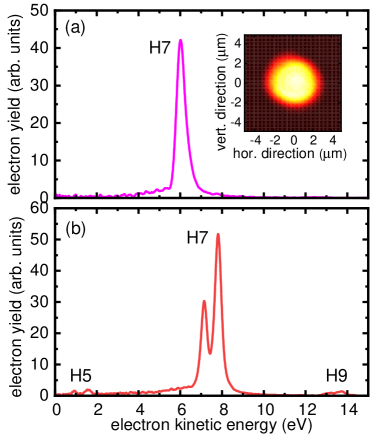
The XUV beam profile recorded at a distance of 50 cm behind the driving laser focus is presented as an inset in Fig. 3(a). The beam radius at this position is about 3 mm, corresponding to an FWHM divergence of 7 mrad, which is similar to the divergence of the violet laser and is a direct consequence of performing the HHG process several Rayleigh lengths away from the focal plane of the driving laser. In Fig. 3(a), a photoelectron spectrum is shown that was obtained after focusing the XUV beam into an Ar gas jet. The spectrum is dominated by the contribution from a single harmonic order with a photon energy of about 22 eV, corresponding to the 7th harmonic of the violet driving laser (i.e. the 14th harmonic of the NIR laser). For comparison, Fig. 3(b) shows a photoelectron spectrum obtained for the ionization of Kr, showing that the contributions from both the 5th and the 9th harmonics are small. The dominance of a single harmonic order was facilitated by the large spectral separation between the harmonic orders ( eV) and by exploiting the filtering characteristics of the Al filter (which has a high transmission for photon energies eV [45]) and the broadband boron carbide focusing mirror (which has a high reflectivity for photon energies eV [46]). The XUV pulse energy at the source was approximately 350 nJ, as measured by an XUV photodiode (AXUV100G, Opto Diode). Taking into account the transmission through the Al filter (40 ) and the reflectivity of the XUV focusing mirror (25 ), an XUV pulse energy of 35 nJ is estimated on target. The XUV pulse duration is estimated as 25 fs, considering that the nonlinear HHG process typically leads to an XUV pulse duration which is approximately half the driving laser pulses duration, see e.g. [47, 48]. An XUV pulse duration of 25 fs was also obtained in HHG simulations when using 800 nm driving pulses [40].
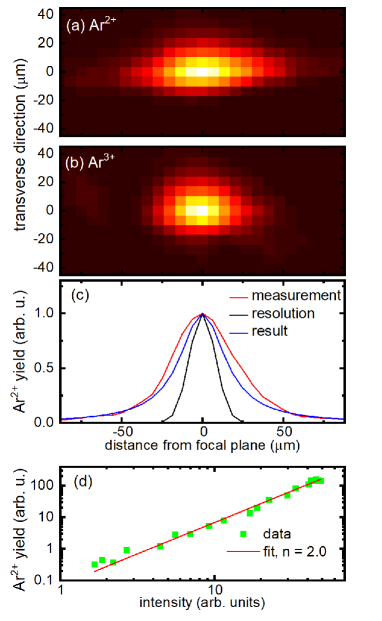
To demonstrate the high intensity of our XUV source, we performed a nonlinear ionization experiment in Ar atoms. This target was chosen, because it allows a comparison with other experiments performed at FELs [1, 3] and using intense HHG sources [4, 6]. To this end, the XUV pulses were focused using a spherical mirror with a focal length of mm. As shown in Fig. 4(a)+(b), both Ar2+ and Ar3+ ions were generated. The energy required to generate Ar2+ is 43.4 eV, and the energy required to generate Ar3+ is 84.1 eV [49], meaning that at least two and four XUV photons need to be absorbed to generate Ar2+ and Ar3+, respectively. In Fig. 4(a)+(b), the spatial distributions of the different ion species are presented as a function of the distance from the XUV focal plane. These distributions are centered at the XUV focal plane, reflecting the nonlinear nature of the ionization process. The narrower distribution of the Ar3+ ion trace compared to the Ar2+ ion trace is a consequence of the higher order of the nonlinearity involved in the former case. The spatially resolved ion traces allow us to determine the relative ion yields at the XUV focal plane, giving an Ar2+ / Ar+ ion yield ratio of 8 and an Ar3+ / Ar2+ ion yield ratio of 17 .
The transversely integrated Ar2+ ion yield shown in Fig. 4(c) (red curve) is used to estimate the XUV Rayleigh length . According to Fig. 4(d), where the Ar2+ yield is shown as a function of the XUV intensity, the generation of Ar2+ is a two-photon process. Therefore, the Ar2+ ion yield as a function of the distance from the XUV focal plane is proportional to [40]. The measured ion distribution is further influenced by the spatial resolution of our method, see black curve in Fig. 4(c). The spatial resolution was estimated by the transverse width of the Ar2+ ion distribution at the XUV focal plane. After performing a deconvolution, the blue curve is obtained, which yields a Rayleigh length of 17 m. This allows us to estimate the XUV beam waist radius according to m, where mm is the estimated XUV beam radius on the focusing mirror and mm is the distance between the mirror and the imaging plane. We note that an imperfect alignment of the spherical mirror leading to astigmatism might affect the estimate of . Using m, an XUV peak intensity of W/cm2 would be obtained. Our results are thus consistent with previous FEL results reported at a photon energy of 20 eV, where Ar3+ was the highest charge state at an intensity of W/cm2 [3]. In our previous experiment performed at an 18-m-long HHG beamline, Ar3+ ions were only observed at intensities of W/cm2 and higher [6]. The higher intensity required for the observation of Ar3+ in that experiment can be attributed to the much shorter XUV pulse duration of about 3 fs in that case [34], meaning that significantly less XUV photons are absorbed before the peak of the XUV intensity is reached. In contrast to the previously mentioned results, Ar3+ was only observed at XUV intensities exceeding W/cm2 using the intense XUV source based on HHG reported in Ref. [4]. This result was attributed to the higher degree of coherence of HHG sources compared to FEL sources, but is not corroborated by the results reported here.
In summary, we have reported on the generation of intense narrowband XUV pulses in a compact and simple setup, which is driven by 395 nm pulses with a moderate pulse energy of 3 mJ. It is straightforward to implement this concept in many laboratories, since no large laboratories or multi-terawatt laser systems are needed, in contrast to many state-of-the-art intense XUV sources based on HHG [30, 16, 4, 5, 6, 31, 32, 38]. Our results could therefore boost research fields that require intense XUV pulses such as XUV strong-field physics and coherent diffractive imaging of nanoscale particles and nanostructures, thereby overcoming the challenges experienced in approaches where several harmonic orders are used [25]. In comparison to setups where a multilayer XUV mirror is used to select a single harmonic order [23], an advantage of the scheme presented here is the higher pulse energy that is contained in each harmonic order. By spectrally tuning either the fundamental laser or the second harmonic, tunable intense XUV pulses can be obtained and can be used to scan across resonances, similar to the way that this can be done at FELs [50]. Instead of the second harmonic, it is also possible to use the third harmonic for HHG [51], allowing access to different harmonic orders and resulting in an even larger spacing between the individual harmonic orders.
References
References
- [1] Wabnitz H, de Castro A R B, Gürtler P, Laarmann T, Laasch W, Schulz J and Möller T 2005 Phys. Rev. Lett. 94(2) 023001 URL https://link.aps.org/doi/10.1103/PhysRevLett.94.023001
- [2] Sorokin A A, Bobashev S V, Feigl T, Tiedtke K, Wabnitz H and Richter M 2007 Phys. Rev. Lett. 99(21) 213002 URL https://link.aps.org/doi/10.1103/PhysRevLett.99.213002
- [3] Motomura K, Fukuzawa H, Foucar L, Liu X J, Prümper G, Ueda K, Saito N, Iwayama H, Nagaya K, Murakami H, Yao M, Belkacem A, Nagasono M, Higashiya A, Yabashi M, Ishikawa T, Ohashi H and Kimura H 2009 J. Phys. B 42 221003 URL https://doi.org/10.1088/0953-4075/42/22/221003
- [4] Nayak A, Orfanos I, Makos I, Dumergue M, Kühn S, Skantzakis E, Bodi B, Varju K, Kalpouzos C, Banks H I B, Emmanouilidou A, Charalambidis D and Tzallas P 2018 Phys. Rev. A 98(2) 023426 URL https://link.aps.org/doi/10.1103/PhysRevA.98.023426
- [5] Bergues B, Rivas D E, Weidman M, Muschet A A, Helml W, Guggenmos A, Pervak V, Kleineberg U, Marcus G, Kienberger R, Charalambidis D, Tzallas P, Schröder H, Krausz F and Veisz L 2018 Optica 5 237–242 URL http://www.osapublishing.org/optica/abstract.cfm?URI=optica-5-3-237
- [6] Senfftleben B, Kretschmar M, Hoffmann A, Sauppe M, Tümmler J, Will I, Nagy T, Vrakking M J J, Rupp D and Schütte B 2020 J. Phys. Photonics 2 034001 URL https://doi.org/10.1088/2515-7647/ab87fd
- [7] Sorokin A A, Bobashev S V, Tiedtke K and Richter M 2006 J. Phys. B 39 L299–L304 URL https://doi.org/10.1088/0953-4075/39/14/l04
- [8] Jiang Y H, Rudenko A, Kurka M, Kühnel K U, Ergler T, Foucar L, Schöffler M, Schössler S, Havermeier T, Smolarski M, Cole K, Dörner R, Düsterer S, Treusch R, Gensch M, Schröter C D, Moshammer R and Ullrich J 2009 Phys. Rev. Lett. 102(12) 123002 URL https://link.aps.org/doi/10.1103/PhysRevLett.102.123002
- [9] Wabnitz H, Bittner L, De Castro A, Döhrmann R, Gürtler P, Laarmann T, Laasch W, Schulz J, Swiderski A, von Haeften K et al. 2002 Nature 420 482
- [10] Iwayama H, Sugishima A, Nagaya K, Yao M, Fukuzawa H, Motomura K, Liu X J, Yamada A, Wang C, Ueda K, Saito N, Nagasono M, Tono K, Yabashi M, Ishikawa T, Ohashi H, Kimura H and Togashi T 2010 J. Phys. B 43 161001 URL https://doi.org/10.1088/0953-4075/43/16/161001
- [11] Schütte B, Arbeiter M, Fennel T, Vrakking M J J and Rouzée A 2014 Phys. Rev. Lett. 112(7) 073003 URL https://link.aps.org/doi/10.1103/PhysRevLett.112.073003
- [12] Helk T, Berger E, Jamnuch S, Hoffmann L, Kabacinski A, Gautier J, Tissandier F, Goddet J P, Chang H T, Oh J, Pemmaraju C D, Pascal T A, Sebban S, Spielmann C and Zuerch M 2021 Science Advances 7 (Preprint https://advances.sciencemag.org/content/7/21/eabe2265.full.pdf) URL https://advances.sciencemag.org/content/7/21/eabe2265
- [13] Ott C, Aufleger L, Ding T, Rebholz M, Magunia A, Hartmann M, Stooß V, Wachs D, Birk P, Borisova G D, Meyer K, Rupprecht P, da Costa Castanheira C, Moshammer R, Attar A R, Gaumnitz T, Loh Z H, Düsterer S, Treusch R, Ullrich J, Jiang Y, Meyer M, Lambropoulos P and Pfeifer T 2019 Phys. Rev. Lett. 123(16) 163201 URL https://link.aps.org/doi/10.1103/PhysRevLett.123.163201
- [14] Ding T, Rebholz M, Aufleger L, Hartmann M, Meyer K, Stooß V, Magunia A, Wachs D, Birk P, Mi Y, Borisova G D, Castanheira C d C, Rupprecht P, Loh Z H, Attar A R, Gaumnitz T, Roling S, Butz M, Zacharias H, Düsterer S, Treusch R, Cavaletto S M, Ott C and Pfeifer T 2019 Phys. Rev. Lett. 123(10) 103001 URL https://link.aps.org/doi/10.1103/PhysRevLett.123.103001
- [15] Tzallas P, Skantzakis E, Nikolopoulos L, Tsakiris G D and Charalambidis D 2011 Nat. Phys. 7 781 URL https://doi.org/10.1038/nphys2033
- [16] Takahashi E J, Lan P, Mücke O D, Nabekawa Y and Midorikawa K 2013 Nat. Commun. 4 2691 URL https://www.nature.com/articles/ncomms3691
- [17] Schnorr K, Senftleben A, Kurka M, Rudenko A, Schmid G, Pfeifer T, Meyer K, Kübel M, Kling M F, Jiang Y H, Treusch R, Düsterer S, Siemer B, Wöstmann M, Zacharias H, Mitzner R, Zouros T J M, Ullrich J, Schröter C D and Moshammer R 2014 Phys. Rev. Lett. 113(7) 073001 URL https://link.aps.org/doi/10.1103/PhysRevLett.113.073001
- [18] Meyer M, Cubaynes D, Richardson V, Costello J T, Radcliffe P, Li W B, Düsterer S, Fritzsche S, Mihelic A, Papamihail K G and Lambropoulos P 2010 Phys. Rev. Lett. 104(21) 213001 URL https://link.aps.org/doi/10.1103/PhysRevLett.104.213001
- [19] Varvarezos L, Düsterer S, Kiselev M D, Boll R, Bomme C, De Fanis A, Erk B, Passow C, Burkov S M, Hartmann G, Ilchen M, Johnsson P, Kelly T J, Manschwetus B, Mazza T, Meyer M, Rompotis D, Zatsarinny O, Gryzlova E V, Grum-Grzhimailo A N and Costello J T 2021 Phys. Rev. A 103(2) 022832 URL https://link.aps.org/doi/10.1103/PhysRevA.103.022832
- [20] Bencivenga F, Cucini R, Capotondi F, Battistoni A, Mincigrucci R, Giangrisostomi E, Gessini A, Manfredda M, Nikolov I, Pedersoli E et al. 2015 Nature 520 205–208 URL https://doi.org/10.1038/nature14341
- [21] Harries J R, Iwayama H, Kuma S, Iizawa M, Suzuki N, Azuma Y, Inoue I, Owada S, Togashi T, Tono K, Yabashi M and Shigemasa E 2018 Phys. Rev. Lett. 121(26) 263201 URL https://link.aps.org/doi/10.1103/PhysRevLett.121.263201
- [22] Flögel M, Durá J, Schütte B, Ivanov M, Rouzée A and Vrakking M J J 2017 Phys. Rev. A 95(2) 021401 URL https://link.aps.org/doi/10.1103/PhysRevA.95.021401
- [23] Ravasio A, Gauthier D, Maia F R N C, Billon M, Caumes J P, Garzella D, Géléoc M, Gobert O, Hergott J F, Pena A M, Perez H, Carré B, Bourhis E, Gierak J, Madouri A, Mailly D, Schiedt B, Fajardo M, Gautier J, Zeitoun P, Bucksbaum P H, Hajdu J and Merdji H 2009 Phys. Rev. Lett. 103(2) 028104 URL https://link.aps.org/doi/10.1103/PhysRevLett.103.028104
- [24] Bostedt C, Eremina E, Rupp D, Adolph M, Thomas H, Hoener M, de Castro A R B, Tiggesbäumker J, Meiwes-Broer K H, Laarmann T, Wabnitz H, Plönjes E, Treusch R, Schneider J R and Möller T 2012 Phys. Rev. Lett. 108(9) 093401 URL https://link.aps.org/doi/10.1103/PhysRevLett.108.093401
- [25] Rupp D, Monserud N, Langbehn B, Sauppe M, Zimmermann J, Ovcharenko Y, Möller T, Frassetto F, Poletto L, Trabattoni A et al. 2017 Nat. Commun. 8 493 URL https://www.nature.com/articles/s41467-017-00287-z
- [26] Ackermann W, Asova G, Ayvazyan V, Azima A, Baboi N, Bähr J, Balandin V, Beutner B, Brandt A, Bolzmann A et al. 2007 Nat. Photon. 1 336–342
- [27] Shintake T, Tanaka H, Hara T, Tanaka T, Togawa K, Yabashi M, Otake Y, Asano Y, Bizen T, Fukui T et al. 2008 Nat. Photon. 2 555–559
- [28] Allaria E, Appio R, Badano L, Barletta W, Bassanese S, Biedron S, Borga A, Busetto E, Castronovo D, Cinquegrana P et al. 2012 Nat. Photon. 6 699–704
- [29] Mashiko H, Suda A and Midorikawa K 2004 Opt. Lett. 29 1927–1929 URL http://ol.osa.org/abstract.cfm?URI=ol-29-16-1927
- [30] Manschwetus B, Rading L, Campi F, Maclot S, Coudert-Alteirac H, Lahl J, Wikmark H, Rudawski P, Heyl C M, Farkas B, Mohamed T, L’Huillier A and Johnsson P 2016 Phys. Rev. A 93(6) 061402 URL https://link.aps.org/doi/10.1103/PhysRevA.93.061402
- [31] Hort O, Albrecht M, Nefedova V E, Finke O, Mai D D, Reyné S, Giambruno F, Frassetto F, Poletto L, Andreasson J, Gautier J, Sebban S and Nejdl J 2019 Opt. Express 27 8871–8883 URL http://www.opticsexpress.org/abstract.cfm?URI=oe-27-6-8871
- [32] Kühn S, Dumergue M, Kahaly S, Mondal S, Füle M, Csizmadia T, Farkas B, Major B, Várallyay Z, Cormier E, Kalashnikov M, Calegari F, Devetta M, Frassetto F, Månsson E, Poletto L, Stagira S, Vozzi C, Nisoli M, Rudawski P, Maclot S, Campi F, Wikmark H, Arnold C L, Heyl C M, Johnsson P, L’Huillier A, Lopez-Martens R, Haessler S, Bocoum M, Boehle F, Vernier A, Iaquaniello G, Skantzakis E, Papadakis N, Kalpouzos C, Tzallas P, Lépine F, Charalambidis D, Varjú K, Osvay K and Sansone G 2017 J. Phys. B 50 132002 URL https://doi.org/10.1088/1361-6455/aa6ee8
- [33] Motoyama H, Iwasaki A, Takei Y, Kume T, Egawa S, Sato T, Yamanouchi K and Mimura H 2019 Appl. Phys. Lett. 114 241102 URL https://doi.org/10.1063/1.5091587
- [34] Major B, Kretschmar M, Ghafur O, Hoffmann A, Kovács K, Varjú K, Senfftleben B, Tümmler J, Will I, Nagy T, Rupp D, Vrakking M J J, Tosa V and Schütte B 2020 J. Phys. Photonics 2 034002 URL https://doi.org/10.1088/2515-7647/ab869d
- [35] Kobayashi Y, Sekikawa T, Nabekawa Y and Watanabe S 1998 Opt. Lett. 23 64–66 URL http://ol.osa.org/abstract.cfm?URI=ol-23-1-64
- [36] Sekikawa T, Kosuge A, Kanai T and Watanabe S 2004 Nature 432 605–608
- [37] Barillot T, Matia-Hernando P, Greening D, Walke D, Witting T, Frasinski L, Marangos J and Tisch J 2017 Chem. Phys. Lett. 683 38 – 42 ISSN 0009-2614 URL http://www.sciencedirect.com/science/article/pii/S0009261417304645
- [38] Li J, Wang Y, Guo T, White J, Weidman M, Wu Y, Hu K, Jager M F, Kaplan C J, Geneaux R et al. 2020 J. Phys. B 53 145602
- [39] Poletto L, Frassetto F, Calegari F, Anumula S, Trabattoni A and Nisoli M 2013 Opt. Express 21 13040–13051 URL http://www.opticsexpress.org/abstract.cfm?URI=oe-21-11-13040
- [40] Major B, Ghafur O, Kovács K, Varjú K, Tosa V, Vrakking M J J and Schütte B 2021 Optica 8 960–965 URL http://www.osapublishing.org/optica/abstract.cfm?URI=optica-8-7-960
- [41] Gademann G, Ple F, Paul P M and Vrakking M J J 2011 Opt. Express 19 24922 URL https://doi.org/10.1364/OE.19.024922
- [42] Irimia D, Dobrikov D, Kortekaas R, Voet H, van den Ende D A, Groen W A and Janssen M H M 2009 Rev. Sci. Instrum. 80 113303 URL https://doi.org/10.1063/1.3263912
- [43] Eppink A T J B and Parker D H 1997 Rev. Sci. Instrum. 68 3477
- [44] Stei M, von Vangerow J, Otto R, Kelkar A H, Carrascosa E, Best T and Wester R 2013 J. Chem. Phys. 138 214201 URL https://doi.org/10.1063/1.4807482
- [45] Henke B L, Gullikson E M and Davis J C 1993 At. Data Nucl. Data Tables 54 181–342
- [46] Larruquert J I and Keski-Kuha R A M 1999 Appl. Opt. 38 1231–1236 URL http://ao.osa.org/abstract.cfm?URI=ao-38-7-1231
- [47] Mauritsson J, Johnsson P, López-Martens R, Varjú K, Kornelis W, Biegert J, Keller U, Gaarde M B, Schafer K J and L’Huillier A 2004 Phys. Rev. A 70(2) 021801 URL https://link.aps.org/doi/10.1103/PhysRevA.70.021801
- [48] Nabekawa Y, Hasegawa H, Takahashi E J and Midorikawa K 2005 Phys. Rev. Lett. 94(4) 043001 URL https://link.aps.org/doi/10.1103/PhysRevLett.94.043001
- [49] Kramida A, Yu Ralchenko, Reader J and and NIST ASD Team 2013 NIST Atomic Spectra Database (ver. 5.1), [Online]. Available: http://physics.nist.gov/asd [2014, September 8]. National Institute of Standards and Technology, Gaithersburg, MD.
- [50] Takanashi T, Golubev N V, Callegari C, Fukuzawa H, Motomura K, Iablonskyi D, Kumagai Y, Mondal S, Tachibana T, Nagaya K, Nishiyama T, Matsunami K, Johnsson P, Piseri P, Sansone G, Dubrouil A, Reduzzi M, Carpeggiani P, Vozzi C, Devetta M, Negro M, Faccialà D, Calegari F, Trabattoni A, Castrovilli M C, Ovcharenko Y, Mudrich M, Stienkemeier F, Coreno M, Alagia M, Schütte B, Berrah N, Plekan O, Finetti P, Spezzani C, Ferrari E, Allaria E, Penco G, Serpico C, De Ninno G, Diviacco B, Di Mitri S, Giannessi L, Jabbari G, Prince K C, Cederbaum L S, Demekhin P V, Kuleff A I and Ueda K 2017 Phys. Rev. Lett. 118(3) 033202 URL https://link.aps.org/doi/10.1103/PhysRevLett.118.033202
- [51] Popmintchev D, Hernández-García C, Dollar F, Mancuso C, Pérez-Hernández J A, Chen M C, Hankla A, Gao X, Shim B, Gaeta A L, Tarazkar M, Romanov D A, Levis R J, Gaffney J A, Foord M, Libby S B, Jaron-Becker A, Becker A, Plaja L, Murnane M M, Kapteyn H C and Popmintchev T 2015 Science 350 1225–1231 ISSN 0036-8075 URL https://science.sciencemag.org/content/350/6265/1225