Attosecond spectroscopy of size-resolved water clusters
Abstract
Electron dynamics in water are of fundamental importance for a broad range of phenomena[1, 2, 3], but their real-time study faces numerous conceptual and methodological challenges[4, 5, 6]. Here, we introduce attosecond size-resolved cluster spectroscopy and build up a molecular-level understanding of the attosecond electron dynamics in water. We measure the effect that the addition of single water molecules has on the photoionization time delays[7, 8, 9, 10, 11] of water clusters. We find a continuous increase of the delay for clusters containing up to 4-5 molecules and little change towards larger clusters. We show that these delays are proportional to the spatial extension of the created electron hole, which first increases with cluster size and then partially localizes through the onset of structural disorder that is characteristic of large clusters and bulk liquid water. These results establish a previously unknown sensitivity of photoionization delays to electron-hole delocalization and reveal a direct link between electronic structure and attosecond photoemission dynamics. Our results offer novel perspectives for studying electron/hole delocalization and its attosecond dynamics.
Electronic dynamics in water play a central role in a broad range of scientific and technological research areas ranging from radiation chemistry to photocatalysis. The dynamics induced by ionization of water are of particular relevance since they initiate the processes underlying radiation damage[2, 3, 12]. The ionization of water is predicted to lead to the formation of a delocalized electron hole, followed by its localization on one water molecule and proton transfer to a neighboring molecule, forming H3O+ and OH[13]. The latter step has been time-resolved only very recently using one-photon extreme-ultraviolet (XUV) photoionization of water clusters[4] and strong-field ionization of liquid water[5]. Both experiments independently determined a 30-50 fs time scale for proton transfer. The formation of the delocalized electron hole, as well as its localization have so far escaped experimental scrutiny because of their sub-femtosecond time scales.
In this work, we access the attosecond time scale of the photoionization dynamics of water on the molecular level by introducing attosecond size-resolved cluster spectroscopy (ASCS). Coupling attosecond interferometry[9, 11, 14, 15] with electron-ion coincidence spectroscopy, we determine photoionization delays for water clusters of increasing size, achieving single-molecule resolution. Photoionization time delays of (H2O)n are found to continuously increase from to . We show that this increase directly reflects the augmenting delocalization of the electron hole created in the ionization process. For these small clusters, we find a linear relationship between the photoionization time delays and the first moment of the electron-hole density created in the ionization process. Beyond the photoionization delays vary little, an effect that we attribute to the partial localization of the electron hole caused by the onset of structural disorder characteristic of larger clusters and bulk liquid water. These assignments are further confirmed by calculations on the O-1s photoionization delays of water clusters, which display these effects even more clearly owing to the atomic character of the orbitals. As we show below, the present results also confirm the interpretation of photoemission delays from liquid water [6].
Our work thus also reveals a possible experimental access to the spatial delocalization of electronic wave functions, which has always been difficult to characterize. Electron delocalization plays a fundamental role in the properties of solids, where the perfect translational symmetry of single crystals creates fully delocalized electronic wave functions (or Bloch waves), which are disrupted by local disorder in a phenomenon known as Anderson localization[16, 17]. The delocalization of electronic wave functions is also central for understanding the aromaticity of molecules, charge transfer between a metal atom and its ligands, or between a solute and a solvent. The electronic structure of water clusters and liquid water has so far mainly been accessible through quantum-chemical calculations, which have predicted partial (de)localization of the electronic wavefunctions[18]. Experimental access to this information has however not been reported so far.
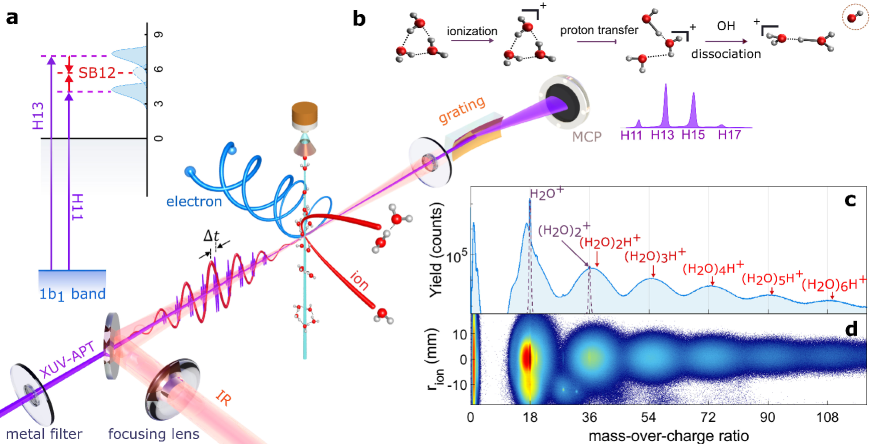
Figure 1 provides a conceptual overview of our measurements. An XUV attosecond pulse train (APT) generated through high-harmonic generation is focused into a supersonic water-cluster beam, where it is spatio-temporally overlapped with a near-infrared (IR) laser pulse. The APT and IR pulses are phase locked through an actively-stabilized Mach-Zehnder interferometer. The three-dimensional momentum distributions of electrons and ions generated from this interaction are detected in coincidence using COLd Target Recoil Ion Momentum Spectroscopy (COLTRIMS)[19, 20, 21] [see Methods for details]. The photoionization time delays of water clusters are measured by recording photoelectron spectra as a function of the time delay between the overlapping APT and IR pulses, in coincidence with each ionic fragment. As shown in the inset of Fig. 1a, single-photon XUV ionization gives rise to the main bands (MB) in the photoelectron spectra, whereas the additional IR interaction creates sidebands (SB).
The unique assignment of the coincident attosecond photoelectron spectra to a specific cluster size is possible because of a dissociative-ionization mechanism that is general for small ( 20 molecules) water clusters at low ionization energies (Fig. 1b). Following outer-valence single ionization, water clusters undergo rapid proton transfer, followed by the loss of a single OH unit on a sub-picosecond time scale[22, 23, 24, 25, 26, 27, 28, 29, 30], such that each detected fragment (H2O)nH+ mainly originates from the neutral (H2O)n+1 precursor for n 6 (see SM Section 1.2 for details). The observed mass spectrum (Fig. 1c) indeed shows a well-resolved progression of broad peaks that is easily assigned to (H2O)nH+ with . The width of the peaks is caused by the kinetic-energy release in the dissociative photoionization, as highlighted in Fig. 1d, which shows the mass spectrum as a function of the detected position radius of the ions on the detector (). The only unprotonated species (H2O+ and (H2O), purple dashed curves) originate from the photoionization of H2O and (H2O)2, respectively. The broad distribution peaking at a MOC of 17 is OH+ originating from the dissociative ionization of H2O+. A fraction of the photoionized dimers remains bound, leading to the sharp (H2O) peak, and the remainder dissociates to produce (H2O)H+. Analogous results have been obtained following the ionization of D2O clusters. The corresponding mass spectra are shown in Fig. S7.
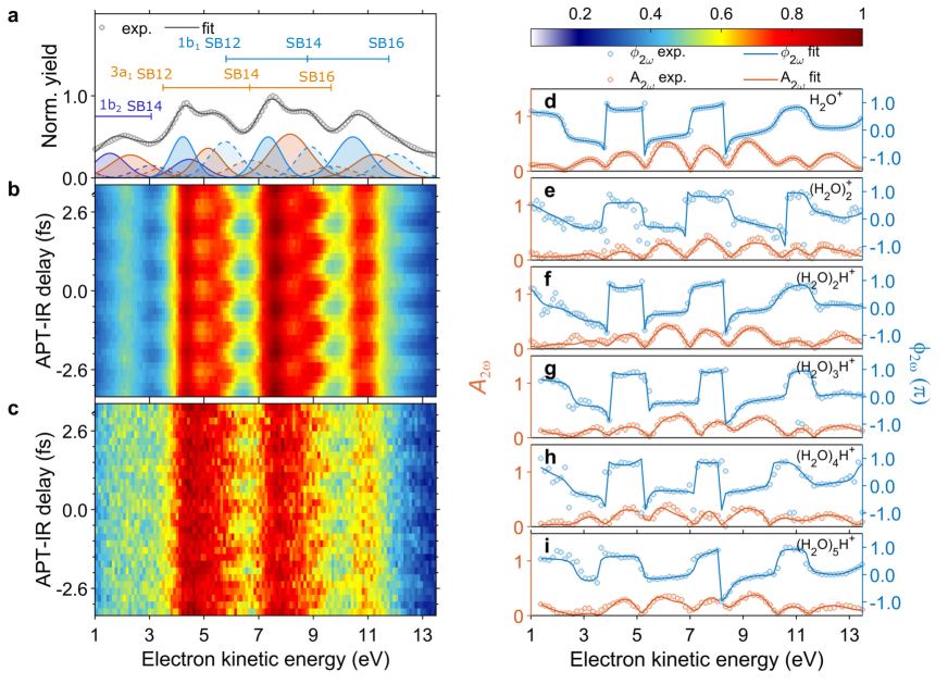
Figure 2 shows the attosecond photoelectron spectra (APS) obtained in coincidence with each cluster size. The APS measured in coincidence with H2O+ (Fig. 2a) is dominated by the contributions of harmonic orders 11, 13, and 15, and ionization from the two outermost (1b1 and 3a1) molecular orbitals of H2O. The black line shows a fit using the literature values of the vertical binding energies. The filled spectra correspond to the decomposition of the APS in MB (full colored lines) and SB (dashed lines) spectra. Figures 2b and 2c show the characteristic oscillations with a period of 1.33 fs in the APS coincident with H2O+ and (H2O)2H+, respectively. The remaining APS are shown in Fig. S3. Analogous results obtained for D2O clusters are shown in Fig. S8. The SB-intensity oscillations take the form , where is the angular IR frequency, is the experimentally varied APT-IR delay, , is the harmonic emission time, and is the system-specific photoemission time delay. Here, we determine relative photoemission delays between water clusters (H2O)n and H2O, as a function of , which cancels the contribution of . Because the ionization energies vary by less than 0.6 eV from , the relative measurement also causes negligible contributions of the continuum-continuum (or Coulomb-laser coupling) delays[31, 32] on the order of 4-6 as for SB12-14.
The main challenge in the determination of photoionization time delays from such measurements is the considerable spectral overlap. We therefore use a general procedure, introduced[33] and validated in our recent work[6], that resolves this challenge. Instead of integrating the APS oscillations over specific spectral regions, our approach fully accounts for the spectral overlap. Briefly, we Fourier transform the APS along the time-delay axis and then fit the complex-valued Fourier transform at the 2 frequency by assigning a specific phase shift to each spectral component of the MB and SB spectra. Details are given in the SM, Section 1.5. We keep the spectral positions and amplitudes fixed to values determined from the delay-integrated spectra (see, e.g., Fig. 2a and S3). The success of this fitting procedure is highlighted by the excellent agreement between the experimental data (circles in Figs. 2d-i) and the fits (full lines). The robustness of the fitting procedure to variations of the initial guesses is shown in Fig. S6.
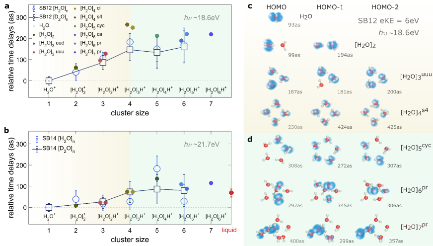
Figures 3a and 3b show the cluster-size-resolved photoionization time delays corresponding to the 1b1 photoelectron bands from monomer to hexamer, relative to the monomer delay, as determined from SB12 and SB14, respectively. The time delays measured in SB12 (18.6 eV photon energy) increase as a function of the cluster size up to the tetramer, followed by little variation. The results for SB14 show a similar behavior, with indications of a slightly slower convergence as a function of cluster size. The latter results can be compared to our recent measurements of bulk liquid water, which yielded a photoemission delay of 6920 as relative to the water monomer for SB14[6], indicated as the red dot in Fig. 3b. The close agreement between the H2O- and D2O-cluster results suggest that nuclear-motion effect are not relevant within the accuracy of the present measurements.
To understand the mechanisms governing these delays, we performed ab-initio quantum-scattering calculations of the photoionization delays (see SM Section 2 and Ref.[34] for details). Starting from the trimer, each water cluster exists in several isomeric forms[35, 36, 37, 38, 39, 40, 41, 42]. At the low temperatures reached in our supersonic expansion, only one or two isomers are thermally populated, as detailed in the SM (Section 1.2 and Table S1). We used the equilibrium geometries of the most stable cluster isomers reported in Ref.[36] to perform electronic-structure calculations with a correlation-consistent valence-triple-zeta (cc-pVTZ) basis set. These served as an input to the photoionization calculations performed by solving the electron/water-cluster-ion scattering problem at the experimentally relevant scattering energies using the iterative Schwinger variational principle[43, 44]. The input orbitals, scattering potentials and scattering wave functions were all represented by single-center expansions using a typical maximal angular-momentum value of , whereby numerical convergence with respect to this parameter was ensured. The photoionization time delays, resolved as a function of photoemission direction in the molecular (cluster) frame and the cluster orientation in the laboratory frame, were obtained and subsequently angularly averaged using the partial photoionization cross sections as weighting factors (for details, see SM Section 2). These calculations yielded angular-integrated one-photon-ionization (or Wigner) delays. We have compared these delays to two-photon (XUV+IR or RABBIT) delays obtained by additionally including the effect of the IR field on the photoionization delays. The results, shown in Figs. S9-S13, establish the close correspondence of the one- and two-photon delays in the case of water clusters, both angle-resolved and angle-integrated, and therefore support our comparison of the angle-averaged one-photon delays with the observables of the present experiment. Using this methodology, photoionization delays were obtained for each of the orbitals of (H2O)n that contribute to the 1b1 band of each water cluster. The cross-section-weighted average of these delays (defined in Eq.(1) in the Methods section) are shown as the large filled symbols. The agreement between theory and experiment is excellent (Figs. 3a and 3b).
Figures 3c and 3d show the densities of the highest-occupied molecular orbitals of the 1b1 band of the most stable isomer of each cluster size, together with their absolute photoionization delays. Figure 3c suggests that the increasing orbital delocalization correlates with the increasing time delay. The HOMO of the dimer has almost the same time delay as the monomer, whereas the delocalization of the HOMO-1 in the dimer leads to an increase of the time delay by nearly 100 as. This comparison also shows the very small effect ( 6 as) of electron scattering on the neutral H2O neighbor in the dimer. Among the trimer orbitals, it is also the most delocalized orbital (HOMO-2) which has the largest photoionization delay (200 as). The tetramer orbitals are perfectly delocalized over all molecules, owing to the S4 symmetry of its most stable isomer, which leads to the largest photoionization delays (up to 425 as) found in our calculations.
Interestingly, a further increase of the cluster size does not increase the delays further. The reason is obvious from Fig. 3d. Most larger clusters than the tetramer have a lower symmetry, many of them having no symmetry elements at all. This leads to a localization of the orbital densities on a small number of typically 2-3 neighboring molecules. This effect is reflected as a stagnation of the associated photoionization delays around values of 300 as in SB12. This observation suggests that the disorder-induced orbital localization in the larger clusters causes the experimentally observed saturation of the measured photoionization delays at the largest cluster sizes measured in this work.
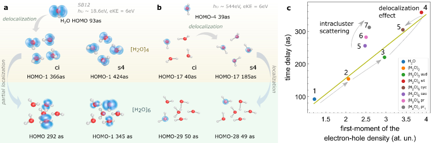
To further verify this surprisingly simple relation between time delays and orbital localization, we performed additional calculations on the oxygen-1s orbitals of the water clusters, with complete results shown in the SM (Figs. S14-S19). The O1s-orbitals have the advantage of remaining essentially atomic in character and not being significantly modified by hydrogen bonding and orbital hybridization. For this reason, they allow us to isolate the effect of orbital (de)localization even more clearly. The results obtained for the O1s-band are shown in Fig. 4b, where they are compared to the results for the 1b1 band (Fig. 4a). This comparison further supports our interpretation. The photoionization delay remains essentially unchanged from the monomer (39 as) to the tetramer ci (40 as), where the O-1s orbital is fully localized, but increases to 185 as in the tetramer s4, where the O-1s orbital is fully delocalized as a consequence of the high symmetry. Increasing the cluster size further results in a complete localization of the O-1s orbital, which leads to a remarkable decrease of the photoemission delay to 49-50 as in the hexamer. The photoionization delays of the O1s orbitals in the hexamer being practically identical with the monomer (39 as) highlights, both, the direct link between photoionization delay and orbital delocalization and the very small effect (a few attoseconds) of electron scattering on the neutral H2O moieties of the clusters.
Finally, we establish a quantitative link between time delays and electron-hole delocalization. Figure 4c shows a correlation plot between photoionization delays of the 1b1 band of the water clusters (H2O)n and the first moment of the electron-hole density in the 1b1 band of the singly-ionized clusters (H2O) (defined in Eqs. (2)-(3) in Methods). The most stable isomers of all clusters from to display a nearly perfect linear relationship between the two quantities, as indicated by the straight line in Fig. 4c (correlation coefficient ). This representation also highlights the continuous increase of the delocalization from to , followed by partial localization at , visible in the fact that the second-most stable isomer of and the most stable isomers of and 7 have nearly the same first moment of the electron-hole density. Interestingly, the delays of the latter three isomers display a continuous, yet very small increase, which we assign to intracluster scattering, quantifying this effect as well.
These results have a range of interesting implications. First, they demonstrate the sensitivity of photoionization delays to orbital delocalization in water clusters, which, to our knowledge, has not been experimentally accessible in any form of matter so far. Second, they reveal the mechanism that is responsible for orbital localization in liquid water on the molecular level, i.e. the onset of structural disorder. This effect is reminiscent of Anderson localization in solids[16]. Whereas perfect crystals with translational symmetry have fully delocalized bands, the presence of defects causes their localization, which has a multitude of interesting consequences in solid-state physics. The orbital localization in liquid water can thus be viewed as a consequence of its structural disorder.
We have introduced a new technique, ASCS, and have applied it to measure photoionization time delays of size-resolved water clusters. This study has revealed an unexpectedly simple relationship between orbital localization and time delays, establishing an experimental pathway to probing electron-hole localization in complex matter. Looking forward, our methods can be used to temporally resolve both local and non-local electronic relaxation dynamics in size-resolved water clusters, such as Auger decay, intermolecular Coulombic decay[45, 46] and electron-transfer-mediated decay[47]. More generally, they will facilitate a molecular-level understanding of attosecond electron dynamics in the liquid phase, with implications for the elementary processes underlying chemical reactivity and biological function.
Methods
Laser Setup and Attosecond-Pulse Generation
The experimental setup is based on a regeneratively amplified Titanium-Sapphire laser system which delivers near-infrared (IR) femtosecond laser pulses with 1.2 mJ energy at 5 kHz repetition rate, a central wavelength of 800 nm and 36 fs pulse duration (full-width at half-maximum in intensity). This laser beam is split with a 70:30 beam splitter and the more intense part is focused into a 3 mm long, xenon-filled gas cell to generate an extreme-ultraviolet attosecond pulse train (XUV-APT) via high-harmonic generation. A coaxial 100-nm aluminum foil on a quartz ring is placed before a nickel-coated toroidal mirror (f = 50 cm) to spectrally filter the XUV spectrum and eliminate the residual IR pulse co-propagating with the XUV beam. The XUV spectrum was characterized with a home-built XUV spectrometer consisting of a 100-nm aluminum film, an aberration-corrected flat-field grating (Shimadzu 1200 lines/mm) and a micro-channel-plate (MCP) detector coupled to a phosphor screen. The XUV-APT is recombined with the remaining part of the IR beam after the toroidal mirror via a perforated silver mirror to constitute a Mach-Zehnder interferometer. The path length difference, i.e. the time delay between the overlapping XUV-APT and IR pulses is controlled via a high-precision direct-current motor (PI, resolution 0.1 m) and a piezoelectric motor (PI, resolution 0.1 nm), constituting a combined delay stage operating on femtosecond and attosecond time scales, respectively.
Coincidence Spectrometer
The phase-locked XUV-APT and IR pulses are collinearly focused into the supersonic gas jet in a COLTRIMS (COLd Target Recoil Ion Momentum Spectroscopy)[19, 20, 21] spectrometer. The electrons and ions created by XUV photoionization are guided by a weak homogeneous electric field (3.20 Vcm-1) and a homogeneous magnetic field (6.70 G) towards two time- and position-sensitive detectors at opposite ends of the spectrometer. The detectors consist of two MCPs (Photonis) in Chevron configuration, followed by a three-layer delay-line anode (HEX) with a crossing angle of 60 degrees between adjacent layers and an active radius of 40 mm manufactured by RoentDek. For the electrons, the length of the extraction region is 7 cm, followed by a 14 cm field-free region. A homogeneous magnetic field is applied over all regions by a set of Helmholtz coils, which are tilted to counteract the earth’s magnetic field. The COLTRIMS gives access to the typical electron-ion coincidence measurement with full three-dimensional momentum resolution in 4 solid angle. The momentum resolution of electrons is = = 0.001 a.u. and = 0.056 a.u., where corresponds to the direction of light propagation, is the direction of the supersonic gas jet and is the time-of-flight direction. The photoelectron kinetic energy is calibrated via the XUV-APT photoelectron spectrum of argon with an ionization potential of 15.8 eV.
Cluster source
The neutral water clusters[48, 49] are formed in a continuous supersonic expansion into vacuum with a water vapor pressure of 0.3 MPa through a 50 m nozzle orifice and pass through two conical skimmers (Beam Dynamics) located 10 mm and 30 mm downstream with a diameter of 200 m and 1 mm, respectively. The liquid water is maintained at 408 K to give rise to a sufficient vapor pressure in a container of 0.7 L to support a stable water-cluster beam for a duration of 120 hours. The water-cluster source is coupled to the COLTRIMS via two differential pumping stages. To maintain the ultrahigh vacuum in the main reaction chamber, a differentially pumped beam dump captures the molecular beam after the interaction region.
Definition of the calculated quantities
The calculation of the orbital-specific photoionization delays is described in the main text and in more detail in Section 2 of the SM. Here, we additionally define the cross-section-averaged delays shown in Figs. 3a, 3b as large empty circles and also in Fig. 4c. Since the contributions of individual orbitals to the 1b1 band of the water clusters cannot be resolved, we introduce the cross-section-average of the time delays over the orbitals constituting the 1b1 band of (H2O)n as follows:
| (1) |
where is the photoionization cross section of orbital of the 1b1 band at the photon energy and is the corresponding photoionization time delay.
In Fig. 4c, we additionally show the first moment of the electron-hole density, calculated using ORBKIT package [50]. In the case of a single orbital (with index ) this is defined as
| (2) |
where is the density of orbital and is its center of charge. In analogy to the time delays, we also define a cross-section average of the orbital delocalization as
| (3) |
Acknowledgment
We thank A. Schneider and M. Seiler for their technical support. Funding We gratefully acknowledge funding from an ERC Consolidator Grant (Project No. 772797-ATTOLIQ), project 200021_172946 as well as the NCCR-MUST, funding instruments of the Swiss National Science Foundation. D. J. thanks the European Union’s Horizon 2020 programme (FP-RESOMUS - MSCA 801459) program for a fellowship and A. Schild for introduction to ORBKIT. The results have been obtained on the ETH Zürich Euler cluster and the NCCR-Cluster supercomputer. Author contributions X.G. and S.H. carried out the experiments and analysed the experimental data. X.G. constructed the experimental apparatus with contributions from S.H., K.Z. and C.P.. D.J. and H.J.W. performed the theoretical calculations. X.G., S.H., and H.J.W wrote the initial manuscript. All authors discussed and reviewed the manuscript. Competing Interests The authors declare no competing interests.
Additional information
Supplementary information is available for this paper.
Correspondence and requests for materials should be addressed to H.J.W.
Data availability statement All data is available from the corresponding author on request.
References
- [1] Sanche, L. Beyond radical thinking. Nature 461, 358–359 (2009).
- [2] Boudaiffa, B., Cloutier, P., Hunting, D., Huels, M. A. & Sanche, L. Resonant formation of dna strand breaks by low-energy (3 to 20 ev) electrons. Science 287, 1658–1660 (2000).
- [3] Garrett, B. C. et al. Role of water in electron-initiated processes and radical chemistry: Issues and scientific advances. Chemical reviews 105, 355–390 (2005).
- [4] Svoboda, V. et al. Real-time observation of water radiolysis and hydrated electron formation induced by extreme-ultraviolet pulses. Science Advances 6, eaaz0385 (2020).
- [5] Loh, Z.-H. et al. Observation of the fastest chemical processes in the radiolysis of water. Science 367, 179–182 (2020).
- [6] Jordan, I. et al. Attosecond spectroscopy of liquid water. Science 369, 974–979 (2020).
- [7] Cavalieri, A. L. et al. Attosecond spectroscopy in condensed matter. Nature 449, 1029 (2007).
- [8] Schultze, M. et al. Delay in photoemission. Science 328, 1658–1662 (2010).
- [9] Klünder, K. et al. Probing single-photon ionization on the attosecond time scale. Phys. Rev. Lett. 106, 143002 (2011).
- [10] Ossiander, M. et al. Attosecond correlation dynamics. Nature Physics 13, 280–285 (2016).
- [11] Huppert, M., Jordan, I., Baykusheva, D., von Conta, A. & Wörner, H. J. Attosecond delays in molecular photoionization. Phys. Rev. Lett. 117 (2016).
- [12] Alizadeh, E., Orlando, T. M. & Sanche, L. Biomolecular damage induced by ionizing radiation: the direct and indirect effects of low-energy electrons on dna. Annual review of physical chemistry 66, 379–398 (2015).
- [13] Marsalek, O. et al. Chasing charge localization and chemical reactivity following photoionization in liquid water. The Journal of chemical physics 135, 224510 (2011).
- [14] Cattaneo, L. et al. Attosecond coupled electron and nuclear dynamics in dissociative ionization of H2. Nature Physics 14, 733–739 (2018).
- [15] Nandi, S. et al. Attosecond timing of electron emission from a molecular shape resonance. Science Advances 6, eaba7762 (2020).
- [16] Anderson, P. W. Absence of diffusion in certain random lattices. Physical Review 109, 1492–1505 (1958).
- [17] Anderson, P. W. Basic notions of condensed matter physics (Westview Press, 1997). URL https://doi.org/10.4324/9780429494116.
- [18] Prendergast, D., Grossman, J. C. & Galli, G. The electronic structure of liquid water within density-functional theory. The Journal of chemical physics 123, 014501 (2005).
- [19] Dörner, R. et al. Cold Target Recoil Ion Momentum Spectroscopy: A ’momentum microscope’ to view atomic collision dynamics. Physics Report 330, 95–192 (2000).
- [20] Jagutzki, O. et al. Multiple hit readout of a microchannel plate detector with a three-layer delay-line anode. IEEE Transactions on Nuclear Science 49 II, 2477–2483 (2002).
- [21] Ullrich, J. et al. Recoil-ion and electron momentum spectroscopy: reaction-microscopes. Reports on Progress in Physics 66, 1463–1545 (2003).
- [22] Dong, F., Heinbuch, S., Rocca, J. J. & Bernstein, E. R. Dynamics and fragmentation of van der Waals clusters: (H 2O) n, (CH 3OH) n, and (NH 3) n upon ionization by a 26.5 eV soft x-ray laser. Journal of Chemical Physics 124, 224319 (2006).
- [23] Shiromaru, H., Shinohara, H., Washida, N., Yoo, H. & Kimura, K. Synchrotron radiation measurements of appearance potentials for (H2O)2+, (H2O)3+,(H2O)2H+ and (H2O)3H+ in supersonic jets. Chemical physics letters 141, 7–11 (1987).
- [24] Shi, Z., Ford, J. V., Wei, S. & Castleman, A. W. Water clusters: Contributions of binding energy and entropy to stability. The Journal of Chemical Physics 99, 8009–8015 (1993).
- [25] Wei, S. & Castleman, A. W. Using reflection time-of-flight mass spectrometer techniques to investigate cluster dynamics and bonding. International Journal of Mass Spectrometry and Ion Processes 131, 233–264 (1994).
- [26] Bobbert, C., Schütte, S., Steinbach, C. & Buck, U. Fragmentation and reliable size distributions of large ammonia and water clusters. The European Physical Journal D 19, 183–192 (2002).
- [27] Tachikawa, H. Ionization Dynamics of the Small-Sized Water Clusters: A Direct Ab Initio Trajectory Study. The Journal of Physical Chemistry A 108, 7853–7862 (2004).
- [28] Belau, L., Wilson, K. R., Leone, S. R. & Ahmed, M. Vacuum ultraviolet (VUV) photoionization of small water clusters. Journal of Physical Chemistry A 111, 10075–10083 (2007).
- [29] Liu, X., Lu, W. C., Wang, C. Z. & Ho, K. M. Energetic and fragmentation stability of water clusters (H2O)n, n = 2-30. Chemical Physics Letters 508, 270–275 (2011).
- [30] Zamith, S., Labastie, P. & Lhermite, J. M. Fragmentation cross sections of protonated water clusters. Journal of Chemical Physics 136, 214301 (2012).
- [31] Dahlström, J. M., L’Huillier, A. & Maquet, A. Introduction to attosecond delays in photoionization. Journal of Physics B: Atomic, Molecular and Optical Physics 45, 183001 (2012).
- [32] Pazourek, R., Nagele, S. & Burgdörfer, J. Attosecond chronoscopy of photoemission. Rev. Mod. Phys. 87, 765–802 (2015).
- [33] Jordan, I., Jain, A., Gaumnitz, T., Ma, J. & Wörner, H. J. Photoelectron spectrometer for liquid and gas-phase attosecond spectroscopy with field-free and magnetic bottle operation modes. Review of Scientific Instruments 89, 053103 (2018).
- [34] Baykusheva, D. & Wörner, H. J. Theory of attosecond delays in molecular photoionization. The Journal of Chemical Physics 146, 124306 (2017).
- [35] Shi, Z., Ford, J. V., Wei, S. & Castleman, A. W. Water clusters: Contributions of binding energy and entropy to stability. The Journal of Chemical Physics 99, 8009–8015 (1993).
- [36] Temelso, B., Archer, K. A. & Shields, G. C. Benchmark structures and binding energies of small water clusters with anharmonicity corrections. Journal of Physical Chemistry A 115, 12034–12046 (2011).
- [37] Malloum, A., Fifen, J. J., Dhaouadi, Z., Nana Engo, S. G. & Conradie, J. Structures, relative stability and binding energies of neutral water clusters,H2On, = 2-30. New Journal of Chemistry 43, 13020–13037 (2019).
- [38] Liu, K. et al. Characterization of a cage form of the water hexamer. Nature 381, 501–503 (1996).
- [39] Liu, K., Cruzan, J. D. & Saykally, R. J. Water clusters. Science 271, 929–933 (1996).
- [40] Xantheas, S. S. Cooperativity and hydrogen bonding network in water clusters. Chemical Physics 258, 225–231 (2000).
- [41] Richardson, J. O. et al. Concerted hydrogen-bond breaking by quantum tunneling in the water hexamer prism. Science 351, 1310–1313 (2016).
- [42] Cvitaš, M. T. & Richardson, J. O. Quantum tunnelling pathways of the water pentamer. Physical Chemistry Chemical Physics 22, 1035–1044 (2020).
- [43] Gianturco, F. A., Lucchese, R. R. & Sanna, N. Calculation of low-energy elastic cross sections for electron-cf4 scattering. J. Chem. Phys. 100, 6464–6471 (1994).
- [44] Natalense, A. P. P. & Lucchese, R. R. Cross section and asymmetry parameter calculation for sulfur 1s photoionization of sf6. J. Chem. Phys. 111, 5344–5348 (1999).
- [45] Jahnke, T. et al. Ultrafast energy transfer between water molecules. Nature Physics 6, 139–142 (2010).
- [46] Mucke, M. et al. A hitherto unrecognized source of low-energy electrons in water. Nature Physics 6, 143–146 (2010).
- [47] Unger, I. et al. Observation of electron-transfer-mediated decay in aqueous solution. Nature Chemistry 9, 708 (2017).
- [48] Hagena, O. F. & Obert, W. Cluster formation in expanding supersonic jets: effect of pressure, temperature, nozzle size, and test gas. The Journal of Chemical Physics 56, 1793–1802 (1972).
- [49] Pradzynski, C. C., Forck, R. M., Zeuch, T., Slavíček, P. & Buck, U. A fully size-resolved perspective on the crystallization of water clusters. Science 337, 1529–1532 (2012).
- [50] Hermann, G. et al. Orbkit: A modular python toolbox for cross-platform postprocessing of quantum chemical wavefunction data. Journal of Computational Chemistry 37, 1511–1520 (2016).