Self-assembly of polyhedral bilayer vesicles from Piezo ion channels
Abstract
Piezo ion channels underlie many forms of mechanosensation in vertebrates, and have been found to bend the membrane into strongly curved dome shapes. We develop here a methodology describing the self-assembly of lipids and Piezo into polyhedral bilayer vesicles. We validate this methodology for bilayer vesicles formed from bacterial mechanosensitive channels of small conductance, for which experiments found a polyhedral arrangement of proteins with snub cube symmetry and a well-defined characteristic vesicle size. On this basis, we calculate the self-assembly diagram for polyhedral bilayer vesicles formed from Piezo. We find that the radius of curvature of the Piezo dome provides a critical control parameter for the self-assembly of Piezo vesicles, with high abundances of Piezo vesicles with octahedral, icosahedral, and snub cube symmetry with increasing Piezo dome radius of curvature.
The ability to sense mechanical stimuli such as touch and changes in fluid pressure is fundamental to life. Piezo ion channels Coste et al. (2010) have recently been found to provide the molecular basis for a wide range of seemingly unrelated forms of mechanosensation in vertebrates Ranade et al. (2015); Honoré et al. (2015); Murthy and Dubin (2017); Wu et al. (2017); Gottlieb (2017); Parpaite and Coste (2017); Douguet et al. (2019); Dance (2020). Structural studies Guo and MacKinnon (2017); Saotome et al. (2018); Zhao et al. (2018); Wang et al. (2019) have demonstrated that Piezo is an unusually large ion channel that locally bends the membrane into the approximate shape of a spherical dome. Only closed-state structures of Piezo, obtained in the absence of transmembrane gradients, are currently available. The interaction of Piezo with the surrounding lipid membrane has been investigated through electron microscopy experiments in which Piezo proteins were embedded in lipid bilayer vesicles Guo and MacKinnon (2017); Lin et al. (2019). The highly curved shape of the (closed-state) Piezo dome yields pronounced shape deformations in the surrounding lipid membrane Guo and MacKinnon (2017); Haselwandter and MacKinnon (2018); Lin et al. (2019). These shape deformations may play an important role in Piezo gating Guo and MacKinnon (2017); Saotome et al. (2018); Zhao et al. (2018); Wang et al. (2019); Haselwandter and MacKinnon (2018); Lin et al. (2019), with transitions from closed to open states of Piezo potentially being accompanied by an increase in the radius of curvature of the Piezo dome.
Electron cryotomography experiments on membrane protein polyhedral nanoparticles (MPPNs) formed from bacterial mechanosensitive channels of small conductance (MscS) Wu et al. (2013); Basta et al. (2014); Bass et al. (2002); Steinbacher et al. (2007) have shown that lipids and membrane proteins can self-assemble into lipid bilayer vesicles with a polyhedral protein arrangement and a well-defined characteristic size, thus facilitating structural studies. Similarly, MPPNs formed from Piezo may permit structural analysis of Piezo in the presence of transmembrane gradients Basta et al. (2014); Zhang et al. (2003); Liu et al. (2004); Cockburn et al. (2004) and aid the further investigation of the interaction of Piezo with the surrounding lipid membrane. In this Letter we develop a model of the self-assembly of MPPNs from Piezo ion channels. Our theoretical approach is based on a previous mean-field model Li et al. (2016, 2017) in which the observed symmetry and size of MPPNs formed from MscS were found to emerge from the interplay of protein-induced lipid membrane deformations, topological defects in protein packing in MPPNs, and thermal effects. This previous model relied on the assumption that membrane proteins in MPPNs only weakly curve the membrane, which is a suitable assumption for MscS Bass et al. (2002); Steinbacher et al. (2007); Phillips et al. (2009) but not Piezo Guo and MacKinnon (2017); Saotome et al. (2018); Zhao et al. (2018); Wang et al. (2019); Lin et al. (2019); Haselwandter and MacKinnon (2018). We first develop a general methodology for predicting the symmetry and size of MPPNs composed of proteins that may induce arbitrarily large membrane shape deformations. We validate this methodology for MPPNs formed from MscS. On this basis, we then calculate the self-assembly diagram for MPPNs formed from Piezo. We find that the radius of curvature of the Piezo dome provides a critical control parameter for the self-assembly of MPPNs from Piezo proteins, with high abundances of MPPNs with octahedral, icosahedral, and snub cube symmetry as the radius of curvature of the Piezo dome is increased. Our analysis suggests that, under suitable conditions, self-assembly of MPPNs from Piezo proteins results in highly symmetric MPPNs with a well-defined characteristic size.
Statistical thermodynamics of MPPN self-assembly. As detailed in Refs. Li et al. (2016, 2017); Ma and Haselwandter (2020), the formalism describing the statistical thermodynamics of micelle and viral capsid self-assembly Ben-Shaul and Gelbart (1994); Safran (2003); Bruinsma et al. (2003) successfully predicts the observed symmetry and size of MPPNs formed from MscS Wu et al. (2013); Basta et al. (2014). We adapt here this formalism to explore the self-assembly of MPPNs formed from Piezo ion channels. In particular, we take MPPNs to be in the thermodynamic equilibrium state minimizing the Helmholtz free energy , where is the internal energy of the system and and are the entropy and temperature of the system, respectively. MPPNs formed from MscS were obtained experimentally Basta et al. (2014); Wu et al. (2013) in dilute aqueous solutions with a protein number fraction , where
| (1) |
in which denotes the total number of proteins bound in MPPNs with proteins each and denotes the total number of solvent (water) molecules in the system. In this dilute limit with no interactions between MPPNs, is given by the mixing entropy Ben-Shaul and Gelbart (1994); Safran (2003)
| (2) |
where is Boltzmann’s constant and the MPPN number fraction . Similarly, we have the MPPN internal energy Ben-Shaul and Gelbart (1994); Safran (2003); Li et al. (2016, 2017); Ma and Haselwandter (2020)
| (3) |
where is the minimum energy of MPPNs with proteins each. We obtain by minimizing the MPPN energy , at each , with respect to the MPPN radius at the bilayer midplane, (see Fig. 1). The dominant symmetry and size of MPPNs observed in experiments on MPPNs formed from MscS are successfully predicted by considering contributions to due to membrane bending deformations, , and topological defects in protein packing in MPPNs, , such that Wu et al. (2013); Basta et al. (2014); Li et al. (2016, 2017). We show below how can be calculated from and for MPPNs with the large membrane curvatures induced by the Piezo dome Guo and MacKinnon (2017); Haselwandter and MacKinnon (2018); Lin et al. (2019).
Minimization of with respect to results in Li et al. (2016, 2017); Ben-Shaul and Gelbart (1994); Safran (2003); Bruinsma et al. (2003)
| (4) |
where the protein chemical potential is determined by the constraint
| (5) |
imposing the fixed protein number fraction in Eq. (1). In our previous work on MPPN self-assembly Li et al. (2016, 2017); Ma and Haselwandter (2020) we restricted to the range . As shown below, the strongly curved shape of the Piezo dome means that the self-assembly diagram for MPPNs formed from Piezo can be dominated by MPPN states with . We allow here for the -range , with the smallest number of proteins per MPPN permitting the formation of a polyhedral structure corresponding to . The MPPN equilibrium distribution is then given by
| (6) |
where is the fraction of MPPNs containing proteins each and is obtained from Eq. (4) with Eqs. (1) and (5).
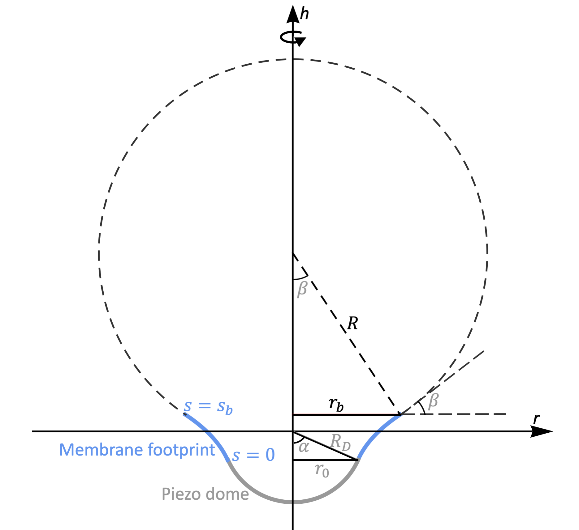
Nonlinear MPPN shape equations. We describe here the Piezo dome as a spherical cap with area and radius of curvature Guo and MacKinnon (2017); Saotome et al. (2018); Zhao et al. (2018); Wang et al. (2019) (Fig. 1). Assuming that MPPNs are under negligible membrane tension Li et al. (2017), the shape and energy of Piezo’s membrane footprint can be estimated Haselwandter and MacKinnon (2018) by minimizing the bending energy of the lipid membrane Canham (1970); Helfrich (1973); Evans (1974),
| (7) |
where is the lipid bilayer bending rigidity, and are the local principal curvatures of the mid-bilayer surface, and the integral runs over Piezo’s membrane footprint. Previous experiments on MPPNs formed from MscS employed lipids with Basta et al. (2014); Wu et al. (2013); Rawicz et al. (2000). We use this value of throughout this Letter.
Membrane-mediated interactions between Piezo proteins are expected to favor approximately hexagonal protein arrangements Góźdź and Gompper (2001); Auth and Gompper (2009); Müller and Deserno (2010); Fournier (1999); Weitz and Destainville (2013). The bending energy of MPPNs can then be estimated from a mean-field approach Góźdź and Gompper (2001); Auth and Gompper (2009); Müller and Deserno (2010); Li et al. (2016, 2017); Ma and Haselwandter (2020) in which the boundary of the hexagonal unit cell of the protein lattice is approximated by a circle. In particular, we divide the surface of MPPNs containing Piezo proteins into identical, circular membrane patches, each with a Piezo dome at its center (Fig. 1). Using the arclength parameterization of surfaces, Eq. (7) can be rewritten as Peterson (1985); Seifert et al. (1991); Jülicher and Seifert (1994); Kühnel (2015)
| (8) | |||||
for each membrane patch, where is the arclength along the profile of Piezo’s membrane footprint, at the inner boundary of Piezo’s membrane footprint (the boundary of the Piezo dome) and at the outer boundary of Piezo’s membrane footprint away from the Piezo dome, denotes the height of Piezo’s membrane footprint along its symmetry axis , denotes the radial coordinate of Piezo’s membrane footprint perpendicular to the -axis, denotes the angle between the tangent to the profile of Piezo’s membrane footprint and the -axis, and the Lagrange parameter functions and enforce the geometric constraints and inherent in the arclength parameterization of surfaces (Fig. 1).
The boundary conditions on Piezo’s membrane footprint at the Piezo dome boundary follow from the assumption that the membrane surface is smooth at Guo and MacKinnon (2017); Haselwandter and MacKinnon (2018) (Fig. 1):
| (9) | |||||
| (10) | |||||
| (11) |
with the membrane-Piezo dome contact angle Weisstein (2017)
| (12) |
Denoting the contact angle at the outer boundary of Piezo’s membrane footprint by and the solid angle associated with each unit cell on the MPPN surface by , we have . Since each unit cell on the MPPN surface contains one protein, we also have , resulting in the boundary condition Auth and Gompper (2009); Li et al. (2016, 2017); Ma and Haselwandter (2020)
| (13) |
at (Fig. 1). Equations (9)–(13) encapsulate the effects of a particular Piezo dome shape and protein number per MPPN on . With the (arbitrary) origin of the - coordinate system fixed via Eqs. (9) and (10) (Fig. 1), we assume that the values of and can be freely varied when finding the extremal functions of Eq. (8) Courant and Hilbert (1953); van Brunt (2004), resulting in the natural boundary conditions
| (14) | |||||
| (15) |
at , where and are the generalized momenta associated with the generalized displacements and , in which the Lagrangian function is given by the integrand in Eq. (8).
To determine the stationary shapes of MPPNs we solve the Hamilton equations associated with the membrane bending energy in Eq. (8) Kibble and Berkshire (2004); Deserno and Bickel (2003); Deserno (2004); Nowak and Chou (2008); Zhang and Nguyen (2008); Hashemi et al. (2014); Foret (2014); Haselwandter and MacKinnon (2018) subject to the boundary conditions on Piezo’s membrane footprint in Eqs. (9)–(15). Compared to the corresponding Euler-Lagrange equations associated with Eq. (8) Courant and Hilbert (1953); van Brunt (2004); Kibble and Berkshire (2004); Peterson (1985); Seifert et al. (1991); Jülicher and Seifert (1994); Agudo-Canalejo and Lipowsky (2016); Bahrami et al. (2016) these Hamilton equations are of first rather than second order in derivatives. The Hamilton equations for Eq. (8) are given by
| (16) | |||||
| (17) | |||||
| (18) | |||||
| (19) | |||||
| (20) | |||||
| (21) |
where is the generalized momentum associated with the generalized displacement . The boundary condition in Eq. (15) and the Hamilton equation in Eq. (21) imply that for . The solutions of the remaining five Hamilton equations in Eqs. (16)–(20) are specified by the five boundary conditions in Eqs. (9)–(14). A numerical difficulty arises here in that some of these boundary conditions are specified at , while others are specified at . We thus solve Eqs. (16)–(20) using a shooting method Peterson (1985); Seifert et al. (1991); Jülicher and Seifert (1994); Agudo-Canalejo and Lipowsky (2016); Bahrami et al. (2016); Deserno and Bickel (2003); Deserno (2004); Nowak and Chou (2008); Zhang and Nguyen (2008); Hashemi et al. (2014); Foret (2014); Haselwandter and MacKinnon (2018); Burden and Faires (2011); Gautschi (2012), for which we introduce the boundary conditions
| (22) | |||||
| (23) |
where and must be adjusted so as to satisfy the boundary conditions in Eqs. (13) and (14). The values of and can be conveniently determined through the FindRoot command in Mathematica mat (2017). We obtain by substituting the solutions of Eq. (16)–(21) into Eq. (8) and (numerically) integrating with respect to .
In the above numerical calculation of , the size of Piezo’s membrane footprint enters through the value of . We note that the MPPN radius is related to the in-plane membrane patch radius via (Fig. 1). In general, different values of yield different values of and, hence, for the stationary membrane footprints. We therefore minimize, at each , with respect to the MPPN size by adjusting so that is minimal, and then determine the values of and associated with this value of . We used here the -range nm, and generally employed a resolution nm for our numerical calculations. However, we found that, within the range nm, may vary rapidly with , and therefore used a finer resolution nm within this range of . We interpolated between the values of considered here using third-order splines. For each Piezo dome shape and protein number per MPPN considered, the value of minimizing thus specifies the MPPN size with minimal bending energy.
Small-gradient approximation for MPPNs. In the Monge parameterization of Eq. (7), is regarded as a single-valued function of , Kühnel (2015). In the small-gradient limit of the Monge parameterization, , the stationary membrane shapes implied by Eq. (7) can be solved for analytically Auth and Gompper (2009), yielding an exact expression for the membrane bending energy in MPPNs. In particular, denoting the obtained for by , we have Auth and Gompper (2009); Li et al. (2016, 2017); Ma and Haselwandter (2020)
| (24) |
Equation (24) was employed previously Li et al. (2016, 2017); Ma and Haselwandter (2020) to construct the MPPN self-assembly diagram for MPPNs formed from MscS proteins, which only weakly curve the membrane. For the purposes of this Letter, the analytic solution in Eq. (24) presents a useful reference point for the fully nonlinear, numerical solutions obtained from Eq. (8).
Topological defects in protein packing. The spherical shape of MPPNs necessitates topological defects in the preferred hexagonal packing of proteins, which incur an -dependent energy penalty. At the mean-field level, deviations from hexagonal protein packing due to the spherical shape of MPPNs can be quantified for a given , in analogy to viral capsids Bruinsma et al. (2003), through the fraction of the surface of a sphere enclosed by identical non-overlapping circles at closest packing Li et al. (2016, 2017); Ma and Haselwandter (2020), . We use here the values of , and associated symmetries of protein packing in MPPNs, compiled in Refs. Clare and Kepert (1986, 1991). Approximating the spring network associated with the energetically preferred hexagonal protein arrangement by a uniform elastic sheet, the leading-order contribution to the MPPN defect energy is thus given by Bruinsma et al. (2003); Li et al. (2016, 2017); Ma and Haselwandter (2020)
| (25) |
where is the stretching modulus of the elastic sheet, is the MPPN surface area, and denotes the optimal packing fraction associated with a uniform hexagonal protein arrangement. As detailed in Refs. Li et al. (2016, 2017); Ma and Haselwandter (2020), the stretching modulus in Eq. (25) is given by
| (26) |
where is the value of that yields, for a given , the minimal .
Most straightforwardly, the MPPN surface area in Eq. (25) can be approximated via Li et al. (2016, 2017); Ma and Haselwandter (2020), where is the surface area associated with a spherical MPPN shape. The approximation breaks down for large enough deviations from a spherical shape, which is expected to be the case for MPPNs formed from Piezo. As an alternative to , we therefore consider here the choice with the area of the deformed MPPN surface, , being given by
| (27) |
where, as noted above, nm2 for the Piezo dome. For MPPNs formed from MscS proteins, we approximate the MPPN surface area occupied by MscS by the spherical cap area in Eq. (27), in which the MscS in-plane radius nm Li et al. (2016); Bass et al. (2002); Steinbacher et al. (2007). Unless indicated otherwise, we use here when evaluating Eq. (25).
Constructing MPPN self-assembly diagrams. We construct MPPN self-assembly diagrams from the fraction of MPPNs containing proteins each, in Eq. (6). To this end, we obtain the minimal MPPN energy, in Eq. (3), at each by minimizing the sum of , calculated from the stationary implied by the arclength or Monge parameterization of Eq. (7), and the corresponding in Eq. (25) with respect to the MPPN radius . We perform this minimization subject to steric constraints due to the finite size of lipids and proteins. To determine the resulting constraints on we note that, at closest packing, the area of a sphere of radius enclosed by non-overlapping circles is given by Clare and Kepert (1986, 1991). Thus, each circular membrane patch occupies an area and, as a result, suspends an angle with respect to the MPPN center. Following Refs. Li et al. (2016, 2017); Ma and Haselwandter (2020) we assume that the MPPN size must be large enough so that the Piezo dome at the center of each membrane patch is surrounded by at least one layer of lipids, resulting in the steric constraint
| (28) |
where denotes the lipid radius. For the lipids employed in experiments on MPPNs formed from MscS Basta et al. (2014); Wu et al. (2013) we have nm Damodaran and Merz (1993), which we use throughout this Letter. Once is calculated, the are conveniently obtained via Eq. (5) for arbitrary protein number fractions . In the arclength parameterization of Eq. (7), changes in or necessitate repeated numerical solution of Eqs. (16)–(20). We determine for a discrete set of values of or and interpolate between these values, using third-order splines, to find the dominant -states of MPPNs showing the largest values of (see below).
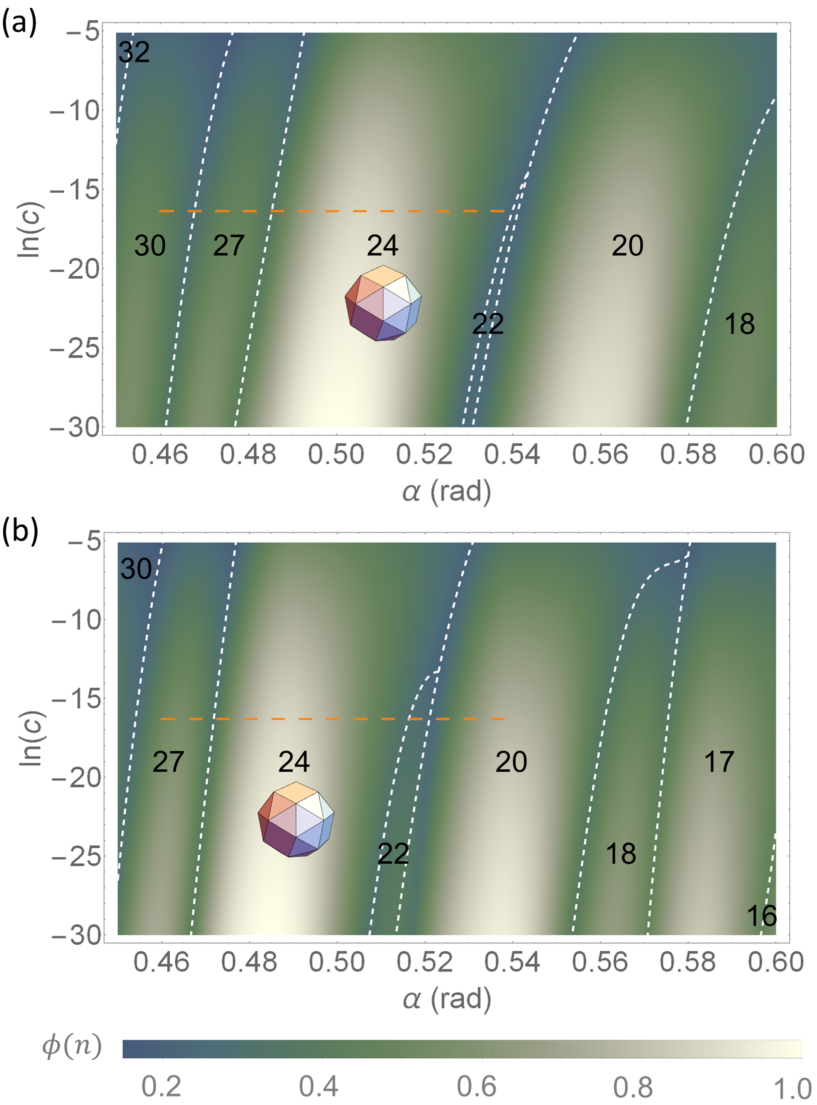
MPPN self-assembly from MscS. Since MscS proteins only weakly curve the membrane Bass et al. (2002); Steinbacher et al. (2007); Phillips et al. (2009), the Monge parameterization of Eq. (7) is expected to yield a good approximation of the self-assembly diagram of MPPNs formed from MscS. Figures 2(a) and 2(b) show the self-assembly diagrams for MPPNs formed from MscS obtained from the arclength and Monge parameterizations of Eq. (7), respectively. Both parameterizations of Eq. (7) predict, with no free parameters, that the snub cube (-symmetry; ) provides the dominant MPPN symmetry for the -range – rad associated with MscS Bass et al. (2002); Steinbacher et al. (2007); Phillips et al. (2009); Li et al. (2016) and the protein number fraction used in experiments on MPPNs formed from MscS Wu et al. (2013); Basta et al. (2014) (dashed horizontal lines in Fig. 2). Furthermore, the arclength and Monge parameterizations of Eq. (7) predict that the dominant MPPNs with snub cube symmetry have a characteristic bilayer midplane radius nm and nm, respectively. These predictions of Eq. (6) are in quantitative agreement with experiments on MPPNs formed from MscS Wu et al. (2013); Basta et al. (2014). A notable discrepancy between the MPPN self-assembly diagrams predicted by the arclength and Monge parameterizations of Eq. (7) is that, for the Monge parameterization, a given dominant MPPN -state tends to appear at slightly smaller values of . For the -range relevant for MscS, this means that subdominant MPPNs tend to have larger in Fig. 2(a) than in Fig. 2(b). Since the Monge parameterization becomes less accurate as is increased, these shifts tend to become more pronounced as is increased.
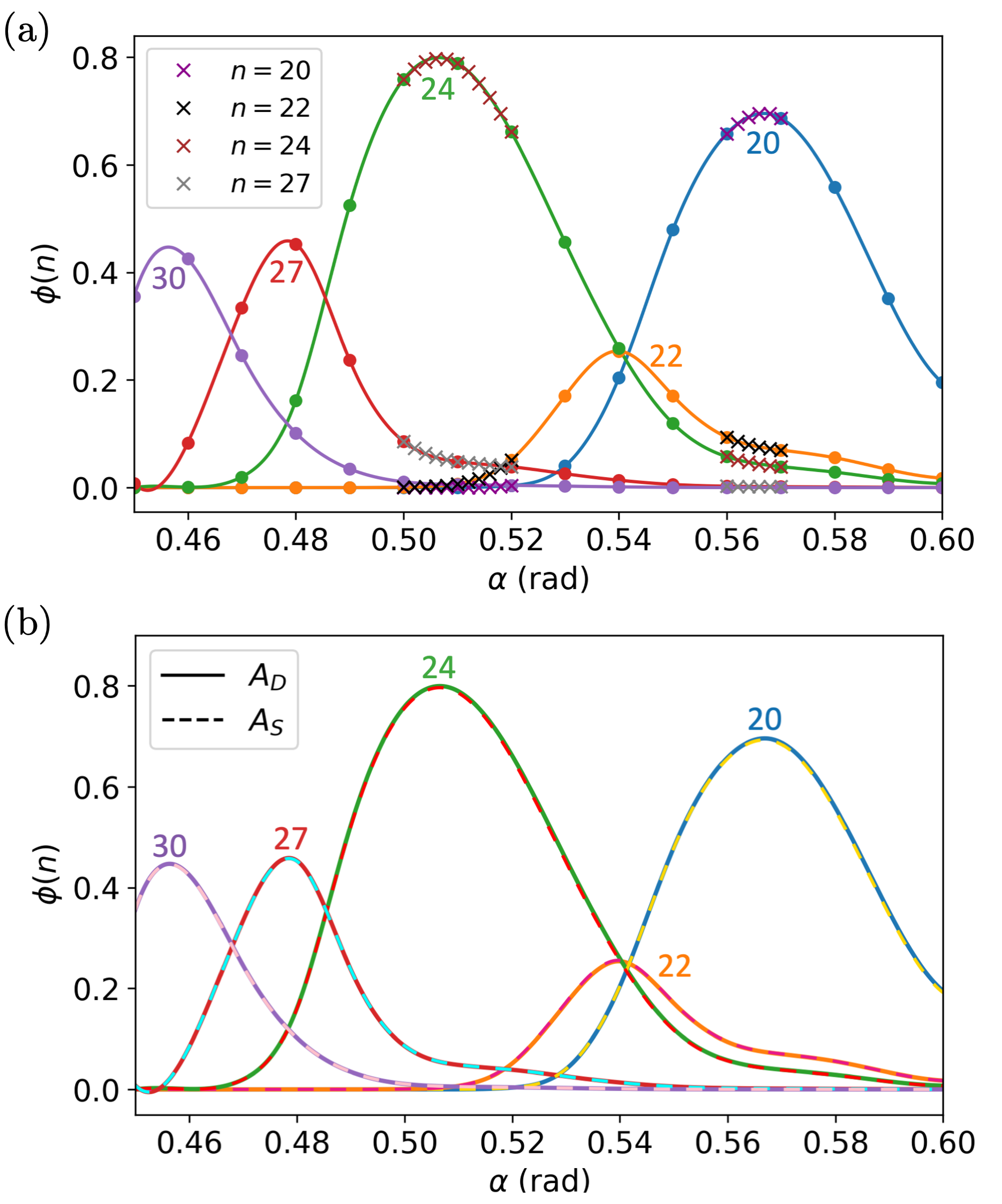
As described above, the self-assembly diagram in Fig. 2(a) was obtained by interpolating between a discrete set of values of . Figure 3(a) compares the interpolated at the resolution rad used for Fig. 2(a) for selected (dominant) MPPN -states at the protein number fraction Wu et al. (2013); Basta et al. (2014) with the corresponding results of calculations done at a finer resolution rad. Figure 3(a) suggests that the interpolation scheme employed here provides accurate estimates of the dominant for continuous . Figure 3(b) compares the results for in Fig. 2(a), obtained with in Eq. (27), with the corresponding results obtained with . As expected, provides a good approximation for the weak membrane curvatures induced by MscS. But is, in general, not expected to give accurate results for membrane proteins such as Piezo that strongly curve the membrane. In the following we therefore focus on in Eq. (27).
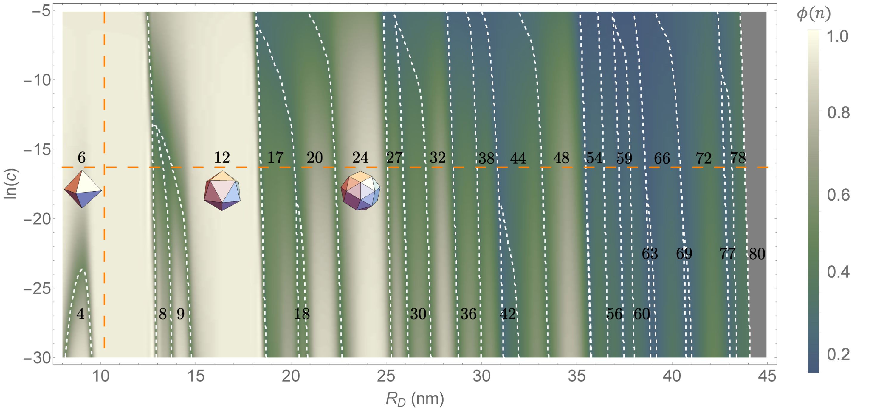
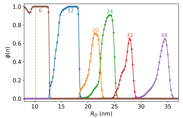
MPPN self-assembly from Piezo. Figure 4 shows the self-assembly diagram for MPPNs formed from Piezo ion channels as a function of the radius of curvature of the Piezo dome and the protein number fraction in solution. We obtained the results in Fig. 4 using the arclength parameterization of Eq. (7) with in Eq. (27). The MPPN self-assembly diagram in Fig. 4 includes highly curved MPPN states with, for instance, a contact angle rad at nm. The vertical dashed line in Fig. 4 indicates the Piezo dome radius of curvature nm observed for a closed state of Piezo Guo and MacKinnon (2017); Saotome et al. (2018); Zhao et al. (2018); Wang et al. (2019) while the horizontal dashed line indicates, for reference, the protein number fraction used in experiments on MPPNs formed from MscS Wu et al. (2013); Basta et al. (2014). Figure 5 shows, for this protein number fraction, the associated with dominant MPPN -states in Fig. 4 as a function . The results in Figs. 4 and 5 were obtained through (third-order spline) interpolation of with respect to , for numerical results at a resolution nm. Similar results are obtained with a finer resolution in .
We find in Fig. 4 that the dominant MPPN -state depends only weakly on the protein number fraction in solution, but strongly on the Piezo dome radius of curvature. As is increased, Piezo’s membrane footprint becomes less curved Haselwandter and MacKinnon (2018), yielding larger and less curved MPPNs that incorporate more Piezo proteins. Most notably, the MPPN self-assembly diagram in Fig. 4 is dominated by highly symmetric MPPN -states with octahedral (; ), icosahedral (; ), or snub cube (; ) symmetry Clare and Kepert (1986, 1991). For the protein number fraction used in experiments on MPPNs formed from MscS Wu et al. (2013); Basta et al. (2014), MPPN octahedra are dominant in Fig. 4 for nm, MPPN icosahedra are dominant for nm, and MPPN snub cubes are dominant for nm. At , the dominant MPPN states with , 12, and 24 in Fig. 4 have the characteristic MPPN radii nm, nm, and nm, with the top of the Piezo dome being located approximately – nm, – nm, and – nm above the spherical surface defined by , respectively (Fig. 1).
Figures 4 and 5 show that, in addition to , 12, and 24, MPPNs with , 32, and 48 can also be abundant at . These MPPN -states have -symmetry (), -symmetry (), and -symmetry () Clare and Kepert (1986, 1991). For , MPPN states with , 32, and 48 are dominant for nm, nm, and nm, and have the characteristic MPPN radii , , and , with the top of the Piezo dome being located approximately – nm, – nm, and – nm above the spherical surface defined by , respectively (Fig. 1). Overall, Figs. 4 and 5 thus predict that self-assembly of MPPNs from Piezo can yield highly symmetric and highly curved MPPN states, with the radius of curvature of the Piezo dome providing the critical control parameter for the symmetry and size of MPPNs formed from Piezo.
Conclusion. We have developed here a methodology for predicting the symmetry and size of MPPNs with arbitrarily large (nonlinear) membrane curvature deformations. For MPPNs formed from MscS Bass et al. (2002); Steinbacher et al. (2007) this methodology predicts, with no adjustable parameters, the observed symmetry and size of MPPNs Wu et al. (2013); Basta et al. (2014). Since MscS proteins only weakly curve the membrane Bass et al. (2002); Steinbacher et al. (2007); Phillips et al. (2009), similar conclusions were reached previously using a small-gradient approximation Li et al. (2016, 2017). In contrast, (closed-state) Piezo proteins have a highly curved structure Guo and MacKinnon (2017); Saotome et al. (2018); Zhao et al. (2018); Wang et al. (2019), and the resulting membrane shape deformations are highly nonlinear Haselwandter and MacKinnon (2018). We find here that the self-assembly diagram for MPPNs formed from Piezo critically depends on the Piezo dome radius of curvature. In particular, for the Piezo dome radius of curvature nm observed for a closed state of Piezo Guo and MacKinnon (2017); Saotome et al. (2018); Zhao et al. (2018); Wang et al. (2019), we generally find MPPNs with six Piezo proteins and octahedral symmetry to be dominant. As the value of is increased, we find dominant MPPN states with icosahedral and snub cube symmetry, composed of 12 and 24 Piezo proteins, respectively. Such highly symmetric MPPN states may allow structural studies of MPPNs formed from Piezo Zhang et al. (2003); Liu et al. (2004); Cockburn et al. (2004) in the presence of transmembrane gradients similar to those found in cellular environments Wu et al. (2013); Basta et al. (2014). Intriguingly, if gating of Piezo is accompanied by an increase in Guo and MacKinnon (2017); Saotome et al. (2018); Zhao et al. (2018); Wang et al. (2019); Lin et al. (2019); Haselwandter and MacKinnon (2018), the distinct MPPN symmetries predicted here may be associated with distinct, biologically relevant conformational states of Piezo.
Acknowledgments. We thank J. Agudo-Canalejo, O. Kahraman, D. Li, R. MacKinnon, and M. H. B. Stowell for useful discussions. This work was supported by NSF award number DMR-1554716 and the USC Center for High-Performance Computing.
References
- Coste et al. (2010) B. Coste, J. Mathur, M. Schmidt, T. J. Earley, S. Ranade, M. J. Petrus, A. E. Dubin, and A. Patapoutian, Science 330, 55 (2010).
- Ranade et al. (2015) S. S. Ranade, R. Syeda, and A. Patapoutian, Neuron 87, 1162 (2015).
- Honoré et al. (2015) E. Honoré, J. R. Martins, D. Penton, A. Patel, and S. Demolombe, in Rev. Physiol., Biochem. and Pharm. Vol. 169 (Springer, 2015) pp. 25–41.
- Murthy and Dubin (2017) S. E. Murthy and A. Dubin, A. E and. Patapoutian, Nat. Rev. Mol. Cell Biol. 18, 771 (2017).
- Wu et al. (2017) J. Wu, A. H. Lewis, and J. Grandl, Trends Biochem. Sci. 42, 57 (2017).
- Gottlieb (2017) P. A. Gottlieb, in Curr. Top. Membr., Vol. 79 (Elsevier, 2017) pp. 1–36.
- Parpaite and Coste (2017) T. Parpaite and B. Coste, Curr. Biol. 27, R250 (2017).
- Douguet et al. (2019) D. Douguet, A. Patel, A. Xu, P. M. Vanhoutte, and E. Honoré, Trends Pharmacol. Sci. 40, 956 (2019).
- Dance (2020) A. Dance, Nature 577, 158 (2020).
- Guo and MacKinnon (2017) Y. R. Guo and R. MacKinnon, eLife 6, e33660 (2017).
- Saotome et al. (2018) K. Saotome, S. E. Murthy, J. M. Kefauver, T. Whitwam, A. Patapoutian, and A. B. Ward, Nature 554, 481 (2018).
- Zhao et al. (2018) Q. Zhao, H. Zhou, S. Chi, Y. Wang, J. Wang, J. Geng, K. Wu, W. Liu, T. Zhang, M.-Q. Dong, J. Wang, X. Li, and B. Xiao, Nature 554, 487 (2018).
- Wang et al. (2019) L. Wang, H. Zhou, M. Zhang, W. Liu, T. Deng, Q. Zhao, Y. Li, J. Lei, X. Li, and B. Xiao, Nature 573, 225 (2019).
- Lin et al. (2019) Y.-C. Lin, Y. R. Guo, A. Miyagi, J. Levring, R. MacKinnon, and S. Scheuring, Nature 573, 230 (2019).
- Haselwandter and MacKinnon (2018) C. A. Haselwandter and R. MacKinnon, eLife 6, e41968 (2018).
- Wu et al. (2013) H.-J. Wu, T. Basta, M. Morphew, D. C. Rees, M. H. B. Stowell, and Y. C. Lee, in The 8th Annual IEEE International Conference on Nano/Micro Engineered and Molecular Systems (2013) pp. 84–87.
- Basta et al. (2014) T. Basta, H. J. Wu, M. K. Morphew, J. Lee, N. Ghosh, J. Lai, J. M. Heumann, K. Wang, Y. C. Lee, D. C. Rees, and M. H. B. Stowell, Proc. Natl. Acad. Sci. U.S.A. 111, 670 (2014).
- Bass et al. (2002) R. B. Bass, P. Strop, M. Barclay, and D. C. Rees, Science 298, 1582 (2002).
- Steinbacher et al. (2007) S. Steinbacher, R. B. Bass, P. Strop, and D. C. Rees, Mechanosensitive Ion Channels, Part A, Curr. Top. Membr., Vol. 58 (Academic Press, New York, 2007) pp. 1–24.
- Zhang et al. (2003) W. Zhang, P. R. Chipman, J. Corver, P. R. Johnson, Y. Zhang, S. Mukhopadhyay, T. S. Baker, J. H. Strauss, M. G. Rossmann, and R. J. Kuhn, Nat. Struct. & Mol. Biol. 10, 907 (2003).
- Liu et al. (2004) Z. Liu, H. Yan, K. Wang, T. Kuang, J. Zhang, L. Gui, X. An, and W. Chang, Nature 428, 287 (2004).
- Cockburn et al. (2004) J. J. B. Cockburn, N. G. A. Abrescia, J. M. Grimes, G. C. Sutton, J. M. Diprose, J. M. Benevides, G. J. Thomas, J. K. H. Bamford, D. H. Bamford, and D. I. Stuart, Nature 432, 122 (2004).
- Li et al. (2016) D. Li, O. Kahraman, and C. A. Haselwandter, Phys. Rev. Lett. 117, 138103 (2016).
- Li et al. (2017) D. Li, O. Kahraman, and C. A. Haselwandter, Europhys. Lett. 117, 58001 (2017).
- Phillips et al. (2009) R. Phillips, T. Ursell, P. Wiggins, and P. Sens, Nature 459, 379 (2009).
- Ma and Haselwandter (2020) M. Ma and C. A. Haselwandter, Phys. Rev. E 102, 042411 (2020).
- Ben-Shaul and Gelbart (1994) A. Ben-Shaul and W. M. Gelbart, in Micelles, Membranes, Microemulsions, and Monolayers (Springer New York, 1994) pp. 1–104.
- Safran (2003) S. A. Safran, Statistical Thermodynamics of Surfaces, Interfaces, and Membranes (Westview Press, Boulder, 2003) pp. 234–238.
- Bruinsma et al. (2003) R. F. Bruinsma, W. M. Gelbart, D. Reguera, J. Rudnick, and R. Zandi, Phys. Rev. Lett. 90, 248101 (2003).
- Canham (1970) P. B. Canham, J. Theor. Biol. 26, 61 (1970).
- Helfrich (1973) W. Helfrich, Z. Naturforsch. C 28, 693 (1973).
- Evans (1974) E. A. Evans, Biophys. J. 14, 923 (1974).
- Rawicz et al. (2000) W. Rawicz, K. C. Olbrich, T. McIntosh, D. Needham, and E. Evans, Biophys. J. 79, 328 (2000).
- Góźdź and Gompper (2001) W. T. Góźdź and G. Gompper, Europhys. Lett. 55, 587 (2001).
- Auth and Gompper (2009) T. Auth and G. Gompper, Phys. Rev. E 80, 031901 (2009).
- Müller and Deserno (2010) M. M. Müller and M. Deserno, Prog. Theor. Phys. Supp. 184, 351 (2010).
- Fournier (1999) J.-B. Fournier, Eur. Phys. J. B - Condensed Matter and Complex Systems 11, 261 (1999).
- Weitz and Destainville (2013) S. Weitz and N. Destainville, Soft Matter 9, 7804 (2013).
- Peterson (1985) M. A. Peterson, J. Appl. Phys. 57, 1739 (1985).
- Seifert et al. (1991) U. Seifert, K. Berndl, and R. Lipowsky, Phys. Rev. A 44, 1182 (1991).
- Jülicher and Seifert (1994) F. Jülicher and U. Seifert, Phys. Rev. E 49, 4728 (1994).
- Kühnel (2015) W. Kühnel, Differential Geometry, 3rd ed., Vol. 77 (American Mathematical Society, 2015).
- Weisstein (2017) E. W. Weisstein, From MathWorld – A Wolfram Web Resource. http://mathworld.wolfram.com/SphericalCap.html (2017).
- Courant and Hilbert (1953) R. Courant and D. Hilbert, Methods of Mathematical Physics, 5th ed. (Interscience Punlishers, Inc., New York, 1953).
- van Brunt (2004) B. van Brunt, The Calculus of Variations (Springer, New York, 2004).
- Kibble and Berkshire (2004) T. W. B. Kibble and F. H. Berkshire, Classical Mechanics, 5th ed. (Imperial College Press, London, 2004).
- Deserno and Bickel (2003) M. Deserno and T. Bickel, Europhys. Lett. 62, 767 (2003).
- Deserno (2004) M. Deserno, Phys. Rev. E 69, 031903 (2004).
- Nowak and Chou (2008) S. A. Nowak and T. Chou, Phys. Rev. E 78, 021908 (2008).
- Zhang and Nguyen (2008) R. Zhang and T. T. Nguyen, Phys. Rev. E 78, 051903 (2008).
- Hashemi et al. (2014) S. M. Hashemi, P. Sens, and F. Mohammad-Rafiee, Journal of The Royal Society Interface 11, 20140769 (2014).
- Foret (2014) L. Foret, The Euro. Phys. J. E 37, 42 (2014).
- Agudo-Canalejo and Lipowsky (2016) J. Agudo-Canalejo and R. Lipowsky, Soft Matter 12, 8155 (2016).
- Bahrami et al. (2016) A. H. Bahrami, R. Lipowsky, and T. R. Weikl, Soft Matter 12, 581 (2016).
- Burden and Faires (2011) R. L. Burden and J. D. Faires, Numerical Analysis, 9th ed. (Brooks/Cole, Cengage Learning, Boston, 2011).
- Gautschi (2012) W. Gautschi, Numerical Analysis, 2nd ed. (Springer Science & Business Media, New York, 2012).
- mat (2017) Mathematica 11.2 (Wolfram Research, Inc., Champaign, IL, 2017).
- Clare and Kepert (1986) B. W. Clare and D. L. Kepert, Proc. R. Soc. London A 405, 329 (1986).
- Clare and Kepert (1991) B. W. Clare and D. L. Kepert, J. Math. Chem. 6, 325 (1991).
- Damodaran and Merz (1993) K. V. Damodaran and K. M. Merz, Langmuir 9, 1179 (1993).