Impact of screening and relaxation onto weakly coupled 2D heterostructures
Abstract
The stacking of different 2D materials provides a promising approach to realize new states of quantum matter. In this combined scanning tunneling microscopy (STM) and density functional theory (DFT) study we show that the structure in weakly bound, purely van der Waals (vdW) interacting systems is strongly influenced by screening and relaxation. We studied in detail the physisorption of lead phthalocyanine (PbPc) molecules on epitaxial monolayer graphene on SiC(0001) as well as on highly ordered pyrolytic graphite (HOPG), resembling truly 2D and anisotropic, semi-infinite 3D supports. Our analysis demonstrates that the different deformation ability of the vdW coupled systems, i.e. their actual thickness and buckling, triggers the molecular morphology and exhibits a proximity coupled band structure. It thus provides important implications for future 2D design concepts.
Heterostructures made layer by layer in a precisely chosen sequence out of 2D materials were suggested to design bulk quantum materials with entirely new functions Geim and Grigorieva (2013). Indeed, proximity coupling reveals superconductivity in twisted bilayer graphene Novoselov et al. (2019); Cao et al. (2018). The absence of dangling bonds in 2D materials is expected to allow a flexible and lego-like epitaxial growth of lattice mismatched materials in random order Koma (1999).
However, as fabricating a 3D stack out of 2D sheets, the same layers may experience a different coupling, e.g. due to modified screening. Coulomb interaction in 2D and 3D is fundamentally different Cudazzo et al. (2011). In contrast to the isotropic 3D case, for 2D the charge is redistributed on a circle around the point charge, i.e. the residual electric field depends on the polar angle, resulting in a non-local screening behavior which leads usually to strong and -dependent renormalization of quasiparticle energies, e.g. excitons Qiu et al. (2013); Ugeda et al. (2014); Qiu et al. (2017) and reduced energy gaps Neaton et al. (2006); Garcia-Lastra et al. (2009); Noori et al. (2019).
Among thousands of feasible 2D materials Mounet et al. (2018), graphene is still the most perfect and flexible one, thus ideal to elucidate principles of proximity coupling. Epitaxial graphene on SiC(0001) provides the flexibility to control the interface and its electronic properties Riedl et al. (2010); Yazdi et al. (2016); Kruskopf et al. (2016): monolayer graphene (MLG) grown on SiC(0001) is -type doped while quasi-free monolayer graphene (QFMLG) on the same substrate is slightly -type doped Riedl et al. (2009a). HOPG, in contrast, is charge neutral and represents the semi-infinite 3D counterpart of graphene Mammadov et al. (2017).
Long-ranged ordered molecular 2D structures can be realized also by physisorption of -conjugated organic molecules on surfaces Gottfried (2015). Their combination with solid state 2D structures proposes advanced stacking sequences with tailored properties. However, the comprehensive understanding of the physisorption process can become a formidable challenge. Usually, the adsorbate layer and surface lattice are not commensurate. Long-range dispersing forces between the molecules provide a possibility for various phases. The complex interaction scheme with the substrate often comes along with charge transfer (between substrate and adsorbate) superimposing the effect of screening.
Here, the shuttlecock-like lead phthalocyanine (PbPc) molecule with a large gap between the highest occupied and lowest unoccupied molecular orbitals, HOMO and LUMO, helps to suppress charge transfer with the substrate. It thus provides an excellent candidate to study implications of screening and proximity coupling in physisorbed systems. It contains four benzene-pyrrole moieties, which are connected via meso-aza nitrogens. The central Pb atom is coordinated to the four adjacent pyrrole nitrogens and is located outside the molecular plane further reducing the interaction with the substrate.
In this Letter, we analyzed the adsorption of PbPc on variously doped epitaxial graphene and HOPG. For QFMLG and MLG, the buckling of the graphene layer promotes a quasi-free, densely packed and chiral PbPc molecular layer structure with almost identical lattice parameters. Only minor 2D screening is observed, so that the molecular states remain almost unaffected. In contrast, large substrate induced dispersion of the HOMO is found on HOPG, where the more distant molecules interact via the substrate predominantly. Here, -stacking leads to proximity coupling of PbPc with deeper graphite layer s and to a strongly -dependent reduction of the molecular gap.
| free layer | MLG | QFMLG | HOPG | |
|---|---|---|---|---|
| b1 (nm) | 1.42 0.05 | 1.40 0.05 | 1.58 0.05 | |
| calc. | 1.35 | 1.37 | 1.37 | 1.54 |
| b2 (nm) | 1.38 0.05 | 1.40 0.05 | 1.49 0.05 | |
| calc. | 1.33 | 1.30 | 1.30 | 1.54 |
| (∘) | 90.3 1.0 | 90.0 1.0 | 90.1 1.0 | |
| calc. | 98.3 | 91.9 | 91.9 | 92.2 |
| (∘) | — | 30 3 | 30 3 | 30 3 |
| (∘) | 39.2 2.0 | 38.5 2.0 | 30.5 2.0 | |
| calc. | — | 39.4 | 39.1 | 30.0 |
| height (Å) | — | 2.5/3.5/4.6 | 2.3/3.5/4.7 | 3.1/3.3/4.7 |
| buckling (Å) | — | 0.51 | 0.67 | 0.02 |
| tilting (∘) | — | 9.2 | 9.5 | |
| — | 68 | 68 | 90 | |
| Eb/C (meV) | — | 29 | 31 | 26 |
| Eb/PbPc (eV) | 1.18 | 1.97 | 2.11 | 2.35 |
| Eb,intra (eV) | 1.18 | 0.25 | 0.24 | 0.01 |
As substrates epitaxial monolayer graphene (MLG) and hydrogen-intercalated quasi-free monolayer graphene (QFMLG) on semi-insulating 6-SiC(0001) as well as highly ordered pyrolytic graphite (HOPG) were used. While HOPG resembles a charge neutral anisotropic 3D material, MLG and QFMLG are both 2D materials but with completely different electrochemical potentials Mammadov et al. (2017). Details about fabrication and characterization of the substrates are reported elsewhere Riedl et al. (2008, 2009b); Momeni Pakdehi et al. (2018, 2019). The adsorption of PbPc molecules was done at 300 K under ultra high vacuum with same adsorption rates in order to allow a direct comparison Nguyen et al. (2019). The atomic structure and positions of molecular energy levels were investigated by low temperature scanning tunneling microscopy (LT-STM, 6 K and 80 K). Scanning tunneling spectroscopy (STS) was recorded using lock-in technique (20 meV, 1 kHz). For the dI/dV spectra an average of at least 10 curves were acquired at various positions across the molecules.
Our experiments are supplemented by DFT calculations using a supercell approach and periodic boundary conditions. For HOPG, molecular layers of PbPc molecules were modeled on 6 layer thick Bernal-stacked graphite. Thereby, the simplicity of the substrate allows a direct modeling in the experimentally observed quasi-square surface unit cell (with =90 atoms per C layer, see Tab. 1). In contrast, for QFMLG and MLG, square unit cells are either incommensurable with the underlying SiC(0001) substrate or the graphene layers. As a result, the unit cell of PbPc on (QF)MLG/SiC(0001) contains at least two molecules. However, the absence of any indications in the STM experiment suggests that the SiC part of the substrate plays a minor role. Thus, the MLG calculations were restricted to a simplified unit cell containing one PbPc, where the interaction with the substrate (68 atoms per C layer) is reduced to the topmost graphene layer plus a partially H decorated buffer layer, whereby the level of doping increases almost linearly with the number of in this way -coordinated C atoms. The doping level, i.e. the position of the Dirac point ED (cf. Fig. 5a), can thus be adjusted via the degree of H decoration. For a 0.4 eV shift determined experimentally Momeni Pakdehi et al. (2019), about 15 % of the C atoms of the buffer layer have to be covalently bound to the underlaying SiC substrate (cf. Fig. 5b), in fair agreement with experiment Emtsev et al. (2008).
Structural relaxation calculations are performed with the Quantum ESPRESSO package using periodic boundary conditions and a 331 -point sampling Giannozzi et al. (2009, 2017). STM images are simulated based on VASP calculations using the Tersoff-Hamann approach to analyse the tunneling current Tersoff and Hamann (1985). Specifically, we use scalar relativistic norm-conserving pseudopotentials and a plane wave basis set with 90 Ry energy cutoff. For structure relaxation the semi-local PBE functional was used to include many-body effects due to exchange and correlation (XC). Afterwards the B3LYP hybrid functional was used to accurately determine the electronic structure for the PBE relaxed structures. The use of B3LYP copes the DFT-underestimization of the molecular HOMO-LUMO gap (see Ref. Baran and Larsson, 2010; Zhang et al., 2007 and Table II), and allows a 1:1 comparison of the resulting density of states (DOS) with the experimental STS spectra. In all calculations the D3 dispersion correction was used for a reasonable description of non-local correlation effects Grimme et al. (2010).
Our experiments and calculations clearly reveal an adsorption of PbPc, where the central Pb atom is pointing upwards on HOPG as well as on epitaxial graphene, as shown in Fig. 1. At least 0.3 eV per molecule (0.72 eV on HOPG) is gained by this preferential adsorption geometry. This is in contrast to Au(111) surfaces, where PbPc molecules show both, up- and down configurations Madhuri et al. (2017). PbPc on Cu(100) and Ag(100) have been found to form a chiral monolayer structure, while on Pd(100) a stable achiral state was reported Mugarza et al. (2010); Cai et al. (2015). The latter adsorption geometry was explained by stronger hybridization between the orbital of the macrocyclic C atoms in PbPc and the orbitals of the Pd substrate.
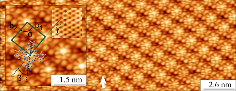
PbPc on all the investigated graphene and graphite templates forms highly-ordered chiral monolayer structures with a
single PbPc molecules in quasi-square unit cells, as shown in Fig. 1 exemplarily for QFMLG Nguyen et al. (2019).
Table 1 summarizes the lattice parameters and molecular orientations
which were deduced from STM images taken across the edges of the PbPc islands (cf. with Ref. Nguyen et al. (2019)).
They are nicely confirmed and rationalized by our DFT simulations, i.e. by minimizing total energy
while varying cell size and shape.
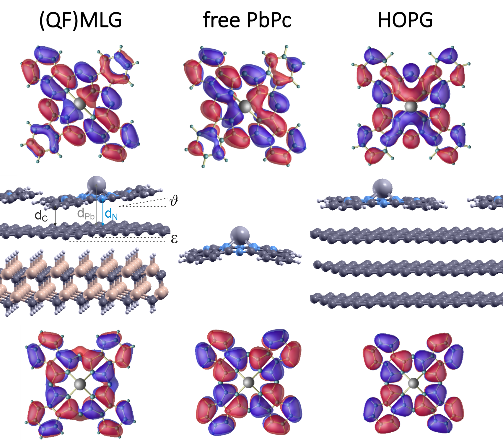
The lattice parameters for PbPc on QFMLG and MLG are similar (even identical in theory), while the parameters found on HOPG are considerably larger by about 10% (cf. Tab. 1). The different lattice parameters come along with specific details of the adsorption structure. The characteristic shuttlecock structure of the free PbPc relaxes upon physisorption on all the three substrates. In case of HOPG, the C4v symmetry of the gas phase PbPc molecules is retained, but all wings are found almost planar, cf. Fig. 2. It maximizes the attractive vdW interaction per molecule with the graphene template and resembles the geometry of isolated physisorbed molecules Kera et al. (2007); Shibuta et al. (2010); Yamamoto et al. (2011). The adsorption height of the C-atoms of about 3.1 Å (cf. Tab. 1) is similar to the interlayer distance in graphite. Together with the planar adsorption geometry this suggests stacking as a predominant driving force. In essence, this stacking gives rise to a proximity-coupled band structure, as we will show below.
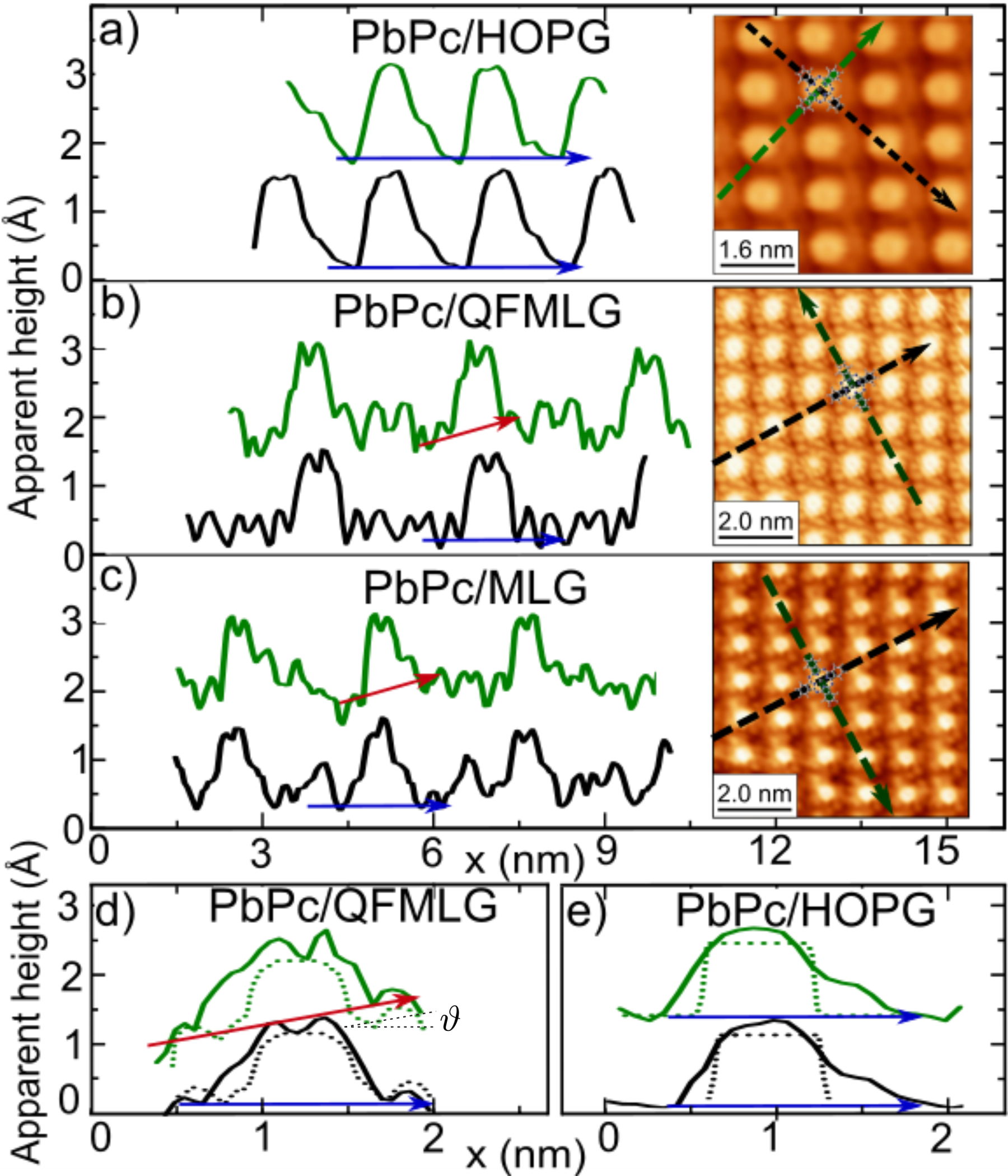
In contrast, the C4v symmetry of the PbPc upon adsorption on (QF)MLG is lifted. Two neighboring benzene-pyrrole units are bended towards the surface, while the others are lifted to different extent (cf. Fig. 2). The formation of a layer of in this way tilted molecules is in agreement with the asymmetry seen in the STM height profiles taken along the two principal axes of the molecules on (QF)MLG (cf. Fig. 3 b,c). Details of the height profiles are shown in the close-up in panel d) and coincide in all cases with profiles obtained from DFT calculated STM images shown in Fig. 4.
The tilted structure on (QF)MLG allows a closer arrangement with strongly increased ( 20) intermolecular coupling, E (see Tab. 1) while providing a maximum binding energy per substrate area (i.e. per C-atom). The resulting lattice constants are about 10% smaller than on HOPG and, notably, comparable to a potential freestanding molecular PbPc layer 111The overall total energy minimum of a free molecular layer is found for a lattice equivalent to a coverage of 1 molecule per 71 C atoms, very close to the (QF)MLG structures (1 PbPc molecule per 68 C atoms, see Table 1)..
What is the driving force behind the different adsorption schemes? — The geometry of PbPc on MLG and QFMLG is very similar, despite their different electrochemical potentials. Obviously, the doping level of the two 2D substrates are of minor relevance Mammadov et al. (2017). According to recent transport measurements Nguyen et al. (2019), charge transfer is also not taking place, in agreement with the present STS and DFT calculations (see below).
A conceivable reason is the corrugation of epitaxial graphene on SiC. The buckling facilitates adsorption of tilted PbPc molecules: The topmost graphene layer is considerably upwards bended towards the PbPc macrocycle compensating the tilting-induced losses in vdW interaction. Although the exfoliation energy for HOPG is one order of magnitude lower than in case of MLG/SiC Wang et al. (2015); Wells et al. (2014) a local deformation of the uppermost C layer in HOPG costs by far more energy (182 meV instead of 86 meV for MLG). Obviously, the inherent corrugation of epitaxial graphene layers (the lateral strain also responsible for the buckling) allows for a flexible adaption of the substrate to the adsorbed molecular structures. Thus, the tilting of the PbPc molecule is a direct consequence of the deformation ability of the 2D support.
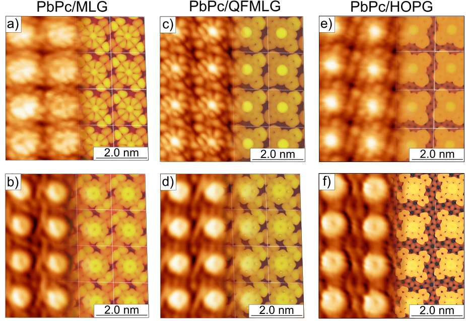
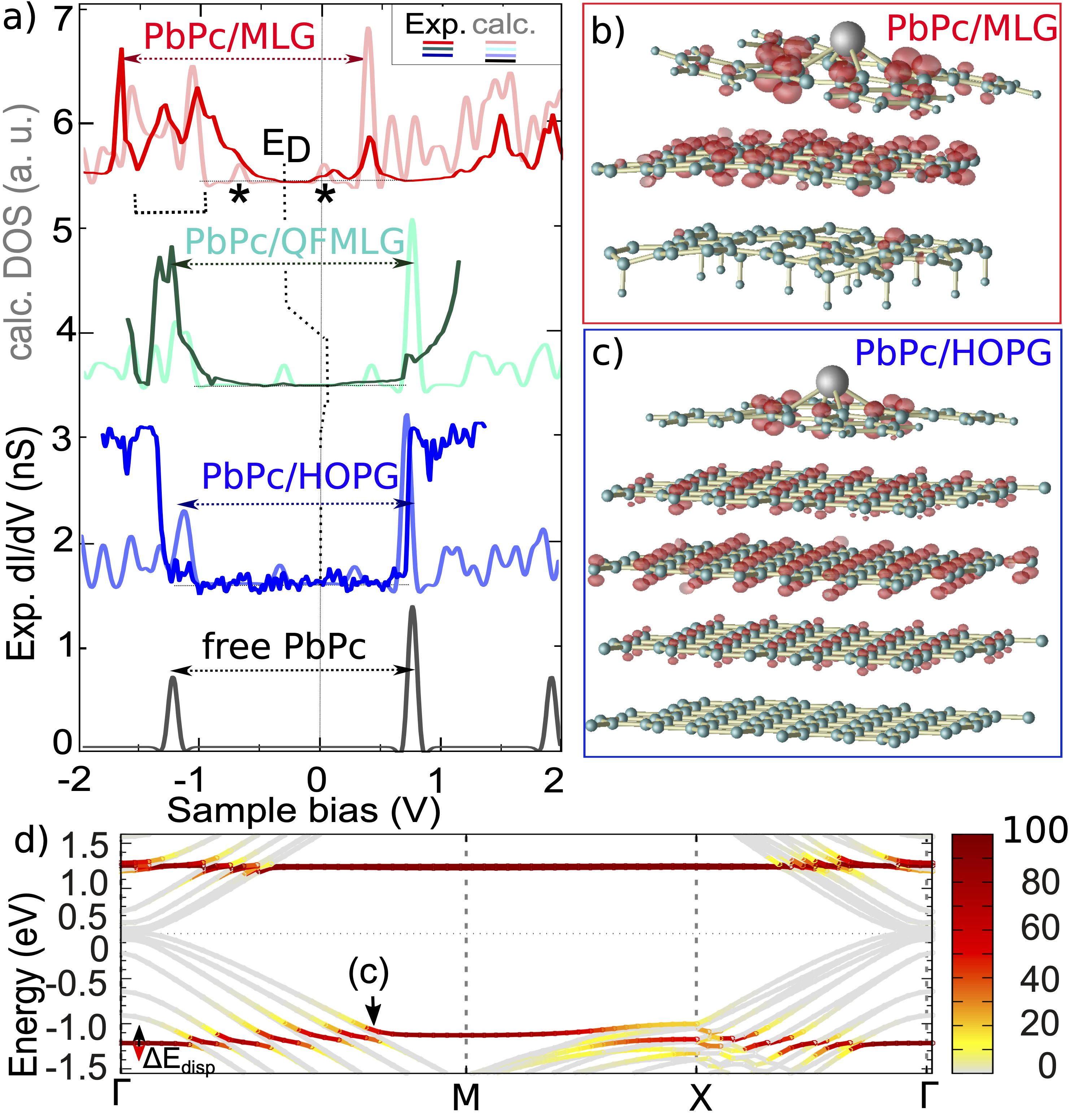
A common STM feature for all three substrates is a donut-like shape of the occupied state in the center of the molecule. It was seen for all investigated substrates and is nicely reproduced by DFT in Fig. 4 Nguyen et al. (2019). This demonstrates that the central Pb atom does not contribute to the HOMO (cf. Fig. 2, 5b,c) for all ivestigated substrates, whereby this spectroscopic fingerprint becomes most obvious at slightly different tunneling voltages, see Fig. 4. For a more detailed analysis, additional STS measurements were performed. In Fig. 5 a) averaged dI/dV-spectra are shown and compared with the B3LYP-D3 calculated density of states (DOS):
| System (XC-funct.) | isolated | gapmax/min | Edisp |
|---|---|---|---|
| PbPc isolated (LDA,PZ-D3) | 1.32 | ||
| PbPc isolated (PBE-D3) | 1.33 | ||
| PbPc isolated (B3LYP-D3) | 2.01 | ||
| freestanding PbPc film (MLG) | 2.03/2.00 | 0.03 | |
| freestanding PbPc film (QFMLG) | 2.01/2.00 | 0.01 | |
| freestanding PbPc film (HOPG) | 2.01/2.01 | 0.00 | |
| PbPc/MLG | 1.98 | 1.98/1.94 | 0.05 |
| PbPc/QFMLG | 1.95 | 1.94/1.85 | 0.09 |
| PbPc/HOPG | 2.00 | 2.00/1.75 | 0.39 |
(i) While the energies for the HOMO and LUMO states of gas phase PbPc Baran and Larsson (2010) and adsorbed on QFMLG as well as HOPG are similar, the spectrum measured on MLG is shifted to lower energies (by 0.4 eV), reflecting the -type doping of MLG. Whereas the position of the LUMO is obvious also in this case, the identification of the HOMO requires theoretical support: Those C atoms (of the buffer layer) covalently bound to the SiC substrate, introduce additional occupied states partially superimposing the HOMO (see bracket in Fig. 5).
(ii) Interestingly, the different doping levels of MLG and QFMLG play no role. Very similar HOMO and only slightly different LUMO STM signatures suggest that both structures experience an almost identical lateral screening behavior. There are also only minor relative shifts of the HOMO and LUMO levels (cf. Tab. 2). This is in line with literature where relevant screening effects onto the molecular electronic structure are restricted to substrate 2D-layer distances clearly below 3 Å Niesner and Fauster (2014); Marks et al. (2014).
Contrary, for HOPG our B3LYP-D3 band structure calculations reveal a large dispersion of the molecular HOMO ( 0.39 eV). The LUMO is affected by much lower extend (cf. Fig. 5 d). Similar to the case of metallic substrates Cai et al. (2015); Kera et al. (2007), the resulting -point dependent renormalization of the molecular HOMO-LUMO gap can be attributed to -stacking of the eight macrocyclic C atoms (those bridging two N atoms) with the substrate C atoms (see Fig. 5 b,c) which is largest for the second graphite layer. Notably, this hybridization effect is not coming along with a gap opening in the substrate bands and, thus, mimics an example of proximity coupling. The concomitant modification of the band structure is strongly -dependent, and the maximum size effect is restricted to rather small regions within the Brillouin zone (Fig. 5 d), explaining the shoulder observed in STS slightly below –1 V sample bias. The fact, that the molecular states of PbPc on HOPG reveal a strongly enhanced dispersion, although the intermolecular distance is larger compared to (QF)MLG), underlines the importance of a substrate mediated interaction.
In summary, we comprehensively studied vdW interacting heterostructures by means of PbPc monolayer structures on 2D graphene, (QF)MLG, and semi-infinite 3D graphite, HOPG. Albeit the surface structure of all templates are the same and charge transfer is not taking place, the molecular layer reveals very different lattice parameters and underwent different relaxation schemes. Formation of almost identical densely packed PbPc molecular layers with strongly tilted molecules were found on both 2D templates, despite their very different work functions, showing that lateral Thomas Fermi screening plays a minor role. Contrary, the interaction with the upper graphite (HOPG) layers, in particular the second, favors an almost planar adsorption of PbPc at the expense of a considerably larger lattice constant. The dispersing molecular states unambiguously demonstrate the presence of a substrate mediated interaction and the band structure exhibits spectral features of proximity coupling. Therefore, the actual thickness of a 3D stack built from 2D sheets appears to be decisive for the vdW heteroepitaxy and impacts recent layer by layer design concepts Koma (1999); Geim and Grigorieva (2013).
Acknowledgement
We thank D. Momeni Pakdehi (PTB Braunschweig) for providing us epitaxially grown graphene samples on SiC(0001).
Financial support by the DFG (through project Te386/17-1 and TRR 142, project number 231447078) is gratefully acknowledged.
The in parts demanding numerical calculations were possible thanks to CPU-time grants of the Paderborn Center for Parallel Computing, (PC)2.
References
- Geim and Grigorieva (2013) A. K. Geim and I. V. Grigorieva, Nature 499, 419 (2013).
- Novoselov et al. (2019) K. S. Novoselov, D. V. Andreeva, W. Ren, and G. Shan, Frontiers of Physics 14, 13301 (2019).
- Cao et al. (2018) Y. Cao, V. Fatemi, S. Fang, K. Watanabe, T. Taniguchi, E. Kaxiras, and P. Jarillo-Herrero, Nature 556, 43 (2018).
- Koma (1999) A. Koma, Journal of Crystal Growth 201-202, 236 (1999).
- Cudazzo et al. (2011) P. Cudazzo, I. V. Tokatly, and A. Rubio, Phys. Rev. B 84, 085406 (2011).
- Qiu et al. (2013) D. Y. Qiu, F. H. da Jornada, and S. G. Louie, Phys. Rev. Lett. 111, 216805 (2013).
- Ugeda et al. (2014) M. M. Ugeda, A. J. Bradley, S.-F. Shi, F. H. da Jornada, Y. Zhang, D. Y. Qiu, W. Ruan, S.-K. Mo, Z. Hussain, Z.-X. Shen, F. Wang, S. G. Louie, and M. F. Crommie, Nature Materials 13, 1091 (2014).
- Qiu et al. (2017) D. Y. Qiu, F. H. da Jornada, and S. G. Louie, Nano Lett. 17, 4706 (2017).
- Neaton et al. (2006) J. B. Neaton, M. S. Hybertsen, and S. G. Louie, Phys. Rev. Lett. 97, 216405 (2006).
- Garcia-Lastra et al. (2009) J. M. Garcia-Lastra, C. Rostgaard, A. Rubio, and K. S. Thygesen, Phys. Rev. B 80, 245427 (2009).
- Noori et al. (2019) K. Noori, N. L. Q. Cheng, F. Xuan, and S. Y. Quek, 2D Materials 6, 035036 (2019).
- Mounet et al. (2018) N. Mounet, M. Gibertini, P. Schwaller, D. Campi, A. Merkys, A. Marrazzo, T. Sohier, I. E. Castelli, A. Cepellotti, G. Pizzi, and N. Marzari, Nature Nanotechnology 13, 246 (2018).
- Riedl et al. (2010) C. Riedl, C. Coletti, and U. Starke, Journal of Physics D: Applied Physics 43, 374009 (2010).
- Yazdi et al. (2016) G. R. Yazdi, T. Iakimov, and R. Yakimova, Crystals 6, 53 (2016).
- Kruskopf et al. (2016) M. Kruskopf, D. M. Pakdehi, K. Pierz, S. Wundrack, R. Stosch, T. Dziomba, M. Götz, J. Baringhaus, J. Aprojanz, C. Tegenkamp, J. Lidzba, T. Seyller, F. Hohls, F. J. Ahlers, and H. W. Schumacher, 2D Materials 3, 041002 (2016).
- Riedl et al. (2009a) C. Riedl, C. Coletti, T. Iwasaki, A. A. Zakharov, and U. Starke, Phys. Rev. Lett. 103, 246804 (2009a).
- Mammadov et al. (2017) S. Mammadov, J. Ristein, J. Krone, C. Raidel, M. Wanke, V. Wiesmann, F. Speck, and T. Seyller, 2D Materials 4, 015043 (2017).
- Gottfried (2015) J. M. Gottfried, Surface Science Reports 70, 259 (2015).
- Riedl et al. (2008) C. Riedl, A. A. Zakharov, and U. Starke, Applied Physics Letters 93, 033106 (2008).
- Riedl et al. (2009b) C. Riedl, C. Coletti, T. Iwasaki, A. A. Zakharov, and U. Starke, Phys. Rev. Lett. 103, 246804 (2009b).
- Momeni Pakdehi et al. (2018) D. Momeni Pakdehi, J. Aprojanz, A. Sinterhauf, K. Pierz, M. Kruskopf, P. Willke, J. Baringhaus, J. P. Stöckmann, G. A. Traeger, F. Hohls, C. Tegenkamp, M. Wenderoth, F. J. Ahlers, and H. W. Schumacher, ACS Applied Materials & Interfaces 10, 6039 (2018), pMID: 29377673.
- Momeni Pakdehi et al. (2019) D. Momeni Pakdehi, K. Pierz, S. Wundrack, J. Aprojanz, T. T. N. Nguyen, T. Dziomba, F. Hohls, A. Bakin, R. Stosch, C. Tegenkamp, F. J. Ahlers, and H. W. Schumacher, ACS Applied Nano Materials 2, 844 (2019).
- Nguyen et al. (2019) T. N. Nguyen, J. Aprojanz, M. Jäger, T. H. Nguyen, and C. Tegenkamp, Surface Science 686, 45 (2019).
- Emtsev et al. (2008) K. V. Emtsev, F. Speck, T. Seyller, L. Ley, and J. D. Riley, Phys. Rev. B 77, 155303 (2008).
- Giannozzi et al. (2009) P. Giannozzi et al., Journal of Physics: Condensed Matter 21, 395502 (19pp) (2009).
- Giannozzi et al. (2017) P. Giannozzi et al., Journal of Physics: Condensed Matter 29, 465901 (2017).
- Tersoff and Hamann (1985) J. Tersoff and D. R. Hamann, Phys. Rev. B 31, 805 (1985).
- Baran and Larsson (2010) J. D. Baran and J. A. Larsson, Phys. Chem. Chem. Phys. 12, 6179 (2010).
- Zhang et al. (2007) Y. Zhang, X. Cai, X. Zhang, H. Xu, Z. Liu, and J. Jiang, Int. J. Quantum Chem. 107, 952 (2007).
- Grimme et al. (2010) S. Grimme, J. Antony, S. Ehrlich, and H. Krieg, The Journal of Chemical Physics 132, 154104 (2010).
- Madhuri et al. (2017) K. P. Madhuri, P. Kaur, M. E. Ali, and N. S. John, J. Phys. Chem. C 121, 9249 (2017).
- Mugarza et al. (2010) A. Mugarza, N. Lorente, P. Ordejón, C. Krull, S. Stepanow, M.-L. Bocquet, J. Fraxedas, G. Ceballos, and P. Gambardella, Phys. Rev. Lett. 105, 115702 (2010).
- Cai et al. (2015) Y. Cai, S. Xu, X. Qiao, L. Wang, Y. Liu, T. Wang, and X. Xu, Phys. Chem. Chem. Phys. 17, 23651 (2015).
- Kera et al. (2007) S. Kera, H. Fukagawa, T. Kataoka, S. Hosoumi, H. Yamane, and N. Ueno, Phys. Rev. B 75, 121305 (2007).
- Shibuta et al. (2010) M. Shibuta, K. Yamamoto, K. Miyakubo, T. Yamada, and T. Munakata, Phys. Rev. B 81, 115426 (2010).
- Yamamoto et al. (2011) R. Yamamoto, I. Yamamoto, M. Mikamori, T. Yamada, K. Miyakubo, and T. Munakata, Surface Science 605, 982 (2011).
- Note (1) The overall total energy minimum of a free molecular layer is found for a lattice equivalent to a coverage of 1 molecule per 71 C atoms, very close to the (QF)MLG structures (1 PbPc molecule per 68 C atoms, see Table 1).
- Wang et al. (2015) W.-H. Wang, C. Gong, W. Wang, S. K. Fullerton-Shirey, A. Seabaugh, and K. Cho, The Journal of Physical Chemistry C 119, 20016 (2015).
- Wells et al. (2014) G. Wells, T. Hopf, K. Vassilevski, E. Escobedo-Cousin, N. Wright, A. Horsfall, J. Goss, A. O’Neill, and M. Hunt, Applied Physics Letters 105, 193109 (2014).
- Niesner and Fauster (2014) D. Niesner and T. Fauster, Journal of Physics: Condensed Matter 26, 393001 (2014).
- Marks et al. (2014) M. Marks, A. Schöll, and U. Höfer, Journal of Electron Spectroscopy and Related Phenomena 195, 263 (2014).