Malignancy Prediction and Lesion Identification from Clinical Dermatological Images
Abstract
We consider machine-learning-based malignancy prediction and lesion identification from clinical dermatological images, which can be indistinctly acquired via smartphone or dermoscopy capture. Additionally, we do not assume that images contain single lesions, thus the framework supports both focal or wide-field images. Specifically, we propose a two-stage approach in which we first identify all lesions present in the image regardless of sub-type or likelihood of malignancy, then it estimates their likelihood of malignancy, and through aggregation, it also generates an image-level likelihood of malignancy that can be used for high-level screening processes. Further, we consider augmenting the proposed approach with clinical covariates (from electronic health records) and publicly available data (the ISIC dataset). Comprehensive experiments validated on an independent test dataset demonstrate that ) the proposed approach outperforms alternative model architectures; ) the model based on images outperforms a pure clinical model by a large margin, and the combination of images and clinical data does not significantly improves over the image-only model; and ) the proposed framework offers comparable performance in terms of malignancy classification relative to three board certified dermatologists with different levels of experience.
keywords:
Dermatology, Medical Imaging, Lesion Identification, Deep Learning1 Introduction
Prior to the COVID-19 pandemic, access to dermatology care was challenging due to limited supply and increasing demand. According to a survey study of dermatologists, the mean standard deviation (SD) waiting time was 3332 days, 64% of the appointments exceeded the criterion cutoff of 3 weeks and 63% of the appointments exceeded 2-week criterion cutoff for established patients. During the COVID-19 pandemic, the number of dermatology consultations were reduced by 80-90% to urgent issues only, leading to delay in care of dermatologic concerns. Moreover, the issue of access is very significant for the growing Medicare population, expected to account for 1 in 5 patients by 2030 [vincent2010next], due to a higher incidence of skin cancer.
Access issues in dermatology are concerning as there has been an increasing incidence of skin cancers, particularly a 3-fold increase in melanoma over the last 40 years [statfacts]. Many of the skin lesions of concern are screened by primary care physicians (PCPs). In fact, up to one third of primary care visits contend with at least one skin problem, and skin tumors are the most common reason for referral to dermatology [lowell2001dermatology]. High volume of referrals places a strain on specialty care, delaying visits for high-risk cases. Given the expected rise in baby boomers, with significantly increased risk of skin cancer, there is an urgent need to equip primary care providers to help screen and risk stratify patients in real time, high quality and cost-conscious fashion. PCPs have variable experience and training in dermatology, causing often low concordance between their evaluation and dermatology [lowell2001dermatology]. A consistent clinical decision support (CDS) system has the potential to mitigate this variability, and to create a powerful risk stratification tool, leveraging the frontline network of providers to enhance access to quality and valuable care. In addition, such a tool can aid tele-dermatology workflows that have emerged during the global pandemic.
Over the last decade, several studies in the field of dermatology have demonstrated the promise of deep learning models such as convolutional neural networks (CNN) in terms of classification of skin lesions [esteva2017dermatologist, haenssle2018man], with dermoscopy-based machine learning (ML) algorithms reaching sensitivities and specificities for melanoma diagnosis at 87.6% (95% CI 72.72-100.0) and 83.5% (95% CI: 60.92-100.0), respectively, by meta-analysis [safran2018machine]. Several authors have reported superior performance of ML algorithms for classification of squamous cell carcinoma (SCC) and basal cell carcinomas (BCC) with larger datasets improving performance [han2018classification, esteva2017dermatologist].
From a machine-learning methods perspective, a common approach for classification with dermoscopy images consists on refining pre-trained CNN architectures such as VGG16 as in [lopez2017skin] or AlexNet after image pre-processing, e.g. background removal, as in [salido2018using]. Alternatively, some approaches consider lesion sub-types independently [polat2020detection], sonified images [dascalu2019skin], or by combining clinical data with images to increase the information available to the model for prediction [tognetti2021new]. However, dermoscopy images are generally of good quality, high resolution and minimal background noise, making them less challenging to recognize compared to clinical, wide-field, images.
Beyond dermoscopy images, similar refinement approaches have been proposed based on architectures such as ResNet152 [han2018classification, fujisawa2019deep], with additional pre-processing (illumination correction) [nasr2016melanoma], by using detection models to account for the non-informative background [jafari2016skin, jinnai2020development], or by first extracting features with CNN-based models, e.g., Inception v2, to then perform feature classification with other machine learning methods [dascalu2019skin]. Moreover, comparative studies [brinker2019convolutional, haenssle2018man] have shown that models based on deep learning architectures can perform similarly to dermatologists on various classification tasks.
However, these ML algorithms are often developed with curated image datasets containing high quality clinical and dermoscopy photographs with limited skin variability, i.e., majority Caucasian or Asian sets in the ISIC dataset (dermoscopy), Asan dataset, Hallym dataset, MED-NODE, Edinburgh dataset [han2018classification]. The use of such algorithms trained on images often acquired from high quality cameras and/or dermatoscopes may be limited to specialty healthcare facilities and research settings, with questionable transmissibility in resource-limited settings and the primary care, thus creating a gap between healthcare providers and patients. Smartphone-based imaging is a promising image capture platform for bridging this gap and offering several advantages including portability, cost-effectiveness and connectivity to electronic medical records for secure image transfer and storage. To democratize screening and triage in primary care setting, an ideal ML-based CDS tool should be trained, validated and tested on smartphone-acquired clinical and dermoscopy images, representative of the clinical setting and patient populations for the greatest usability and validity.
While there are challenges to consumer grade smartphone image quality such as variability in angles, lighting, distance from lesion of interest and blurriness, they show promise to improve clinical workflows. Herein, we propose a two-stage approach to detect skin lesions of interest in wide-field images taken from consumer grade smartphone devices, followed by binary lesion classification into two groups: Malignant vs. Benign, for all skin cancers (melanoma, basal cell carcinoma and squamous cell carcinoma) and most common benign tumors. Ground truth malignancy was ascertained via biopsy, as apposed to consensus adjudication. As a result, the proposed approach can be integrated and generalized into primary care and dermatology clinical workflows. Importantly, our work also differs from existing approaches in that our framework can detect lesions from both wide-field clinical and dermoscopy images acquired with smartphones.
This paper is organized as follows: in Section 2 we present the problem formulation and the proposed approach. In Section 3 we describe the data used, the implementation details and quantitative and qualitative experimental results. Finally, in Section LABEL:sc:discussion we conclude with a discussion of the proposed approach and acknowledge some limitations of the study.

2 Problem Formulation
We represent a set of annotated images as , where is the number of instances in the dataset, denotes a color (RBG) image of size (width height) pixels, is a non-empty set of annotations , with elements corresponding to the -th region of interest (ROI) represented as a bounding box with coordinates (horizontal center, vertical center, width, height) and ROI labels , where is the number of ROIs in image . Further, is used to indicate the global image label.
In our specific use case, the images in are a combination of smartphone-acquired wide-field and dermoscopy images with ROIs of 8 different biopsy-confirmed lesion types (ROI labels): Melanoma, Melanocytic Nevus, Basal Cell Carcinoma, Actinic Keratosis/Bowen’s Disease, Benign Keratosis, Dermatofibroma, Vascular Lesions and Other Benign lesions. The location of different lesions was obtained by manual annotation as described below in Section LABEL:sc:dataset. For malignancy prediction, the set of malignant lesions denoted as is defined as Melanoma, Basal Cell Carcinoma, and Actinic Keratosis/Bowen’s Disease/Squamous cell carcinoma while the set of benign lesions contains all the other lesion types. For the global image label , a whole image (smartphone or dermoscopy) is deemed as malignant if at least one of its ROI labels are in the malignant set, .
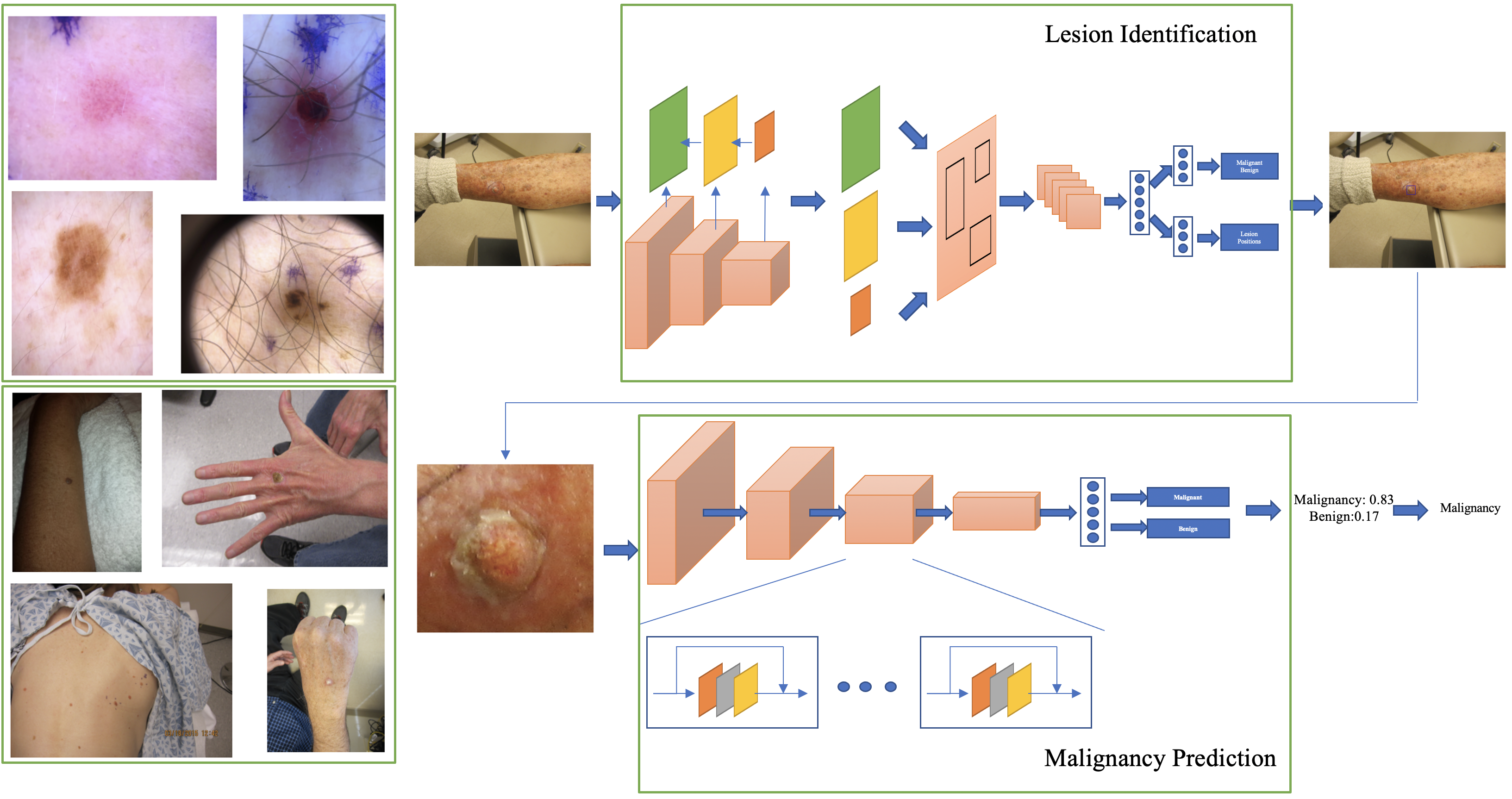
Below, we introduce deep-learning-based models for malignancy prediction, lesion identification and image-level classification for end-to-end processing. An illustration of the two-step malignancy prediction and lesion identification framework is presented in Figure 1.
2.1 Malignancy Prediction
Assuming we know the position of the ROIs, i.e., are always available, the problem of predicting whether a lesion is malignant can be formulated as a binary classification task. Specifically, we specify a function parameterized by whose output is the probability that a single lesion is consistent with a malignancy pathohistological finding in the area, i.e.,
| (1) |
where is a convolutional neural network that takes the region of defined in as input. In practice, we use a ResNet-50 architecture [he2016deep] with additional details described in Section LABEL:sc:model_details.
2.2 Lesion identification
Above we assume that the location (ROI) of the lesions is known, which may be the case in dermoscopy images as illustrated in Figure 1. However, in general, wide-field dermatology images are likely to contain multiple lesions, while their locations are not known or recorded as part of clinical practice. Fortunately, if lesion locations are available for a set of images (via manual annotation), the task can be formulated as a supervised object detection problem, in which the model takes the whole image as input and outputs a collection of predicted ROIs along with their likelihood of belonging to a specific group. Formally,
| (2) |
where is the likelihood that the predicted region belongs to one of groups of interest, i.e., . In our case, we consider three possible choices for , namely, ) denoted as one-class where the model seeks to identify any lesion regardless of type; ) denoted as malignancy in which the model seeks to separately identify malignant and benign lesions; and ) denoted as sub-type, thus the model is aware of all lesion types of interest.
Note that we are mainly interested in finding malignant lesions among all lesions present in an image as opposed to identifying the type of all lesions in the image. Nevertheless, it may be beneficial for the model to be aware that different types of lesions may have common characteristics which may be leveraged for improved detection. Alternatively, provided that some lesion types are substantially rarer than others (e.g., dermatofibroma and vascular lesions only constitutes 1% each of all the lesions in the dataset described in Section LABEL:sc:dataset), seeking to identity all lesion types may be detrimental for the overall detection performance. This label granularity trade-off will be explored in the experiments. In practice, we use a Faster-RCNN (region-based convolutional neural network) [NIPS2015_14bfa6bb] with a feature pyramid network (FPN) [lin2017feature] and a ResNet-50 [he2016deep] backbone as object detection architecture. Implementation details can be found in Section LABEL:sc:model_details.
2.3 Image classification
For screening purposes, one may be interested in estimating whether an image is likely to contain a malignant lesion so the case can be directed to the appropriate dermatology specialist. In such case, the task can be formulated as a whole-image classification problem
| (3) |
where is the likelihood that image contains a malignant lesion.
The model in (3) can be implemented in a variety of different ways. Here we consider three options, two of which leverage the malignancy prediction and lesion identification models described above.
Direct image-level classification
is specified as a convolutional neural network, e.g., ResNet-50 [he2016deep] in our experiments, to which the whole image is fed as input. Though this is a very simple model that has advantages from an implementation perspective, it lacks the context provided by (likely) ROIs that will make it less susceptible to interference from background non-informative variation, thus negatively impacting classification performance.
Two-stage approach
is specified as the combination of the one-class lesion identification and the malignancy prediction models, in which detected lesions are assigned a likelihood of malignancy using (1). This is illustrated in Figure 1(Right). Then we obtain
| (4) |
where we have replaced the ground truth location in (1) with the predicted locations from (2), and is a permutation-invariant aggregation function. In the experiments we consider two simple parameter-free options:
| (5) | |||||
| (6) | |||||
| (Noisy OR) | (7) |
Other more sophisticated (parametric) options such as noisy AND [kraus2016classifying], and attention mechanisms [NIPS2017_3f5ee243], may further improve performance but are left as interesting future work.
One-step approach
is specified directly from the sub-types lesion identification model in (2) as
| (8) |
From the options described above, the direct image-level classification approach is conceptually simpler and easier to implement but it does not provide explanation (lesion locations) to its predictions. The one-step approach is a more principled end-to-end system that directly estimates lesion locations, lesion sub-type likelihood, and overall likelihood of malignancy, however, it may not be suitable in situations where the availability of labeled sub-type lesions may be limited, in which case, one may also consider replacing the sub-type detection model with the simpler malignancy detection model. Akin to this simplified one-step approach, the two-stage approach provides a balanced trade-off between the ability of estimating the location of the lesions and the need to identify lesion sub-types. All these options will be quantitatively compared in the experiments below.
3 Experiments
Comprehensive experiments to analyze the performance of the proposed approach were performed. First, we describe the details of the dataset and the models being considered, and present evaluation metrics for each task, while comparing various design choices described in the previous section. Then, we study the effects of adding clinical covariates and using an auxiliary publicly available dataset for data augmentation. Lastly, we present some visualization of the proposed model predictions for qualitative analysis.