Also at ]NIST Boulder Laboratories, Boulder, CO 80305 Also at ]NIST Boulder Laboratories, Boulder, CO 80305
New Experimentally Observable Gamma-ray Emissions from 241Am Nuclear Decay
Abstract
With the high resolution of microcalorimeter detectors, previously unresolvable gamma-ray lines are now clearly resolvable. A careful measurement of 241Am decay with a large microcalorimeter array has yielded never before seen or predicted gamma lines at keV and keV. These have been made possible because of new microwave-multiplexing readout and improved analysis algorithms for microcalorimeters.
I Introduction
This letter presents two new gamma-ray emissions (spectral lines) for 241Am and the possible energy level candidates from which these gamma rays might originate. Plutonium and americium photon emissions around 208 keV are used for the non-destructive assay of plutonium mass by IAEA inspectors and material control and accountability (MC&A) technicians working in Department of Energy laboratories [1]. A better understanding of these emissions may lead to higher fidelity plutonium assays.
These peaks were previously not resolvable due to the relatively low energy resolution of HPGe detectors compared to microcalorimeters. With a recently commissioned spectrometer, SOFIA (Spectrometer Optimized for Facility Integrated Applications), measurements of Pu and Am samples showed two previously unknown gamma peaks at 207.72 and 208.21 keV. SOFIA is a 256-pixel superconducting transition-edge sensor microcalorimeter array combined with high-bandwidth microwave frequency-division multiplexed readout, providing high efficiency and high count rate capability [2]. These capabilities combined with improved analysis algorithms for co-adding pixel data allowed these peaks to be clearly resolved.
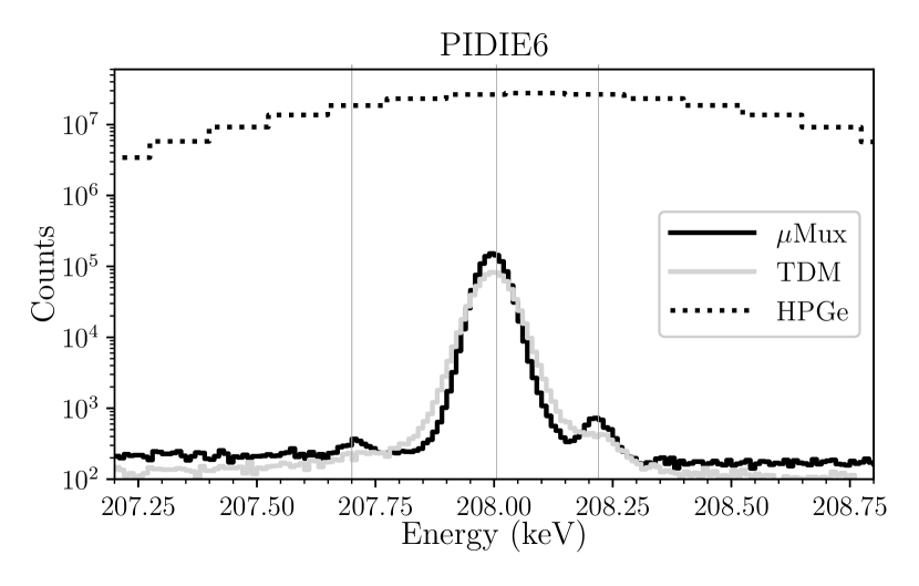
In this work, SOFIA used the SLEDGEHAMMER (Spectrometer to Leverage Extensive Development of Gamma-ray TESs for Huge Arrays using Microwave Multiplexed Enabled Readout) detector array, developed by the University of Colorado as the first large microwave-multiplexed gamma microcalorimeter array [2]. SLEDGEHAMMER was operated in multiple cryostats before it was installed in SOFIA and is well characterized. Typical spectral resolution with SOFIA is 73 eV at 208.0 keV. For these spectra, 1.5 mm of Cd was used to attenuate the high intensity 59.9 keV 241Am transition. Spectral features show up at 207.7 keV and 208.2 keV in all plutonium spectra, yet become more prominent as the ratio of 241Am/241Pu increases. The features can be seen prominently in PIDIE-6 (reactor grade plutonium with a large percentage of 241Am) shown in Figure 1.
Although SOFIA has 256 pixels, only half (128) were run due to readout limitations. In order to get the best quality data, additional pixels were removed from analysis for having too low a resolution, for not enough pulses surviving cuts [3], and for being poorly co-added in the final spectrum (i.e. not all peaks were aligned between a single pixel and the final co-added spectrum). The spectrum from each pixel is co-added with a master pixel (chosen primarily for its linearity) using a cubic spline transformation to match all peaks in the spectrum above a given threshold [4]. This differs from previous approaches in pixel co-adding which relied on co-adding energy-calibrated spectra, where energy calibration only used calibration points. Peak shapes from this new method of co-adding have been shown to be more uniform and Gaussian (Figure 1, black) than peak shapes from the prior co-adding method (Figure 1, grey). In this energy-calibration-first method of co-adding, peak centroids may shift by up to 10 eV from known values accompanied by slight peak shape degradation [5, 6]. Even a small peak shape degradation is enough to make these small 241Am peaks disappear into the background, which is why this research team has not observed these peaks until now.

II Sn X-ray Escape Hypothesis
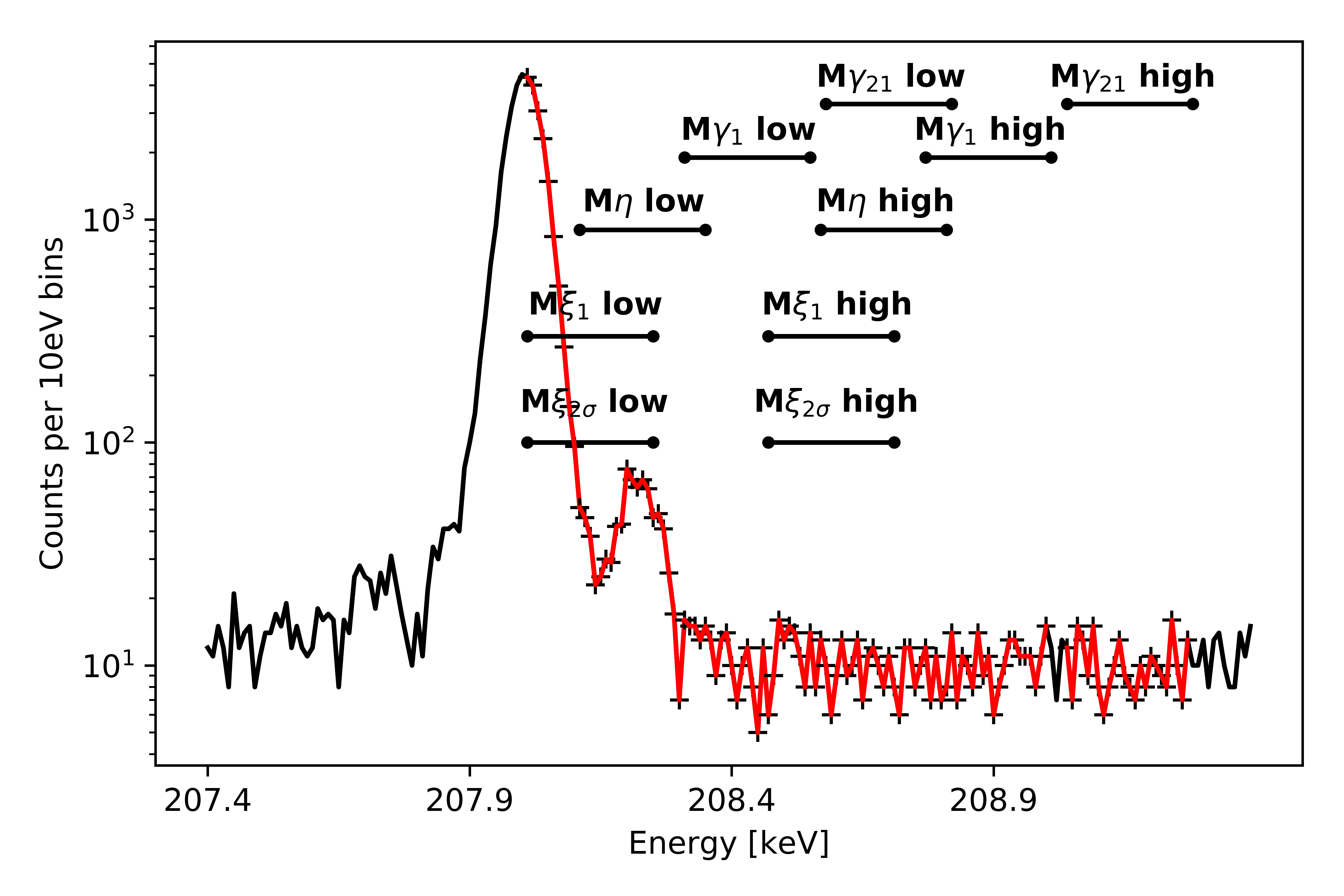
One possible explanation for the 207.7 keV and 208.2 keV peaks is that they are Sn escape peaks, a detector artifact. Here we carefully refute that hypothesis. A gamma ray (with energy ) interacting within the microcalorimeter Sn absorber has a probability of creating a Sn X-ray (with energy ), which may escape the Sn absorber resulting in a thermalized energy of . The probability of this happening is referred to as the X-ray escape fraction and defined as , where is the number of counts in the escape X-ray peak (at energy ) and is the number of counts in the full energy photopeak (at energy ). For the 59.54 keV 241Am peak, the total escape fraction from all K Sn X-rays was measured to be [7]. The most probable X-ray escape comes from the higher energy X-rays, like the K and K X-rays, which together account for 83% of the escape probability. To address the possibility of the satellites of the 208.0 keV peak being a result of Sn escape events, all above-background counts are summed in regions corresponding to two FWHM around the sum of 207.7 keV (or 208.2 keV) and a Sn X-ray. These regions are highlighted in Figure 2.
Using the pure 241Am spectrum, background levels are individually calculated for the L, K, and K regions. The total net counts from those regions assuming the 207.7 keV peak is an escape is . The total net counts from those regions assuming the 208.2 keV peak is an escape is . The total counts in the 207.7 keV peak and the 208.2 keV peak are and , respectively. This clearly indicates that neither the 207.7 keV peak nor the 208.2 keV peaks are likely to be an escape peak from these regions; if they were, this would imply escape probabilities greater than 99% and 60% from the respective regions, contrary to all our observations of known escape peaks. Even using the most conservative of calculations, these peaks are clearly not the products of Sn L or K X-ray escapes.
Let us now consider that these peaks may be a result of an M X-ray escape. Figure 2 (Bottom) shows the regions from which a Sn M X-ray would escape in order to create the satellite features. For the 208.2 keV peak, the region that would provide M X-ray escapes has counts, which is consistent with no counts above background. This is clearly not sufficient to make the 208.2 keV peak an M X-ray escape peak. However, the 207.7 keV is lower in energy than a very high intensity 208.0 keV peak. The total number of counts in the region that would provide M X-ray escape populating the 207.7 keV peak is . If all the counts in the 207.7 keV peak are M X-ray escapes, the M X-ray escape fraction is . This value is only half the escape probability of previously measured K X-ray escapes in Sn [7] and the 207.7 features is not the right distance from the centroid of the 208.0 keV peak. The counts in the M region are off center from the large 208.0 keV peak. Further, no other high intensity peaks show this low energy satellite from an M X-ray escape peak corresponding to this escape fraction, so we conclude that neither the 207.7 keV peak nor the 208.2 keV peak are the products of Sn X-ray escapes.
III Nuclear Transition Possibilities
De-excitation of the 241Am alpha-decay daughter product, 237Np, proceeds via gamma emission or conversion electrons. There are several combinations of 237Np levels whose energy differences agree within uncertainty with the measured centroids at 207.72(2) keV and 208.21(1) keV. Table 1 shows this agreement between experimentally determined centroids and theoretical photon emission energies. Propagated uncertainties in Table 1 consist of centroid least-squares fit uncertainties and the uncertainties in the tabulated 237Np nuclear level energies.
| Centroid [keV] | 207.72(2) | 207.72(2) | 208.21(1) |
|---|---|---|---|
| Transition () | |||
| [keV] | 805.77(12) | 721.96(1) | 434.12(5) |
| [keV] | 597.99(9) | 514.19(4) | 225.96(2) |
| [keV] | 207.78(15) | 207.77(4) | 208.16(5) |
| -0.4 | -1.1 | 0.9 | |
| % | % | % | |
| 241Am BR to | 0.0007 | 0.0004 | |
| 241Am BR to and above | |||
| from (ENSDF) | |||
| from (DDEP) |
Based on the energy levels of 237Np [8, 9], there is only one candidate for a transition that could produce a 208.2 keV photon. The transition from 434.12 keV () to 225.96 keV () would be an electric-monopole (E0) transition. Since single gamma-ray emissions from E0 transitions are forbidden, it is possible that the tentatively assigned angular momentum of the 434.12 keV energy level is wrong.
There were three candidates for the transition producing a 207.7 keV photon. One transition (799.82 keV () to 592.33 keV () is ignored as the transition energy is not well-matched to the experimentally measured value. The transition from 721.96 keV () to 514.19 keV () has a transition energy of 207.77 keV and is a magnetic-dipole (M1) transition, which is not unreasonable given the low intensity of this peak. The transition from 805.77 keV () to 597.99 keV () has an energy difference of 207.78 keV would predominantly be an M1 or M2 transition, depending on the actual angular momentum of the 805.77 keV energy level. A quadrupole transition is much less likely than a dipole transition, but neither are unreasonable given the low experimental branching ratio of this peak [10]. Based on this, there is one likely transition candidate for the 208.2 keV peak and two candidates for the 207.7 keV peak.
IV Branching Ratios
Branching fractions were determined using the measured net peak areas at 207.7 keV, 208.0 keV, and 208.2 keV from several Pu standards such as those used for recent Pu and Am branching ratio measurements [11] as well as a combined sample with three 241Am sources, referred to as a pure 241Am source although it is known to contain 243Am. Each of the three peaks is fit with two gaussians—one representing the single pixel response and one representing the effect of uncorrected drift and co-adding uncertainty—and a background step function. Similar double-Gaussian fitting functions have been successfully used in microcalorimeter spectra such as in [5]. The branching ratio calculation assumes the tabulated 241Am branching fraction at 208.0 keV of photons per 100 disintegrations. Each of these determinations and their combined value with uncertainty is given in Figure 3. The 241Am content for the two CRM samples is taken from a recent intercomparison exercise analysis of certified reference materials [12], whereas other values come from the certificate values (given in Table 2). Uncertainties (67% confidence intervals) taken into account include declared 241Pu and 241Am mass fractions for Pu items, 241Pu and 241Am branching fractions at 208.0 keV, half-lives, and net peak area least-squares fit uncertainties. A combined Type B on Bias methodology with a normal distribution was used to determine the branching ratio and combined uncertainty [13, 14]. In this case, each material type was treated as a separate methodology.
| Item | Mass [g] | Cert. date | 238Pu | 239Pu | 240Pu | 241Pu | 241Am | Meas. date | () | Am% |
|---|---|---|---|---|---|---|---|---|---|---|
| CBNM84 | 6.6 oxide | 20-06-1986 | 0.0703(3) | 84.338(4) | 14.207(4) | 1.0275(9) | 0.217(1) | 31-08-19 | 8.6 | 19.5(4) |
| CBNM70 | 6.6 oxide | 20-06-1986 | 0.8458(9) | 73.319(5) | 18.295(4) | 5.463(2) | 1.171(6) | 02-09-19 | 14.5 | 19.6(4) |
| PIDIE6 | 0.5 oxide | 01-01-1988 | 0.930(6) | 66.34(1) | 23.89(1) | 5.28(2) | 3.8(2) | 10-01-19 | 99.8 | 24.3(6) |
| CRM136 | 0.250 sulfate | 01-10-1987 | 0.222(4) | 84.925(8) | 12.366(8) | 1.902(3) | 7.92(4)* | 17-10-18 | 14 | 31.1(6) |
| CRM137 | 0.250 sulfate | 01-10-1987 | 0.267(3) | 77.55(1) | 18.79(1) | 2.168(3) | 7.71(6)* | 19-09-19 | 20 | 31.5(6) |
| 02-10-19 | 18 | 31.6(6) | ||||||||
| 03-10-19 | 45 | 31.6(6) | ||||||||
| Pure 241Am | 16-03-20 | 27 | 100 |
-
*
The 241Am/241Pu comes from [12] and has separation date of 28-4-2016, different from the certificate value for the Pu isotopics.


Table 1 gives the combined branching ratio from Figure 3 for each peak and shows that 241Am decays more than often enough to elevated states of 237Np to account for these branching fractions. Branching ratios from both ENSDF [8, 9] and DDEP databases [15, 16] are compared. The probabilities of transitioning from the state () with the additional probability from the proposed new transition () is still smaller than the probability of an 241Am alpha decay to populate an energy level at or higher, so this transition is not ruled out on this basis.
V Conclusion
Photon signatures have been measured in various plutonium materials. The agreement displayed in Figure 3 between differing materials with significantly different 241Am/241Pu ratios and different sizes strongly implies that the observed spectral lines are not artifacts from signal processing or co-adding. Evidence has been presented that these signatures originate from 241Am decay. Evidence includes (1) measured branching ratios from a variety of Pu standards with varying 241Am/241Pu mass fraction that agree with the determined branching ratio from the pure Am source, (2) measured peak centroids agree within uncertainty with tabulated differences in 237Np nuclear energies, and (3) measured branching fractions are much lower than the total net fraction of 241Am decays resulting in 237Np excited states above the probable excited state from which the transition originated.
VI Acknowledgements
This work was supported by the G. T. Seaborg Institute, the US Department of Energy (DOE) Nuclear Energy’s Fuel Cycle Research and Development (FCR&D), Materials Protection, Accounting and Control Technologies (MPACT) Campaign and Nuclear Energy University Program (NEUP), and the NIST Innovations in Measurement Science program.
References
- Reilly et al. [1991] D. Reilly, N. Ensslin, H. Smith Jr, and S. Kreiner, Passive nondestructive assay of nuclear materials (panda) manual, Los Alamos National Laboratory LA-UR-90-732 (1991).
- Croce et al. [2019] M. P. Croce, K. E. Koehler, M. H. Carpenter, M. D. Yoho, S. E. Kossmann, S. E. Garner, M. W. Rabin, D. T. Becker, D. A. Bennett, J. D. Gard, J. A. B. Mates, N. J. Ortiz, D. R. Schmidt, A. L. Wessels, J. Ullom, and J. U. M. P. Croce, K. E. Koehler, M. H. Carpenter, M. D. Yoho, S. E. Kossmann, S. E. Garner, M. W. Rabin, D. T. Becker, D. A. Bennett, J. D. Gard, J. A. B. Mates, N. J. Ortiz, D. R. Schmidt, A. L. Wessels, Practical Microcalorimeter Spectrometers, in 60th INMM Annual Meeting (Palm Desert, CA, 2019).
- Becker et al. [2019] D. T. Becker, J. A. B. Mates, G. C. O’Neil, M. W. Rabin, C. D. Reintsema, D. R. Schmidt, D. S. Swetz, P. Szypryt, L. R. Vale, A. L. Wessels, J. N. Ullom, B. K. Alpert, D. A. Bennett, M. P. Croce, J. W. Fowler, J. D. Gard, A. S. Hoover, Y. I. Joe, K. E. Koehler, J. A. B. Mates, G. C. O’Neil, M. W. Rabin, C. D. Reintsema, D. R. Schmidt, D. S. Swetz, P. Szypryt, L. R. Vale, A. L. Wessels, and J. N. Ullom, Advances in Analysis of Microcalorimeter Gamma-Ray Spectra, IEEE Transactions on Nuclear Science 66, 2355 (2019).
- Yoho et al. [2020a] M. Yoho, K. Koehler, S. Garner, D. Vo, and M. Croce, Automated co-adding and energy calibration of large array microcalorimeter data with zero sample knowledge, Nuclear Instruments and Methods in Physics Research Section A: Accelerators, Spectrometers, Detectors and Associated Equipment 969, 164056 (2020a).
- Hoover et al. [2013] A. S. Hoover, R. Winkler, M. W. Rabin, D. T. Vo, J. N. Ullom, D. A. Bennett, W. B. Doriese, J. W. Fowler, R. D. Horansky, D. R. Schmidt, L. R. Vale, and K. Schaffer, Determination of Plutonium Isotopic Content by Microcalorimeter Gamma-Ray Spectroscopy, IEEE Transactions on Nuclear Science 60, 681 (2013).
- Winkler et al. [2015] R. Winkler, A. S. Hoover, M. W. Rabin, D. A. Bennett, W. B. Doriese, J. W. Fowler, J. Hays-Wehle, R. D. Horansky, C. D. Reintsema, D. R. Schmidt, L. R. Vale, and J. N. Ullom, 256-Pixel microcalorimeter array for high-resolution -ray spectroscopy of mixed-actinide materials, Nuclear Instruments and Methods in Physics Research, Section A: Accelerators, Spectrometers, Detectors and Associated Equipment 770, 203 (2015).
- Hoover et al. [2009] A. S. Hoover, M. K. Bacrania, P. J. Karpius, M. W. Rabin, C. R. Rudy, D. T. Vo, J. A. Beall, W. B. Doriese, G. C. Hilton, R. D. Horansky, K. D. Irwin, J. N. Ullom, and L. R. Vale, Application of GEANT4 to the simulation of high energy-resolution microcalorimeter detectors, in IEEE Transactions on Nuclear Science, Vol. 56 (2009) pp. 2294–2298.
- Basunia [2006] M. Basunia, Nuclear Data Sheets for A = 237, Nuclear Data Sheets 107, 2323 (2006).
- [9] Evaluated Nuclear Structure Data File.
- Krane [1987] K. S. Krane, Introductory Nuclear Physics (Wiley-VCH, 1987) pp. 334–335.
- Yoho et al. [2020b] M. Yoho, K. Koehler, D. Becker, D. Bennett, M. Carpenter, M. Croce, J. Gard, J. Mates, D. Mercer, N. Ortiz, D. Schmidt, C. Smith, D. Swetz, A. Tollefson, J. Ullom, L. Vale, A. Wessels, and D. Vo, Improved plutonium and americium photon branching ratios from microcalorimeter gamma spectroscopy, Nuclear Instruments and Methods in Physics Research Section A: Accelerators, Spectrometers, Detectors and Associated Equipment 977, 164307 (2020b).
- Mathew et al. [2019] K. Mathew, T. Kayzar-Boggs, Z. Varga, A. Gaffney, J. Denton, J. Fulwyler, K. Garduno, A. Gaunt, J. Inglis, R. Keller, W. Kinman, D. Labotka, E. Lujan, J. Maassen, T. Mastren, I. May, K. Mayer, A. Nicholl, C. Ottenfeld, T. Parsons-Davis, D. Porterfield, J. Rim, J. Rolison, F. Stanley, R. Steiner, L. Tandon, M. Thomas, R. Torres, K. Treinen, M. Wallenius, A. Wende, R. Williams, and J. Wimpenny, Intercomparison of the Radio-Chronometric Ages of Plutonium-Certified Reference Materials with Distinct Isotopic Compositions, Analytical Chemistry 91, 11643 (2019).
- Levenson et al. [2000] M. S. Levenson, D. L. Banks, K. R. Eberhardt, L. M. Gill, W. F. Guthrie, H. K. Liu, M. G. Vangel, J. H. Yen, and N. F. Zhang, An Approach to Combining Results from Multiple Methods Motivated by the ISO GUM, Journal of Research of the National Institute of Standards and Technology 105, 571 (2000).
- Joint Committee for Guides in Metrology [2008] Joint Committee for Guides in Metrology, Guide to the Expression of Uncertainty in Measurement,(1995), with Supplement 1, Evaluation of measurement data, JCGM 101: 2008, Tech. Rep. (Organization for Standardization, Geneva, Switzerland, 2008).
- Bé et al. [2010] M.-M. Bé, V. Chisté, C. Dulieu, X. Mougeot, E. Browne, V. Chechev, N. Kuzmenko, F. Kondev, A. Luca, M. Galán, A. Nichols, A. Arinc, and X. Huang, Table of Radionuclides, Monographie BIPM-5, Vol. 5 (Bureau International des Poids et Mesures, Pavillon de Breteuil, F-92310 Sèvres, France, 2010).
- [16] Decay Data Evaluation Project, http://www.lnhb.fr/nuclear-data/nuclear-data-table/, last accessed on 05/14/20.