Order from disorder with intrinsically disordered peptide amphiphiles
Abstract
Amphiphilic molecules and their self-assembled structures have long been the target of extensive research due to their potential applications in fields ranging from materials design to biomedical and cosmetic applications. Increasing demands for functional complexity have been met with challenges in biochemical engineering, driving researchers to innovate in the design of new amphiphiles. An emerging class of molecules, namely, peptide amphiphiles, combines key advantages and circumvents some of the disadvantages of conventional phospholipids and block-copolymers. Herein, we present new peptide amphiphiles comprised of an intrinsically disordered peptide conjugated to two variants of hydrophobic dendritic domains. These molecules termed intrinsically disordered peptide amphiphiles (IDPA), exhibit a sharp pH-induced micellar phase-transition from low-dispersity spheres to extremely elongated worm-like micelles. We present an experimental characterization of the transition and propose a theoretical model to describe the pH-response. We also present the potential of the shape transition to serve as a mechanism for the design of a cargo hold-and-release application. Such amphiphilic systems demonstrate the power of tailoring the interactions between disordered peptides for various stimuli-responsive biomedical applications.
keywords:
American Chemical Society, LaTeX[biosoft]The Center for Physics and Chemistry of Living Systems, Tel Aviv University, Israel \alsoaffiliation[nano]The Center for NanoTechnology and NanoScience, Tel Aviv Univeristy, Israel \alsoaffiliation[biosoft]The Center for Physics and Chemistry of Living Systems, Tel Aviv University, Israel \alsoaffiliation[nano]The Center for NanoTechnology and NanoScience, Tel Aviv Univeristy, Israel \alsoaffiliation[LMU] Faculty of Physics and Center for NanoScience, Ludwig-Maximilians-Universität, München, Germany \alsoaffiliation[biosoft]The Center for Physics and Chemistry of Living Systems, Tel Aviv University, Israel \alsoaffiliation[nano]The Center for NanoTechnology and NanoScience, Tel Aviv Univeristy, Israel technion]CryoEM Laboratory of Soft Matter, Faculty of Biotechnology and Food Engineering, Technion-Israel Institute of Technology, Haifa 3200003, Israel \alsoaffiliation[biosoft]The Center for Physics and Chemistry of Living Systems, Tel Aviv University, Israel \alsoaffiliation[nano]The Center for NanoTechnology and NanoScience, Tel Aviv Univeristy, Israel technion]CryoEM Laboratory of Soft Matter, Faculty of Biotechnology and Food Engineering, Technion-Israel Institute of Technology, Haifa 3200003, Israel \alsoaffiliation[china]Guangdong-Technion Israel Institute of Technology, Shantou, Guangdong Province, 515063 China \alsoaffiliation[biosoft]The Center for Physics and Chemistry of Living Systems, Tel Aviv University, Israel \alsoaffiliation[biosoft]The Center for Physics and Chemistry of Living Systems, Tel Aviv University, Israel \alsoaffiliation[nano]The Center for NanoTechnology and NanoScience, Tel Aviv Univeristy, Israel \alsoaffiliation[biosoft]The Center for Physics and Chemistry of Living Systems, Tel Aviv University, Israel \alsoaffiliation[nano]The Center for NanoTechnology and NanoScience, Tel Aviv Univeristy, Israel \abbreviationsIR,NMR,UV
![[Uncaptioned image]](/html/2103.10131/assets/TOC_v01.png)
1 Introduction
Self-assembly of amphiphilic molecules holds great interest from a fundamental scientific point of view, as well as for their potential for creating nanocarriers for various applications, ranging from drugs and nucleic acids in medicine to fragrances and other small chemicals in the food and cosmetics industries 1. The functionality of nanocarriers’ design will depend on many factors, including the mechanism for cargo hold-and-release, bio-compatibility, uniformity, and tunability, all of which present challenging obstacles in engineering efficient nanocarriers. Natural lipids and synthetic block-copolymers are two of the most widely used amphiphiles in designing such systems, each prevailing due to its specific advantages 2, 3, 4, 5, 6, 7, 8, 9, 10, 11, 12
An emerging class of synthetic amphiphiles, namely, peptide amphiphiles (PA), is designed to self-assemble into functional structures by building upon the advantageous characteristics of lipids and block-copolymers 13, 14, 15. The molecules’ hydrophilic domain, usually a bio-inspired peptide engineered to fulfill one or more active roles, is chemically conjugated to a hydrophobic tail group, usually single or double chain fatty acids like those found in lipids. Due to their highly flexible design scheme, PA self-assemblies can act as organic scaffolds for bone-like mineralization 16, anisotropic actuators mimicking skeletal muscle 17, produce new versatile soft materials 18, and enhance neural progenitor cell differentiation into neurons 19. PA can contain spacers or linkers in their design, connecting the hydrophobic tails to the functional hydrophilic domain 20, 21, or conjugating to an ”external” functional group, such as an MRI contrast agent 22. Comprehensive research is done to understand the properties and roles of the different molecular domains and their contribution to the specific self-assembled mesophase 23, 24. In many cases, the bio-inspired peptides are designed or derived from proteins with some degree of secondary structure 25. Nonetheless, PA hydrophilic domains are not exclusively bio-inspired and can be made of any polypeptide sequence, including intrinsically disordered (i.e., unfolded) peptide sequences.
An increasingly large number of proteins have been found to lack a fixed or ordered structure 26. As such, these proteins have been termed intrinsically disordered proteins (IDPs). A more operational definition of an IDP is a protein that does not possess only a single functional conformation, but rather it can fold into an ensemble of functional conformations depending on the setting. This structural plasticity is, in many cases, the result of relatively weaker and transient interactions between proteins segments 27, 28, 29, 30. The nature of their interactions makes IDPs better suited for specific roles than structured proteins as they can interact with multiple partners or retain in liquid condensed phase 31, 32.
Envisioning systems with intricate and precise self-organization potential, as sought after in applications, requires the fabrication and processing of unique nanostructures. A primary limitation of current block-copolymer synthesis is the lack of general methods for producing precise chain structure (i.e., sequence control) to facilitate multiple desired functions. Natural lipids also have limited functionality due to their relatively small hydrophilic domain. In contrast, intrinsically disordered peptide amphiphiles (IDPAs) benefit from the use of relatively short sequences that are easier to synthesize while still retaining rich functionality. PAs can combine the functionality and flexibility of peptides; since there are 20 natural amino acids, there are practically numerous () possible sequences, even for short (18mers) peptides such as the one studied in this work. Naturally, the design of IDP based hydrophilic domains can be inspired by biology, utilizing the immense pre-existing knowledge base of proteins. To date, only few examples demonstrated exciting functionality using disordered domain in PA 17, 33, 19, 20, 34.
Here, we study the self-assembly and encapsulation capabilities of two IDPAs, composed of hydrophilic domain inspired by the neurofilament-low disordered tail domain and a dendritic branching unit used for conjugating the lipophilic tails35, 28, 36. Using turbidity, Small-angle X-ray Scattering (SAXS) and cryogenic transmission electron microscopy (cryo-TEM) measurements, we found that the IDPAs self-assembled into well-defined, low-dispersity nanoparticles. In addition, we show that the IDPAs’ sensitivity to pH leads to a tunable and robust organization of the self-assembled nanoparticles. We further show that minor alterations in the peptide sequence can lead to alteration in IDPA-IDPA interaction and the macroscopic arrangement. Last, we demonstrate the potential of using the pH induced shape transition as a release mechanism for the design of nano carriers.
2 Results and discussion
2.1 Synthesis and the structure of IDPAs
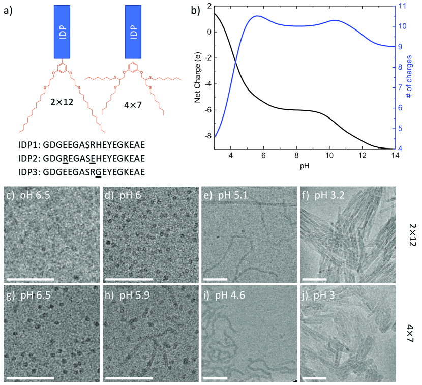
IDP sequences were synthesized on an automated solid-phase peptide synthesizer using Fmoc-protected amino acids. The hydrophilic peptide domain sequence is an 18mer amino-acid polyampholyte, inspired by the intrinsically disordered carboxy domain of the protein neurofilament-low (NF-L) 36, 28, 29, 37. Once the IDP sequences synthesis was completed, an aromatic branching unit containing two allyl or propargyl functionalities and a carboxylic acid was used to cap the N-terminus of the IDP sequence. After the branching units were conjugated, the capped peptides were cleaved from the resin using TFA, and hydrophobic end-groups containing thiols (dodecane-thiol and heptane-thiol) were conjugated to the allyl or propargyl moieties through thiol-ene or -yne click reactions, respectively.38, 39, 40, 41 We term the tail-group variants by 212 and 47, to represent the number and length of the alkyl chains and the amphiphiles as IDPA1 (Fig. 1a). The one letter amino acid sequence of IDPA1 is GDGEEGASRHEYEGKEAE.
Notably, the peptide’s sequence includes 11 protonatable residues, allowing for the net charge of the peptide to vary significantly as a function of pH (Fig. 1b). Specifically, at approximately pH 5.5 there is a decrease in the net charge and in the number of charged residues due to Aspartic Acid and Glutamic Acid residues’ protonation. The critical micelle concentrations (CMC) of 11 M were determined using the solvatochromic dye Nile Red (Supplementary Figure S1). A CMC of 5 M is in the typical micro-molar range for peptide amphiphiles 21, 42, 43, 44.
The peptide’s degree of disorder was experimentally verified by measuring the circular dichroism (CD) spectrum of samples of the peptide (unconjugated) and the two IDPA1 variants (Supplementary Figure S2). In addition, the peptide sequence displays a high probability for disorder and the absence of regular secondary structure using the NetSurfP-2.0 bioinformatic algorithm (Supplementary Figure S3) 45.
2.2 Micellar nanostructures at high pH
We expected IDPA1 to show little to no sensitivity at high pH where the peptides’ net charge state remains constant (Fig. 1b). Indeed, above pH6, we find that both tail-variants assemble into nanoscopic spherical micelles, which were visualized via cryo-TEM (Fig. 1c-j). At this slightly acidic pH, the micelles showed repulsion and remained miscible at a relatively high IDPA concentration (10 mg/ml). SAXS revealed low-dispersity spherical nanostructures, typical for structured particles. A core-shell form-factor was used to fit the SAXS data and provided the radius of the hydrophobic core nm, the width of the peptide shell (hydrophilic domain region surrounding the core) nm, and the respective average electron densities, and (Fig. 2b). Using the SAXS fit and the values from Harpaz et al. 46, we estimate the number of monomers for 47 and 212 IDPA1 at pH 7.5 to be approximately 13 and 40, respectively.
The SAXS measurements further revealed the composite dimensionality of the spherical micelles. Using the Kratky analysis 47, we found a bell-shaped curve at lower , which corresponds to the 3D nature of the micelle at larger length-scales. However, the linear increase at larger suggests an unfolded state of the peptides at smaller length-scales (Fig. 2c). Approaching pH6, we find a mild alteration in the SAXS signal, indicating a structural rearrangement at lower pH that we discuss next.
2.3 pH induced phase-transition
The peptide sequence is a strong polyampholyte; hence, the self-assembly of IDPA1 is expected to depend on pH. Therefore, lowering the pH towards the pI can facilitate a structural phase-transition that depends on the IDP charge density and the amphiphiles’ electrostatic interaction. Turbidity measurements performed on both IDPAs revealed a clear difference in sample translucency above and below pH 6 (Fig. 2a). This transition indicates a macro-molecular aggregation of the self-assembled structures at low pH. Below pH 6, the nanostructures interact to produce larger and more ordered aggregates. Figure 2a showcases the transition from translucent to opaque solutions. The turbidity of both variants changes abruptly at pH 6 and 4.5, for the 47 and 212, respectively.
Cryo-TEM micrographs of IDPA1 variants in the pH range 3-6.5 confirm the microscopic transition hinted at by the turbidity measurements (Fig. 1c-j). The micrographs show a transition from spherical micelles at pH 6.5 to elongated worm-like micelles lower than pH 5.5. Furthermore, at lower pHs the worm-like micelles are strongly interacting and aggregating. In the case of 47, the image taken at pH 5.9 nicely shows the coexistence of spherical and worm-like micelles (Fig. 1h). In addition, the sphere-to-rod transition and micellar growth is earlier for the 47 variant and is consistent with its higher hydrophobicity and turbidity measurements.
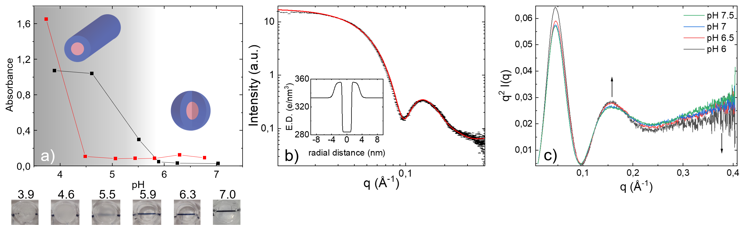
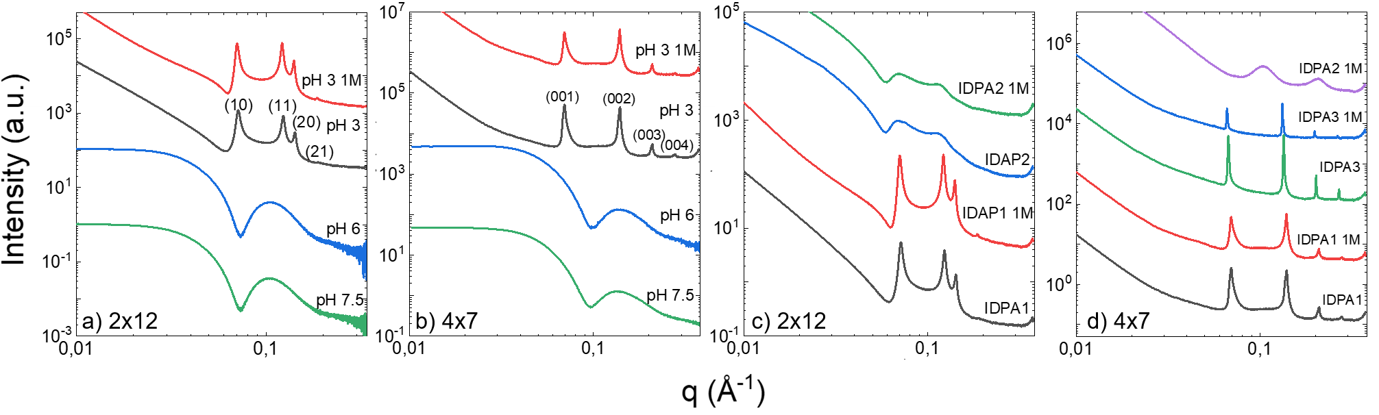
Further verification of the phase transition is clearly shown using SAXS (Figs. 3a,b). At low pH, the scattering is qualitatively different and is no longer a sum of independent spherical scatterers producing a form-factor SAXS signal. Instead, the SAXS signal now includes an additional structure-factor signal produced by the inter-particles’ correlation. The structure-factor peaks position match a 2D hexagonal lattice for 212 and a 1D lattice for 47 with the corresponding unit-cell spacing of nm and nm. The highly-dense packing is also evident bythe cryo-TEM micrographs showing organization of the micellar rods at low pHs (Fig. 1f,j).
2.4 Engineering the self-assembly by point mutation
The added value of using peptides as the hydrophilic domain is the possibility to tune the interactions via small alterations in the sequence, such as a single mutation. We recently demonstrated that a similar sequence peptide alters its self-interaction via a single point mutation 48. Short-ranged transient interactions are also present in the original neurofilament disordered protein 28, 29, 49, 50, 51, 52, presumably due to the oppositely charged amino-acids along the polyampholytic intrinsically disordered C-terminus domain. We synthesized a sequence variant by changing the Glutamic Acid at position 4 with the Arginine at position 9, termed IDPA2. By doing so, we are conserving the net charge and the functional relation between pH and charge but altering the charge distribution at the hydrophilic peptide domain. Such alteration is expected to alter the ionic bridging between the IDPs 52, 28, 49.
In addition, when considering the observed structural pH sensitivity, with a phase-transition being located somewhere between pH 4 and 6 (Figs. 1c,d and 3), a possible origin for the transition can be the charging state of the Histidine amino-acid. To verify this hypothesis, we designed a slightly modified hydrophilic domain sequence, termed IDPA3, that replaces the Histidine at position 10 with a neutral and pH insensitive Glycine residue. Notably, the two new IDPA variants are still considered highly disordered and are assigned the random coil conformation by the predictors (Supplementary Figure S4).
Indeed, we found that the hydrophilic domain (i.e., the disordered peptide) and its interactions in the assembly control the complex aggregations at low pH. SAXS experiments show that replacing the Histidine with a Glycine served to strengthen the interaction between the worm-like micelles (Fig. 3d). However, slightly changing the sequence’s order, namely the IDPA2 variant, noticeably changes the complex aggregation (Fig. 3c). For the 212 variant, we saw a significant weakening of micelle-micelle interactions in the worm-like phase, at 150 mM and 1 M salt concentration, demonstrated by the diminished structure-factor scattering (Fig. 3a). However, the 47 variant showed a similar weakening of the interactions only at 1 M salt pointing towards non-trivial electrostatic interaction 28, 52, 27 between the IDPs (Fig. 3b).
2.5 Cargo encapsulation and release
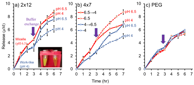
After the pH-depended self-assembly into micelles and worm-like micelles was characterized, we wanted to evaluate how the shape transition will affect encapsulated cargo release. We chose butyl ester of 7-(Diethylamino)coumarin-3-carboxylic acid as the hydrophobic cargo and used a dialysis set-up to study the hydrophobic dye’s release from the assembled structures. Solutions containing micelles of 212 and 47 IDPAs (pH 6.5) were mixed with a stock solution of the dye, followed by filtration of the residual un-encapsulated dyes. Next, the samples’ pH was adjusted to pH 4 by adding a few L of HCl to transform the spherical micellar assemblies into the worm-like micelles. As expected, while the micelle solution was clear, the worm-like micelle solution became highly turbid (Fig. 4).
Next, the two solutions were transferred into dialysis tubes and placed in buffer solutions (pH 6.5 or 4 for the micelles and worm-like micelles, respectively) containing bovine serum albumin (BSA). The BSA’s role was to scavenge the released dyes and avoid their aggregation and dissolution due to their poor aqueous solubility. The solutions were then placed in a shaking incubator at 37∘C, and samples were taken periodically from the outer solution and characterized by a spectrophotometer to determine the released dye’s concentration.
We find that the spherical micelles’ release was faster than the release from the worm-like assemblies (Fig. 4). To quantify how the phase-transition affects the release, we continued measuring the release after the solutions’ pH was adjusted from 6.5 to 4 or vice-versa, and the samples were placed in suitable fresh buffers. As controls, we used samples kept under the same initial pH and placed them into fresh buffers. It was fascinating to see the change in release rate due to the pH-induced shape transition as worm-like micelles that were transformed into spherical micelles (solid blue lines in Fig. 4a,b) started to release the dyes faster, showing a similar rate as the non-altered micelle control (dashed red lines in Fig. 4a,b). Simultaneously, a slower release was observed for spherical micelles, which were transformed into worm-like micelles (solid red lines and dashed blue lines in Fig. 4a,b). The changes in release rate due to the pH-induced change in mesophases were observed for both 212 and 47 IDPAs, with the effect being more significant for the latter. To ensure that the release rates were genuinely affected by the change in the assemblies’ shape, we prepared a non-responsive amphiphile with two dodecane alkyl chains by replacing the hydrophilic IDP with a PEG chain of similar molecular weight. The PEG-based amphiphile (PEG-212) self-assembled into micelles with a diameter of hydration of around 10 nm at both high and low pH (Supplementary Figure S5), demonstrating that its assembly is not responsive to pH at the tested range. We were encouraged to see that the control amphiphile release rates were not affected by the pH-jump after 3 hours. These results indicate that it is indeed the change in the shape of the IDPA assemblies due to altered interaction between the hydrophilic peptide domains, which affects the release rate.
2.6 Discussion
We demonstrated the IDPA could serve as a valuable platform having stimuli-responsive self-assembly. Importantly, these nanoscopic assemblies are well-ordered, although originating from disordered peptides. When modeling the IDPA interactions and phase-transitions presented here, it is essential to understand whether the dominant interactions are non-specific, e.g., based on the total charge-density, or specific, e.g., conformational-based interactions. A generic model for a spherical-to-cylindrical micelle phase-transition will be agnostic to the charged peptide domain’s details, aside from the charge distribution 53. Such a model, detailed for brevity in the supplementary information, describes the free-energy balance between the energy associated with the hydrophilic region’s charge-density and the energetic price paid for inducing a curvature that deviates from the intrinsic one. Essentially, lowering the pH results in a lower net charge that allows the hydrophilic peptide to compress. Assuming that the intrinsic curvature favors cylindrical morphology, this can facilitate the decrease in bending energy with a denser hydrophilic region.
However, the mutated variants’ experimental finding suggests that we must also consider the sequence’s details and charges distribution along the peptide chain. Therefore, a sequence-dependent interaction term must be included in the free-energy calculation, describing the interaction between the amino-acids along two parallel and offset peptides 52, 36, 28 (Supplementary Information).
The experimental results on the IDPA2 variant show that indeed such a small change significantly affects the inter-micelle interactions, and as a result, the aggregation phase was dramatically changed. Therefore, the minute alteration between the original sequence and its variants suggests sequence-specific interactions. Moreover, the entire IDPA architecture, including the hydrophobic domain, also plays a role in the pH-response and the macroscopic aggregated mesophase (i.e., at low pH). For the 47 hydrophobic domain, there is a clear structural difference between the IDPA2 and the other variants at high salt concentration. For the 212 hydrophobic domain, while the condensed phase structural correlation weakens for the IDPA2 variants, they are salt-independent between 0.15-1 M. Indeed, as suggested by our theoretical modeling, the IDPA3 condensed phase (at pH=3) is somewhat similar to the original variant (see supplementary materials).
3 Conclusions
We investigated the conjugation of disordered polypeptide domains with hydrocarbon dendrimers into IDPA. We find that the interactions between the peptides lead to tunable self-assembled nanostructures. The IDPA hydrophilic domains are weakly interacting and disordered by nature, generally associated with transient behavior. Nonetheless, the IDPA self-assembly is remarkably forming ordered nanoparticles.
Moreover, the IDPA system shows pH response and structural phase-transition. It can be useful as a trigger for cargo release, similar to already demonstrated amphiphilic polymer-based nanoparticle systems with drug release enzymatic response. 54
We further demonstrated that minute alteration in the disordered peptide sequence and dendrimer architecture could significantly impact the nanoscopic and macroscopic length-scales. This platform also enables in-situ modifications of the IDPs and the study of the mutual interaction between IDPs. Last, as we showed here, modification of the IDP domain, and its interaction with the surrounding, plays a critical role in the IDPA’s self-assembly and structural transformation from external cues. We expect that this platform can be furthered explored for targeted drug-delivery where tailored biological signals will induce phase-transition and expedite release.
4 Materials and methods
4.1 Synthesis and purification
All peptides were synthesized at the Blavatnik Center for Drug Discovery (BCDD) at Tel Aviv University using automated Fmoc solid-phase peptide synthesis using The Liberty BlueTM automated microwave peptide synthesizer (CEM, Matthews, NC, USA). After the coupling of the last amino acid, either 3,5-bis(allyloxy)benzoic acid or 3,5-bis(prop-2-yn-1-yloxy)benzoic acid 55 were coupled to the N-termius of the peptide. The capped peptides were cleaved from the resin usign standard conditions (95% trifluoroacetic acid (TFA) (v/v), 2.5% H2O (v/v), and 2.5% triisopropylsilane (v/v) for 3 hours).The cleaved di-allyl or di-propargyl were purified by Waters AutoPurification systemTM (MS directed LC) and were further reacted in thiol-ene or thiol-yne reactions with 1-dodecanethiol or 1-heptanethiol, respectively, as described below to yield IDPAs 2 12 and 4 7, respectively.
IDPA1-diallyl: Exact Mass 2164.88. Detected Mass: ES mode +: 723.28 [(M+3H)/3] and 1084.21 [(M+2H)/2].
IDPA1-212: 54 mg (0.025 mmol) of peptide 2 was dissolved with 350l of phosphate buffer (pH 7.4, 100mM) using gentle hitting, then 1 ml of DMF was added. 5.1 mg of DMPA (0.02 mmol) was separately solubilized with 100 l of DMF and added to the peptide solution flowed by addition of 239.5 l 1-dodecanethiol (0.997 mmol). The solution was purged with N2 for 15 minutes and then stirred under UV light for 2 hours. Next, the crud mixture was placed in dialysis membrane with MWCO of 3,000 Da and dialyzed against DI water for 12 hr. The solution was lyophilized and final purification was performed using preparative-scale reversed-phase HPLC (Waters AutoPurification system). The product was confirmed by LC/MS. ACN was removed by rotary evaporation and the solution was further lyophilized yielding a white solid product 31.26 mg (48% yield). Exact mass 2569.23. Detected mass: ES mode +: 857.98 [(M+3H)/3] and 1286.40 [(M+2H)/2].
IDPA1-dipropargyl: Exact Mass 2160.85. Detected mass: ES mode +: 721.91 [(M+3H)/3] and 1082.23 [(M+2H)/2].
IDPA1-47: 58 mg (0.027 mmol) of peptide 1 were dissolved with 350l of phosphate buffer (pH 7.4, 100mM) using gentle hitting, followed by addition o f 1ml of DMF. 5.48 mg of 2,2-Dimethoxy-2-phenylacetophenone (DMPA, 0.021 mmol) was separately solubilized with 100 l of DMF and added to the peptide solution flowed by addition of 338 l 1-heptanethiol (2.1 mmol). The solution was purged with N2 for 15 minutes and then stirred under UV light for 2 hours. Next, the crud mixture was placed in dialysis membrane with MWCO of 3,000 Da and dialyzed against DI water for 12 hr. The solution was lyophilized and the product was purified using reparative-scale reversed-phase HPLC (Waters AutoPurification system). The product was confirmed by LC/MS. ACN was removed by rotary evaporation and further lyophilized yielding a white solid product 19.2 mg (26% yield). Exact Mass 2689.24. Detected Mass: ES mode +: 898.17 [(M+3H)/3] and 1346.72 [(M+2H)/2].
IDPA2-diallyl: Exact Mass 2164.88. Detected Mass: ES mode +: 723.26 [(M+3H)/3] and 1084.19 [(M+2H)/2].
IDPA2-212: Was synthesized similarly to IDPA1. 51 mg (0.024 mmol) of IDP2- were mixed with 4.8 mg of DMPA and 226 l of 1-dodecanethiol and reacted and purified as was described for the synthesis of IDPA1 to yield 30.7 mg (50% yield). MS analysis: Exact Mass: 2569.23 Detected Mass: ES mode +: 858.17 [(M+3H)/3] and 1286.78 [(M+2H)/2].
IDPA2-dipropargyl: Exact Mass 2160.85. Detected Mass: ES mode +: 721.89 [(M+3H)/3] and 1082.14 [(M+2H)/2].
IDPA2-47: Was synthesized and purified similarly to IDPA1. 54 mg (0.025 mmol) of IDP2-dipropargyl were mixed with 5.1 mg of DMPA and 315 l of 1-heptanethiol and reacted and purified as was described for the synthesis of IDPA1 to yield 26.8 mg (40% yield). MS analysis: Exact Mass: 2689.24 Detected Mass: ES mode +: 1346.78 [(M+2H)/2] and 898.22 [(M+3H)/3].
IDPA3-dipropargyl: Exact Mass 2080.81. Detected Mass: ES mode +: 695.06 [(M+3H)/3] and 1041.95 [(M+2H)/2].
IDPA3-47: Was synthesized and purified similarly to IDPA1. 14 mg (0.0067 mmol) of IDP3-dipropargyl were mixed with 1.36 mg of DMPA and 84 l of 1-heptanethiol, and reacted and purified as was described for the synthesis of IDPA1 to yield 9 mg (51%). MS analysis: Exact Mass: 2609.20 Detected Mass: ES mode +: 1306.83 [(M+2H)/2] and 871.47 [(M+3H)/3].
PEG-Dendron (212) synthesis: Dendron: 600 mg (2.5 mmol) of 3,5-(allyloxy) benzoic acid,55 3.11 g of 1-dodecanethiol (15 mmol) and 38.4 mg of 2,2-Dimethoxy-2- phenylacetophenone (DMPA; 0.15 mmol) were dissolved in 800l of DMF. The solution was purged with N2 for 15 minutes and then stirred under UV light for 2 hours. Next, crud mixture was loaded on silica column, thiol excess was washed with 10:90 ethyl acetate and hexane (v/v) and 212 dendron compound was eluted with 30:70 ethyl acetate and hexane (v/v), The fractions that contained the product were unified,evaporated and dried under high vacuum obtaining 1.5 g yellowish oily compound (92% yield). 1H NMR (400 MHz, Chloroform-d) 7.23 (d, J = 2.2 Hz, 2H), 6.69 (t, J = 2.3 Hz, 1H), 4.10 (t, J = 6.1 Hz, 4H), 2.70 (t, J = 7.1 Hz, 4H), 2.52 (t, J = 7.0 Hz, 4H), 2.07 (p, J = 6.5 Hz, 4H), 1.59 (p, J = 7.3 Hz, 4H),1.26-1.38 (m, 37H), 0.88 (t, J = 6.8 Hz, 6H). PEG-Dendron (212): 70 mg of 2kDa PEG-amine 56 were dissolved in 100 l of DCM and 67.3 mg of 212 dendron, and 40 mg of (2-(1H-benzotriazol-1-yl)-1,1,3,3-tetramethyluronium hexafluorophosphate HBTU were dissolved in DCM:DMF 1:1 (1mL) followed by addition of DIPEA 60 l were added to the PEG-amine solution and allowed to stir for 3 hour at room temperature. The crude mixture was loaded on a MeOH based LH20 SEC column. The fractions that contained the product were unified and the MeOH was evaporated in vacuum to obtain 76 mg (82%) of the PEG based amphiphile. 1H NMR (400 MHz, Chloroform-d) 6.91 (d, J = 2.2 Hz, 2H), 6.69 (t, J = 5.8 Hz, 1H), 6.57 (t, J = 2.2 Hz, 1H), 4.08 (t, J = 6.1 Hz, 4H), 3.83-3.45 (PEG backbone), 3.38 (s, 3H), 2.77 (t, J = 6.5 Hz, 2H), 2.70-2.63 (m, 6H), 2.52 (t, J = 7.4 Hz, 4H), 2.05 (p, J = 6.6 Hz, H), 1.88 (p, J = 6.6 Hz, 2H), 1.62 – 1.52 (m, 4H), 1.45 – 1.26 (m, 40H), 0.88 (t, J = 6.7 Hz, 6H).
4.2 SAXS and cryo-TEM sample preparation
The IDPA or peptide powder was first fluidized in purified water (Milli-Q) at twice the desired concentration. The solution was then titrated with NaOH to a pH where the solution became more homogeneous (preferably a pH where the IDPAs are soluble in water). Titration was monitored using a pH probe (Sentek P13 pH Electrode). Following titration, 50 of the solution was combined with 50 of 2X buffer of choice to achieve a pH in the vicinity to the desired one. The 2X buffers Acetic Acid (pH 3-4.5), MES pH (5-6.5), and MOPS (pH 7-7.5) were prepared at 200 mM to achieve final buffer molarity of 100 mM after mixing with IDPA or peptide solution 1:1 (vol:vol).
4.3 SAXS
For solubilizing conditions (above the transition pH, generally above pH 6), samples were measured at three synchrotron facilities: Beamline B21, Diamond Light Source, UK, beamline 12.3.1, SIBYLS, Advanced Light Source, Berkeley, USA and beamline SWING, SOLEIL synchrotron facility, Paris, France.
For phase-separating samples that display sediment (below the transition pH, generally pH 3-5.5), measurements were performed using an in-house X-ray scattering system, with a Genix3D (Xenocs) low divergence Cu radiation source (wavelength of Å) with a Pilatus 300K (Dectris) detector and scatterless slits setup57, as well as beamline I22 at Diamond Light Source. Here, samples were measured inside 1.5 mm quartz capillaries (Hilgenberg).
4.3.1 Peptide’s SAXS analysis
The unconjugated peptide by itself was measured using SAXS at different pH levels to test the effect on its ensemble-averaged structure. The peptide at each pH was measured at four different concentrations to extrapolate to the non-interacting ”zero-concentration” peptides scatterings. From the low momentum transfer () regime of the extrapolated zero-concentration scattering curves, we extracted the radius of gyration () and the forward scattering, , using the Guinier analysis (Supplementary Figure S6). When examining the values of from high to low pH, it seems to remain a constant until pH 5.5 and gradually increase below it. However, this increase in size can be explained by a simultaneous increase in the effective mass of the peptides (increase in forwarding scattering) due to a decrease in inter-peptide repulsion near the isoelectric point (pI).
4.4 Cryo-TEM
Cryo-TEM specimen preparation was performed by applying a 6 drop of the studied solution to a perforated carbon film supported on a 200-mesh TEM copper grid, thinning (blotting), and removing excess solution. Next, the sample was vitrified in liquid ethane at its freezing point (-183 ∘C). The procedure was carried out at a controlled temperature (25 ∘C) and water saturation. The vitrified specimens were stored under liquid nitrogen (-196 ∘C) until examination. The samples were then examined using a Tecnai T12 G2 (FEI, The Netherlands) TEM operated at an accelerating voltage of 120kV, keeping specimen temperature below -170 ∘C during transfer and observation. Images were digitally recorded on a Gatan Ultrascan 1000 cooled CCD camera using the Gatan Digital Micrograph software package. Images were recorded using methodologies we developed 58 under low-electron-dose conditions to minimize electron beam radiation damage.
4.5 Turbidity
All measurements were recorded on a TECAN Infinite M200Pro device. The amphiphiles were treated and prepared in the same manner as previously described to achieve a final concentration of 5 mg ml-1. 100 of each solution was loaded onto a 96 wells plate. The absorbance at 600 nm was scanned for each well.
4.6 CD
Circular dichroism (CD) measurements were performed using a commercial CD spectrometer (Applied Photophysics Chirascan). Both IDPAs, 212 and 47, and the unconjugated IDP, were placed in a glass cuvette with a 10 mm path length. The IDPAs and peptide were mixed with a phosphate buffer to achieve a concentration of 0.05 and 0.1 mg ml-1, respectively. Measurements were performed using phosphate buffer since the buffers used for the X-ray scattering experiments (mainly MOPS and MES) have high absorbance in the relevant CD wavelengths. The 190-260 nm wavelength range was probed in 1 nm steps, with 0.5 secs at each point. Three measurements were performed for each and averaged.
4.7 CMC
The amphiphile was dissolved in the diluent (15ml MOPS buffer solution (pH 7.4), 7.5L of Nile red stock solution (2.5 mM in Ethanol) were added and mixed to give a final concentration of 1.25M) to give the final concentration of 400M IDPA and then sonicated for 5 minutes. This solution was repeatedly diluted by a factor of 1.5 with diluent. 100L of each solution were loaded onto a 96 wells plate. The fluorescence emission intensity was scanned for each well (550 nm Emission intensity scan: 580-800 nm) using TECAN Infinite M200Pro plate reader. Maximum emission intensity was plotted vs. concentration in order to determine the CMC. This procedure was repeated thrice.
4.8 Cargo release experiment
Release experiments of hydrophobic dye (butyl-coumarin) were performed using a dialysis tube (Mini GeBAflex-Kit, 8 kDa MWCO), volume range 10-250L. A volume of 80L of the micelles and worm-like micelle solutions were placed in the dialysis tubes, and each tube was immersed into 8 ml buffer at pH 6.5 or 4 with 0.5 mg/ml of BSA, respectively.
We thank Diamond Light Source for time on Beamlines B21 (SM24693) and I22 (SM21971), Advanced Light Source for time on Beamline SIBYLS 12.3.1 (SB-00941), and the SOLEIL synchrotron facility for time on Beamline SWING (20170798). The work was supported by the Israel Science Foundation (grant numbers 1454/20, 1553/18, 3292/19, 1117/16), Deutsche Forschungsgemeinschaft through SFB 958, NWU-TAU, the LMU-TAU collaboration initiatives, and DFG project GR1030/14-1. We thank Dr. Elvira Haimov and Dr. Boris Redko from the Blavatnik Center for Drug Discovery for their help with the synthesis and purification of the IDPAs. We also acknowledge fruitful discussions with Nathan Gianneschi, Joachim Rädler, Ram Avinery, and technical support of Mingming Zhang.
The Supporting Information is available free of charge at XXX.
References
- Kulkarni 2012 Kulkarni, C. V. Nanoscale 2012, 4, 5779–91
- Dengler et al. 2013 others,, et al. Journal of Controlled Release 2013, 168, 209–224
- Rodriguez et al. 2005 Rodriguez, N.; Pincet, F.; Cribier, S. Colloids and Surfaces B: Biointerfaces 2005, 42, 125–130
- Gunatillake and Adhikari 2003 Gunatillake, P. A.; Adhikari, R. Eur Cell Mater 2003, 5, 1–16
- Segal et al. 2020 Segal, M.; Ozery, L.; Slor, G.; Wagle, S. S.; Ehm, T.; Beck, R.; Amir, R. J. Biomacromolecules 2020, 21, 4076–4086
- Harnoy et al. 2014 Harnoy, A. J.; Rosenbaum, I.; Tirosh, E.; Ebenstein, Y.; Shaharabani, R.; Beck, R.; Amir, R. J. Journal of the American Chemical Society 2014, 136, 7531–7534
- Peer et al. 2007 Peer, D.; Karp, J. M.; Hong, S.; Farokhzad, O. C.; Margalit, R.; Langer, R. Nature Nanotechnology 2007, 2, 751–760
- Mai and Eisenberg 2012 Mai, Y.; Eisenberg, A. Chemical Society Reviews 2012, 41, 5969
- Carlsen and Lecommandoux 2009 Carlsen, A.; Lecommandoux, S. Current Opinion in Colloid and Interface Science 2009, 14, 329–339
- Zhuang et al. 2013 Zhuang, J.; Gordon, M. R.; Ventura, J.; Li, L.; Thayumanavan, S. Chemical Society reviews 2013, 42, 7421–35
- Miyata et al. 2011 Miyata, K.; Christie, R. J.; Kataoka, K. Reactive and Functional Polymers 2011, 71, 227–234
- Segal et al. 2017 Segal, M.; Avinery, R.; Buzhor, M.; Shaharabani, R.; Harnoy, A. J.; Tirosh, E.; Beck, R.; Amir, R. J. Journal of the American Chemical Society 2017, 139, 803–810
- Uversky 2009 Uversky, V. N. BioMed Research International 2009, 2010
- He et al. 2009 He, B.; Wang, K.; Liu, Y.; Xue, B.; Uversky, V. N.; Dunker, A. K. Cell research 2009, 19, 929–949
- Uversky 2017 Uversky, V. N. Current opinion in structural biology 2017, 44, 18–30
- Hartgerink et al. 2001 Hartgerink, J. D.; Beniash, E.; Stupp, S. I. Science 2001, 294, 1684–1688
- Chin et al. 2018 Chin, S. M.; Synatschke, C. V.; Liu, S.; Nap, R. J.; Sather, N. A.; Wang, Q.; Álvarez, Z.; Edelbrock, A. N.; Fyrner, T.; Palmer, L. C.; Szleifer, I.; Olvera De La Cruz, M.; Stupp, S. I. Nature Communications 2018, 9, 1–11
- Hartgerink et al. 2002 Hartgerink, J. D.; Beniash, E.; Stupp, S. I. Proceedings of the National Academy of Sciences 2002, 99, 5133–5138
- Silva et al. 2004 Silva, G. A.; Czeisler, C.; Niece, K. L.; Beniash, E.; Harrington, D. A.; Kessler, J. A.; Stupp, S. I. Science 2004, 303, 1352–1355
- Peters et al. 2009 Peters, D.; Kastantin, M.; Kotamraju, V. R.; Karmali, P. P.; Gujraty, K.; Tirrell, M.; Ruoslahti, E. Proceedings of the National Academy of Sciences 2009, 106, 9815–9819
- Acar et al. 2017 Acar, H.; Samaeekia, R.; Schnorenberg, M. R.; Sasmal, D. K.; Huang, J.; Tirrell, M. V.; LaBelle, J. L. Bioconjugate Chemistry 2017, 28, 2316–2326
- Bull et al. 2005 Bull, S. R.; Guler, M. O.; Bras, R. E.; Meade, T. J.; Stupp, S. I. Nano Letters 2005, 5, 1–4
- Tovar et al. 2005 Tovar, J. D.; Claussen, R. C.; Stupp, S. I. Journal of the American Chemical Society 2005, 127, 7337–7345
- Paramonov et al. 2006 Paramonov, S. E.; Jun, H. W.; Hartgerink, J. D. Journal of the American Chemical Society 2006, 128, 7291–7298
- Trent et al. 2011 Trent, A.; Marullo, R.; Lin, B.; Black, M.; Tirrell, M. Soft Matter 2011, 7, 9572–9582
- Oates et al. 2013 Oates, M. E.; Romero, P.; Ishida, T.; Ghalwash, M.; Mizianty, M. J.; Xue, B.; Dosztányi, Z.; Uversky, V. N.; Obradovic, Z.; Kurgan, L.; Dunker, A. K.; Gough, J. Nucleic Acids Research 2013, 41, 508–516
- Kornreich et al. 2015 Kornreich, M.; Avinery, R.; Malka-Gibor, E.; Laser-Azogui, A.; Beck, R. FEBS Letters 2015, 589, 2464–2476
- Kornreich et al. 2016 Kornreich, M.; Malka-Gibor, E.; Zuker, B.; Laser-Azogui, A.; Beck, R. Physical Review Letters 2016, 117, 148101
- Malka-Gibor et al. 2017 Malka-Gibor, E.; Kornreich, M.; Laser-Azogui, A.; Doron, O.; Zingerman-Koladko, I.; Harapin, J.; Medalia, O.; Beck, R. Biophysical Journal 2017, 112, 892–900
- DeForte and Uversky 2016 DeForte, S.; Uversky, V. N. Molecules 2016, 21, 1090
- Wei et al. 2017 Wei, M. T.; Elbaum-Garfinkle, S.; Holehouse, A. S.; Chen, C. C. H.; Feric, M.; Arnold, C. B.; Priestley, R. D.; Pappu, R. V.; Brangwynne, C. P. Nature Chemistry 2017, 9, 1118–1125
- Shin and Brangwynne 2017 Shin, Y.; Brangwynne, C. P. Science 2017, 357, eaaf4382
- Dzuricky et al. 2020 Dzuricky, M.; Rogers, B. A.; Shahid, A.; Cremer, P. S.; Chilkoti, A. Nature Chemistry 2020, 12, 814–825
- Mozhdehi et al. 2018 others,, et al. Nature chemistry 2018, 10, 496–505
- Uversky and Dunker 2010 Uversky, V. N.; Dunker, A. K. Biochimica et Biophysica Acta - Proteins and Proteomics 2010, 1804, 1231–1264
- Kornreich et al. 2015 Kornreich, M.; Malka-Gibor, E.; Laser-Azogui, A.; Doron, O.; Herrmann, H.; Beck, R. Soft Matter 2015, 11, 5839–5849
- Laser-Azogui et al. 2015 Laser-Azogui, A.; Kornreich, M.; Malka-Gibor, E.; Beck, R. Current Opinion in Cell Biology 2015, 32, 92–101
- Hoyle and Bowman 2010 Hoyle, C. E.; Bowman, C. N. Angewandte Chemie International Edition 2010, 49, 1540–1573
- Kade et al. 2010 Kade, M. J.; Burke, D. J.; Hawker, C. J. Journal of Polymer Science Part A: Polymer Chemistry 2010, 48, 743–750
- Konkolewicz et al. 2009 Konkolewicz, D.; Gray-Weale, A.; Perrier, S. Journal of the American Chemical Society 2009, 131, 18075–18077
- Lowe 2014 Lowe, A. B. Polymer 2014, 55, 5517–5549
- Vincenzi et al. 2015 Vincenzi, M.; Accardo, A.; Costantini, S.; Scala, S.; Portella, L.; Trotta, A.; Ronga, L.; Guillon, J.; Leone, M.; Colonna, G.; Rossi, F.; Tesauro, D. Molecular BioSystems 2015, 11, 2925–2932
- Klass et al. 2019 Klass, S. H.; Smith, M. J.; Fiala, T. A.; Lee, J. P.; Omole, A. O.; Han, B. G.; Downing, K. H.; Kumar, S.; Francis, M. B. Journal of the American Chemical Society 2019, 141, 4291–4299
- Black et al. 2012 Black, M.; Trent, A.; Kostenko, Y.; Lee, J. S.; Olive, C.; Tirrell, M. Advanced Materials 2012, 24, 3845–3849
- Klausen et al. 2019 Klausen, M. S.; Jespersen, M. C.; Nielsen, H.; Jensen, K. K.; Jurtz, V. I.; Sønderby, C. K.; Sommer, M. O. A.; Winther, O.; Nielsen, M.; Petersen, B.; Marcatili, P. Proteins: Structure, Function, and Bioinformatics 2019, 87, 520–527
- Harpaz et al. 1994 Harpaz, Y.; Gerstein, M.; Chothia, C. Structure 1994, 2, 641–649
- Orthaber et al. 2000 Orthaber, D.; Bergmann, A.; Glatter, O. Journal of Applied Crystallography 2000, 33, 218–225
- Chakraborty et al. 2019 Chakraborty, I.; Rahamim, G.; Avinery, R.; Roichman, Y.; Beck, R. Nano Letters 2019, 19, 6524–6534
- Kornreich et al. 2015 Kornreich, M.; Malka-Gibor, E.; Laser-Azogui, A.; Doron, O.; Herrmann, H.; Beck, R. Soft Matter 2015, 11, 5839–5849
- Pregent et al. 2015 Pregent, S.; Lichtenstein, A.; Avinery, R.; Laser-Azogui, A.; Patolsky, F.; Beck, R. Nano Letters 2015, 15, 3080–3087
- Morgan et al. 2020 Morgan, I. L.; Avinery, R.; Rahamim, G.; Beck, R.; Saleh, O. A. Physical Review Letters 2020, 125, 058001
- Beck et al. 2010 Beck, R.; Deek, J.; Jones, J. B.; Safinya, C. R. Nature Materials 2010, 9, 40–46
- Lerche et al. 1987 Lerche, D.; Kozlov, M. M.; Markin, V. S. Biorheology 1987, 24, 23–34
- Daniel et al. 2016 Daniel, K. B.; Callmann, C. E.; Gianneschi, N. C.; Cohen, S. M. Chemical Communications 2016, 52, 2126–2128
- Harnoy et al. 2018 Harnoy, A. J.; Papo, N.; Slor, G.; Amir, R. J. Synlett 2018, 29, 2582–2587
- Harnoy et al. 2014 Harnoy, A. J.; Rosenbaum, I.; Tirosh, E.; Ebenstein, Y.; Shaharabani, R.; Beck, R.; Amir, R. J. Journal of the American Chemical Society 2014, 136, 7531–7534
- Li et al. 2008 Li, Y.; Beck, R.; Huang, T.; Choi, M. C.; Divinagracia, M. Journal of Applied Crystallography 2008, 41, 1134–1139
- Danino 2012 Danino, D. Current Opinion in Colloid and Interface Science 2012, 17, 316–329