Spectroscopic evidence for a new type of surface resonance at noble metal surfaces
Abstract
We investigate the surface- and bulk-like properties of the pristine (110)-surface of silver using threshold photoemission by excitation with light of . Using a momentum microscope, we identified two distinct transitions along the -direction of the crystal. The first one is a so far unknown surface resonance for the (110) noble metal surface, exhibiting an exceptionally large bulk character, that has so far been elusive in surface sensitive experiments. The second one stems from the well known bulk-like Mahan cone oriented along the -direction inside the crystal but projected onto the (110)-surface cut. The existence of the new state is confirmed by photocurrent calculations and its character analyzed.
In solid-state systems, all electronic and optical properties are intrinsically linked to the band structure of the material. This fundamental connection has started the quest to discover novel electronic states in conventional or exotic materials and to uncover their role for the material properties. As a result, it is nowadays widely accepted that most properties of solids can be attributed to the dispersion and the orbital character of either bulk bands or surface states[1]. For instance, bulk bands are responsible for the optical and transport properties of materials while surface states dominate the surface reactivity[2] or the catalytic properties of the surfaces of bulk materials.
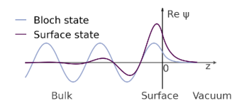
From a fundamental point of view, the distinct separation between bulk and surface states is intrinsically rooted in their different spatial expansion perpendicular to the surface. Bulk electrons strictly follow the transition symmetry of the crystal in three dimensions and are hence described by Bloch states as shown in Fig. 1. These Bloch states are characterized by the crystal momentum of the electrons parallel and perpendicular to the surface and are invariant with respect to the translation of the crystal momentum by any vector of the reciprocal crystal lattice. Accordingly, the energy spectrum of bulk states, i.e., the band structure, is described by and and hence reveals a strong band dispersion perpendicular to the surface. In contrast, the wave functions of surface states are strongly localized in the first surface layers (see Fig. 1) and their wave function and band structure only depend on the momentum of the electrons parallel to the surface. This is most prominently known for the Shockley surface states of noble metal surfaces[3, 4, 5, 6, 7, 8] or surface states of topological insulators[9, 10, 11, 12, 13, 14, 15, 16].
In a vast variety of materials, the electronic surface band structure is even richer and exhibits so-called surface resonances. They are hybrids between surface and bulk states with different surface and bulk character depending on the hybridization strength between their constituents[17]. So far however, only few studies focused on surface resonances, mainly in metals[18], topological insulators[19, 20] and black phosphorus[21, 22]. In these cases, the observed surface resonances are largely dominated by the contributions of the surface states.
In this letter, we revisit the surface band structure of a pristine (110)-surface of a silver single crystal, which has been of interest in other recent photoemission studies[23]. For this surface, a variety of different surface states and surface resonances with dominant surface character have already been reported[24, 25, 26, 27, 28, 29, 30, 31, 32]. The band dispersion of these states is independent on the electron momentum and they exhibit predominant -like orbital character. Both properties are reflected in unique spectroscopic signatures of such crystal-derived surface states, namely the photon energy independent band dispersions and significantly stronger photoemission yield for optical excitations using p-polarized light. In our work, we provide compelling evidence for the existence of a new type of surface resonance in the silver surface band structure, which we refer to as surface scattering resonance. It arises from additional surface contributions to the Fourier sum of the bulk Bloch states and has a strongly light polarization dependent emission pattern as well as a clear band dispersion along . A previous photoemission study[23] mentioned its observation, but did not disclose its origin and character.
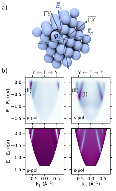
For the experimental part of our study, we employ a photoemission electron microscope combined with a time-of-flight delayline detector installed in an ultrahigh vacuum chamber. The PEEM is operated in momentum space mode and records three-dimensional datasets () in a single data acquisition with a resolution of and . The sample is illuminated with linearly polarized light under near normal incidence (NI, ) or grazing incidence (GI, ) with respect to the surface normal. The orientation of the polarization of the incoming photons with respect to the high symmetry directions of the crystal surface is shown in Fig. 2a). The orientation of the crystal was assessed by low-electron energy diffraction (LEED). Here, the -direction (-direction) corresponds to the (001)-direction ((10)-direction) in real space, which we define as the x-axis (y-axis). In the grazing incidence geometry (), the in-plane electric field component of p-polarized light is rotated by with respect to the -axis. In contrast, for the normal incidence geometry, the in-plane component of the electric field can be changed to any arbitrary angle using a half wave plate.
For our light source, we use photons (fourth harmonic of a Ti:Sa laser oscillator) suitable for observing one-photon photoemission at noble metal surfaces while maintaining the bulk-sensitivity of the experiment.
In conjunction with our experiment, we performed self-consistent electronic structure calculations within the ab-initio framework of density functional theory to support the interpretation of the experimental data. For this, we use the Vosko, Wilk, and Nusair parametrization to supply the exchange and correlation potential[33]. We calculate the electronic structure in a fully relativistic mode by solving the corresponding Dirac equation, using the relativistic multiple-scattering or KKR formalism in the TB-KKR mode[34, 35, 36]. Finally, we base the photocurrent calculations on the resulting half-space electronic structure represented by single-site scattering matrices for the different layers including the wave functions for the corresponding initial and final state energies as additional input quantities. Most importantly, we were able to vary the bulk sensitivity of the calculated photocurrent by altering the lifetimes of the initial and final states, which effectively influences the inelastic mean free path (IMFP). Additional details can be found in the supplemental material.
.
We start our discussion with an overview of near threshold angle resolved photoemission data of the Ag(110) surface. The top row of Fig 2b) shows the experimental energy distribution maps (EDM) extracted along the -direction for p- (left) and s-polarized light (right), respectively. The bottom row shows the corresponding theoretical EDMs calculated for the identical experiment geometry and conditions, which agree well with the experimental data. Minor deviations are attributed to slight differences of the theoretical and experimental Fermi energy due to a well-known shortcoming of the local density approximation (LDA)[37].
For both light polarizations we detect two signatures with strong bulk character, which we refer to as features (I) and (II) as shown in Fig. 2b). In the following discussion, we will provide clear evidence that these states can be assigned to a Mahan-cone-like bulk transition (I) and a new surface resonance (II). The Mahan-cone (I) reveals a strong intensity dependence on the polarization of the exciting light. It is only visible for s-polarized excitation and suppressed for p-polarized excitation. The second feature (II) can be clearly recognized for both light polarizations. The different polarization dependency of both transitions (I) and (II) is further reflected in the photocurrent obtained in the normal incidence geometry.
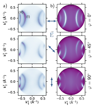
Figure 3 shows constant binding energy maps (CBMs) at the Fermi energy for the angles , with the experimental and theoretical data in the left and right column, respectively (CBMs for additional angles can be found in the supplemental material). In the CBMs, the features (I) and (II) appear as parabolic emission patterns with their center points located on the -axis. Both features (I) and (II) show a polarization dependent photoemission pattern, characterized by inhomogeneities of the intensity for angles between the axes as evident for as well as a local minima for in the case of . For , the calculated intensity pattern fits quantitatively for feature (II) whereas feature (I) appears mirrored at the horizontal axis in contrast to the measurement. Furthermore, the intensity of feature (I) decreases for while being the brightest for . On the other hand feature (II) gains more visibility rotating the polarization from to . These characteristic changes of the photoemission patterns clearly point to a different microscopic origin of both features.
In the next step, we investigate the photon energy dependent photocurrent of the Ag(110) surface band structure. This information can be extracted from our one-step photoemission simulations, which was able to describe the main aspects of our experimental photoemission data with high accuracy. We calculated CBMs for an initial state energy of , a polarization angle of and photon energies between . The corresponding 1D intensity profiles extracted at are shown in Fig. 4a). The spectroscopic features (I) and (II) reveal two well-defined maxima with a photon energy dependent photoemission yield. While the intensity of feature (II) increases with increasing photon energy, the one of feature (I) decreases and almost vanishes at a photon energy of . This points to a different cross section of both features and hence again to a different microscopic origin of both states. More importantly, the maxima of both features (I) and (II) change their position in momentum space when changing the photon energy. This behavior is fully in line with a significant band dispersion of both features for and finally proves the strong bulk-character of both features.
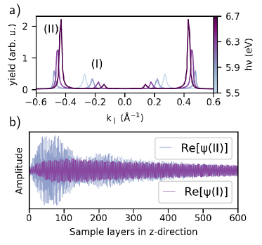
The different character of both features can be unambiguously resolved by extracting the depth dependent real part of the initial state wave function from our calculation. The real part of the wave function of feature (I) is shown as a purple line in Fig. 4b), the one of feature (II) as a blue line. closely resembles the shape of a Bloch wave penetrating far into the bulk of the material (large number of layers) with (almost) constant amplitude. The marginal decrease of the amplitude of can be attributed to the very small but finite life times that had to be considered in our calculations. Accordingly, feature (I) can be attributed to an optical transition between bulk states, which we will identify as the so-called Mahan cone transition[38, 39, 40]. In contrast, the amplitude of the wave function of feature (II) is largest in the region close to the surface and decays towards the interior of the Ag crystal. Crucially, the amplitude of does not decay within a few layers to zero as expected for a Shockley like surface state (see Fig. 1), but still exhibits a non-vanishing magnitude after silver layers. This spatial distribution of reveals all characteristic signatures of a surface resonance. In particular, the large amplitude of inside the Ag bulk is responsible for the extraordinary large bulk character of this surface resonance.
After unambiguously assigning feature (I) and (II) to a bulk transition and a surface resonance with dominated bulk-contribution, we now provide a semi-quantitative explanation of the microscopic origin of both spectroscopic features. For this, we turn to the three-step model of photoemission and construct both transitions in 3D momentum space using constant energy surfaces for the initial and final states. So far, Mahan cone transitions have only been observed for the noble metal surfaces cut in the (111)-direction.
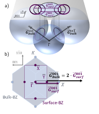
In the following, we will show that these transitions can also be observed for fcc(110) surfaces. We employ the nearly-free electron approximation and present the initial and final state of the Mahan cone transition as constant energy spheres in Fig. 5a). Their respective radii are determined by the total momentum of the electron, with the effective mass . The positions of the spheres of the initial and final state are given by the momentum conservation , where is an arbitrary lattice vector of the reciprocal crystal lattice. Due to energy and momentum conservation, photoelectrons at constant energy can only be excited at the intersection of the initial and final state spheres in this momentum space construction which is highlighted by the purple circles in Fig. 5a). By projecting these circles onto the Ag(110) surface, they form the parabolic features (bright purple) in the accessible field of view (grey circle) that reproduce the experimentally observed feature (I). We can hence conclude that feature (I) is the Mahan cone transition along the L-direction of bulk silver, projected onto the (110)-surface plane. Additionally, we included a construction based on our LDA calculations in the supporting information.
The surface resonance with large bulk character can be explained by additional contributions of surface associated reciprocal lattice vectors to the Bloch states of bulk electrons of the fcc lattice. These additional arise due to the mismatch between the surface projected Brillouin zone of the fcc bulk crystal and the surface Brillouin zone of the fcc(110) surface illustrated in Fig. 5b). The momentum conversation transforms into . This surface-induced modification of the momentum conservation allows us to consider another translation of the surface projected Mahan Cone feature by exactly one reciprocal surface lattice vectors, in this case , where is the surface lattice constant. This results in a new state located in the surface following the momentum dispersion of the bulk state, which we will refer to as surface scattering resonance. Crucially, this surface scattering resonance is not simply a replica of the bulk Mahan Cone scattering at the surface as it reveals its own light polarization and photon energy dependent photocurrent. We hence propose that the surface scattering resonance is truly a new type of surface resonance, which arises from additional surface contributions to the Fourier sum of the bulk Bloch states depending on .
Based on our assignment of features (I) and (II) to the Mahan cone and the surface scattering resonance, we propose an empirical rule for the polarization dependent emission pattern of both features discussed in Fig. 3. The measured photoemission signal can be reproduced by including two simple rules
-
(I)
-
(II)
regarding the angle between and in the geometrical construction, with being constrained within the surface layer for feature (II). This phenomenological model further support our identification of both features as the photoemission yield of the Mahan Cone can be explained by the orientation of the light polarization with respect to the 3D Brillouin zone while the one of the surface scattering resonance is only sensitive to the relative orientation of the light polarization vector and the crystal momentum of the 2D surface Brillouin zone.
In conclusion, our work uncovers clear spectroscopic evidence for a new type of surface resonance with exceptionally large bulk contribution in the surface band structure of an Ag(110) single crystal. This so called surface scattering resonance arises due to a lattice mismatch between the surface projected bulk Brillouin zone and the surface Brillouin zone of the Ag(110) surface leading to additional surface-related contributions to the bulk Bloch states of bulk transitions. We propose that these types of surface resonances can be observed for a vast majority of surfaces since similar lattices mismatches between surface projected bulk Brillouin zone and the surface Brillouin zone are the rule rather than the exception for most single crystal surfaces. This will potentially set the stage to discover new, possibly exotic, surface resonances with exceptional bulk character in a huge variety of quantum or topological materials.
Acknowledgements.
The research leading to these results was funded by the Deutsche Forschungsgemeinschaft (DFG, German Research Foundation) – TRR 173 – 268565370 (project A02) and EB154/37.References
- Speer et al. [2009] N. J. Speer, M. K. Brinkley, Y. Liu, C. M. Wei, T. Miller, and T.-C. Chiang, EPL 88, 67004 (2009).
- Forster et al. [2008] F. Forster, A. Bendounan, J. Ziroff, and F. Reinert, Phys. Rev. B 78, 161408 (2008).
- Gartland and Slagsvold [1975] P. O. Gartland and B. J. Slagsvold, Phys. Rev. B 12, 4047 (1975).
- Nicolay et al. [2000] G. Nicolay, F. Reinert, S. Schmidt, D. Ehm, P. Steiner, and S. Hüfner, Phys. Rev. B 62, 1631 (2000).
- Ünal et al. [2011] A. A. Ünal, C. Tusche, S. Ouazi, S. Wedekind, C.-T. Chiang, A. Winkelmann, D. Sander, J. Henk, and J. Kirschner, Phys. Rev. B 84, 073107 (2011).
- Tamai et al. [2013] A. Tamai, W. Meevasana, P. D. C. King, C. W. Nicholson, A. de La Torre, E. Rozbicki, and F. Baumberger, Phys. Rev. B 87, 075113 (2013).
- Yan et al. [2015] B. Yan, B. Stadtmüller, N. Haag, S. Jakobs, J. Seidel, D. Jungkenn, S. Mathias, M. Cinchetti, M. Aeschlimann, and C. Felser, Nature communications 6, 10167 (2015).
- Yaji et al. [2018] K. Yaji, A. Harasawa, K. Kuroda, R. Li, B. Yan, F. Komori, and S. Shin, Phys. Rev. B 98, 78 (2018).
- Fu et al. [2007] L. Fu, C. L. Kane, and E. J. Mele, Phys. Rev. Lett. 98, 106803 (2007).
- Fu and Kane [2007] L. Fu and C. L. Kane, Phys. Rev. B 76, 045302 (2007).
- Kane [2008] C. L. Kane, Nature Physics 4, 348 (2008).
- Xia et al. [2009] Y. Xia, D. Qian, D. Hsieh, L. Wray, A. Pal, H. Lin, A. Bansil, D. Grauer, Y. S. Hor, R. J. Cava, and M. Z. Hasan, Nature Physics 5, 398 (2009).
- Chen et al. [2009] Y. L. Chen, J. G. Analytis, J.-H. Chu, Z. K. Liu, S.-K. Mo, X. L. Qi, H. J. Zhang, D. H. Lu, X. Dai, Z. Fang, S. C. Zhang, I. R. Fisher, Z. Hussain, and Z.-X. Shen, Science 325, 178 (2009).
- Hasan and Kane [2010] M. Z. Hasan and C. L. Kane, Rev. Mod. Phys. 82, 3045 (2010).
- Jozwiak et al. [2013] C. Jozwiak, C.-H. Park, K. Gotlieb, C. Hwang, D.-H. Lee, S. G. Louie, J. D. Denlinger, C. R. Rotundu, R. J. Birgeneau, Z. Hussain, and A. Lanzara, Nature Physics 9, 293 (2013).
- Seibel et al. [2015] C. Seibel, H. Bentmann, J. Braun, J. Minár, H. Maa, K. Sakamoto, M. Arita, K. Shimada, H. Ebert, and F. Reinert, Phys. Rev. Lett. 114, 066802 (2015).
- Hans Lüth [2010] Hans Lüth, Solid Surfaces, Interfaces and Thin Films, 4th ed. (Springer Berlin Heidelberg, 2010).
- Braun et al. [2014] J. Braun, K. Miyamoto, A. Kimura, T. Okuda, M. Donath, H. Ebert, and J. Minár, New Journal of Physics 16, 015005 (2014).
- Jozwiak et al. [2016] C. Jozwiak, J. A. Sobota, K. Gotlieb, A. F. Kemper, C. R. Rotundu, R. J. Birgeneau, Z. Hussain, D.-H. Lee, Z.-X. Shen, and A. Lanzara, Nature communications 7, 13143 (2016).
- Sánchez-Barriga et al. [2017] J. Sánchez-Barriga, M. Battiato, M. Krivenkov, E. Golias, A. Varykhalov, A. Romualdi, L. V. Yashina, J. Minár, O. Kornilov, H. Ebert, K. Held, and J. Braun, Phys. Rev. B 95, 125405 (2017).
- Golias et al. [2016] E. Golias, M. Krivenkov, and J. Sánchez-Barriga, Phys. Rev. B 93, 075207 (2016).
- Ehlen et al. [2018] N. Ehlen, A. Sanna, B. V. Senkovskiy, L. Petaccia, A. V. Fedorov, G. Profeta, and A. Grüneis, Phys. Rev. B 97, 045143 (2018).
- Andi Li et al. [2020] Andi Li, Namitha Ann James, Tianyi Wang, Zehua Wang, Hrvoje Petek, and Marcel Reutzel, New Journal of Physics (2020).
- Ho et al. [1980] K.-M. Ho, B. N. Harmon, and S. H. Liu, Phys. Rev. Lett. 44, 1531 (1980).
- Reihl et al. [1984] B. Reihl, R. R. Schlittler, and H. Neff, Phys. Rev. Lett. 52, 1826 (1984).
- A. Goldmann et al. [1985] A. Goldmann, V. Dose, and G. Borstel, Phys. Rev. B 32 (1985).
- Bartynski and Gustafsson [1986] R. A. Bartynski and T. Gustafsson, Phys. Rev. B 33, 6588 (1986).
- Altmann et al. [1986] W. Altmann, V. Dose, and A. Goldmann, Zeitschrift für Physik B Condensed Matter 65, 171 (1986).
- L. E. Urbach et al. [1992] L. E. Urbach, K. L. Percival, J. M. Hicks, E. W. Plummer, and and H.-L. Dai, Phys. Rev. B 45, 3769 (1992).
- Knoesel et al. [1996] E. Knoesel, A. Hotzel, T. Hertel, M. Wolf, and G. Ertl, Surface Science 368, 76 (1996).
- A. Gerlach et al. [1999] A. Gerlach, G. Meister, R. Matzdorf, and A. Goldmann, Surface Science 443, 221 (1999).
- Pascual et al. [2001] J. I. Pascual, Z. Song, J. J. Jackiw, K. Horn, and H.-P. Rust, Phys. Rev. B 63, 960 (2001).
- S. H. Vosko et al. [1980] S. H. Vosko, L. Wilk, and M. Nusair, Canadian Journal of Physics 58, 1200 (1980).
- H. Ebert et al. [2020] H. Ebert et al., The Munich SPR-KKR package, version 8.5, https://www.ebert.cup.uni-muenchen.de/en/software-en/13-sprkkr (2020).
- Ebert et al. [2011] H. Ebert, D. Ködderitzsch, and J. Minár, Reports on Progress in Physics 74, 096501 (2011).
- Ebert [2000] H. Ebert, in Electronic Structure and Physical Properies of Solids, edited by H. Dreyssé (Springer Berlin Heidelberg, Berlin, Heidelberg, 2000) pp. 191–246.
- H Eckardt et al. [1984] H Eckardt, L Fritsche, and J Noffke, Journal of Physics F: Metal Physics 14, 97 (1984).
- Mahan [1970] G. D. Mahan, Phys. Rev. B 2, 4334 (1970).
- Stefan Hüfner [2003] Stefan Hüfner, Photoelectron Spectroscopy: Principles and Applications, Advanced Texts in Physics (Springer-Verlag Berlin Heidelberg, 2003).
- Winkelmann et al. [2012] A. Winkelmann, A. Akin Ünal, C. Tusche, M. Ellguth, C.-T. Chiang, and J. Kirschner, New Journal of Physics 14, 083027 (2012).