Domain Adaptation for Medical Image Analysis: A Survey
Abstract
Machine learning techniques used in computer-aided medical image analysis usually suffer from the domain shift problem caused by different distributions between source/reference data and target data. As a promising solution, domain adaptation has attracted considerable attention in recent years. The aim of this paper is to survey the recent advances of domain adaptation methods in medical image analysis. We first present the motivation of introducing domain adaptation techniques to tackle domain heterogeneity issues for medical image analysis. Then we provide a review of recent domain adaptation models in various medical image analysis tasks. We categorize the existing methods into shallow and deep models, and each of them is further divided into supervised, semi-supervised and unsupervised methods. We also provide a brief summary of the benchmark medical image datasets that support current domain adaptation research. This survey will enable researchers to gain a better understanding of the current status, challenges and future directions of this energetic research field.
Index Terms:
Domain adaptation, domain shift, machine learning, deep learning, medical image analysisI Introduction
Machine learning has been widely used in medical image analysis, and typically assume that the training dataset (source/reference domain) and test dataset (target domain) share the same data distribution [1]. However, this assumption is too strong and may not hold in real-world practice. Previous studies have revealed that the test error generally increases in proportion to the distribution difference between training and test datasets [2, 3]. This is referred to as the well-known “domain shift” problem [4]. Even in the deep learning era, deep Convolutional Neural Networks (CNNs) trained on large-scale image datasets may still suffer from domain shift [5]. Thus, how to handle domain shift is a crucial issue to effectively apply machine learning methods to medical image analysis.
Unlike natural image analysis with large-scale labeled datasets such as ImageNet [6, 7], in medical image analysis, a major challenge in constructing reliable and robust machine learning models is the lack of labeled data. Labeling medical images is generally expensive, time-consuming and tedious, requiring labor-intensive participation of physicians, radiologists and other experts. An intuitive solution is to reuse pre-trained models for some related domains [8]. However, the domain shift problem is still widespread among different medical image datasets due to different scanners, scanning parameters, and subject cohorts, etc. As a promising solution to tackle the domain shift/heterogeneity among medical image datasets, domain adaptation has attracted increasing attention in the medical image analysis community, aiming to minimize distribution differences among different but related domains Many researchers have engaged in leveraging domain adaptation methods to solve various tasks in medical image analysis.
There have been a number of surveys on domain adaptation [9, 10, 11, 12, 13, 14, 15] and transfer learning [16, 17, 18, 19, 20, 21] with natural images. However, there are only very limited reviews related to domain adaptation and their applications in medical image analysis which is a broad and important research area. Cheplygina et al. [22] provide a broad survey covering semi-supervised, multi-instance, and transfer learning for medical image analysis. Due to the wide scope, they only review general transfer learning methods in medical imaging applications without focusing on domain adaptation. Morid et al. [23] review transfer learning methods based on pre-training on ImageNet [6] that can benefit from knowledge from large-scale labeled natural images. However, many recent research works tend to design 3D CNN-based domain adaptation models for medical image analysis which are not included in their survey.
In this paper, we review and discuss recent advances and challenges of domain adaptation for medical image analysis. We systematically summarize the existing methods according to their characteristics. Specifically, we categorize different methods into two groups: 1) shallow models, and 2) deep models. Also, each group is further divided into three categories, i.e., supervised, semi-supervised and unsupervised methods. We also provide a summary of benchmark medical imaging datasets used in different domain adaptation research works.
The rest of this paper is organized as follows. We firstly introduce some background knowledge in Section II. In Section III and Section IV, we review the recent advances of domain adaptation methods in medical image analysis. We then present benchmark medical image datasets in Section V. Challenges and future research directions are discussed in Section VI. Finally, the conclusion is given in Section VII.
II Background
II-A Domain Shift in Medical Image Analysis
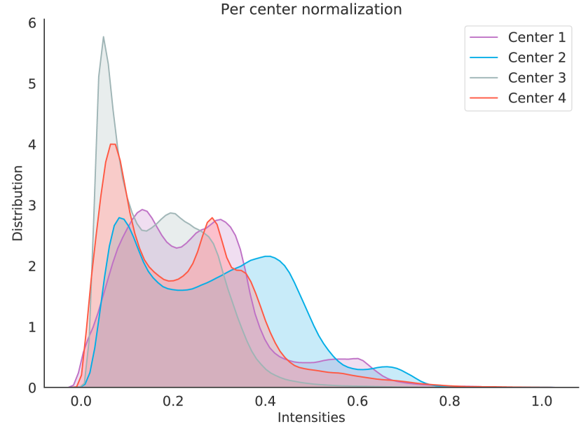
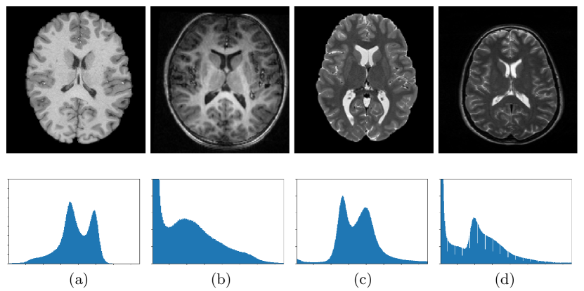
For a machine learning model, domain shift [4, 25, 26, 27] refers to the change of data distribution between its training dataset (source/reference domain) and test dataset (target domain). The domain shift problem is very common in practical applications of various machine learning algorithms and may cause significant performance degradation. Especially for multi-center studies, domain shift is widespread among different imaging centers/sites that may use different scanners, scanning protocols, and subject populations, etc. Fig. 1 illustrates the problem of inter-center domain shift in terms of the intensity distribution of structural magnetic resonance imaging (MRI) from four independent sites (i.e., UCL, Montreal, Zurich, Vanderbilt) in the Gray Matter segmentation challenge [24, 28]. Fig. 2 illustrates the image-level distribution heterogeneity caused by different scanners [29]. From Figs. 1-2, one can observe that there are clear distribution shifts in these medical imaging data. However, many conventional machine learning methods ignore this problem, which would lead to performance degradation [26, 30]. Recently, domain adaptation has attracted increasing interests and attention of researchers, and become an important research topic in machine learning based medical image analysis [31, 32, 33, 22].
II-B Domain Adaptation and Transfer Learning
This survey focuses on domain adaptation for medical image analysis. Since domain adaptation can be regarded as a special type of transfer learning, we first review their definitions to provide a clear understanding of their differences. In a typical transfer learning setting, there are two concepts: “domain” and “task” [16, 17, 18]. A domain relates to the feature space of a specific dataset and the marginal probability distribution of features. A task relates to the label space of a dataset and an objective predictive function. The goal of transfer learning is to transfer the knowledge learned from the task on domain to the task on domain . Note that either the domain and the task may change during the transfer learning process.
Domain adaptation, the focus of this survey, is a particular and popular type of transfer learning [9, 10, 11, 12, 13]. For domain adaptation, it is assumed that the domain feature spaces and tasks remain the same while the marginal distributions are different between the source and target domains (datasets). It can be mathematically depicted as follows. Let represent the joint feature space and the corresponding label space, respectively. A source domain S and a target domain T are defined on , with different distributions and , respectively. Suppose we have labeled samples (subjects) in the source domain, i.e., , and also have samples (with or without labels) in the target domain, i.e., . Then the goal of domain adaptation (DA) is to transfer knowledge learned from S to T to perform a specific task on T, and this task is shared by S and T.
II-C Different Settings of Domain Adaptation
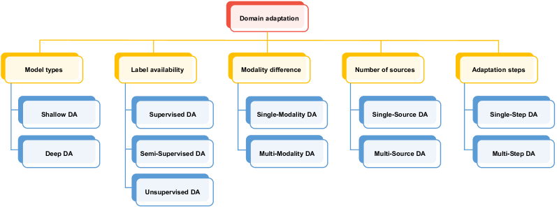
In DA, the source domain and target domain share the same learning tasks. In practice, DA can be categorized into different groups according to different scenarios, constrains, and algorithms. In Fig. 3, we summarize different categories of DA methods for medical image analysis based on six problem settings, i.e., model type, label availability, modality difference, number of sources, and adaptation steps.
-
•
Model Type: Shallow DA & Deep DA. In terms of whether the learning model is shallow or deep, the DA methods can be divided into shallow DA and deep DA [13, 10, 9]. Shallow DA methods usually rely on human-engineered imaging features and conventional machine learning models. In contrast, deep DA methods (especially those with CNN architectures) generally integrate feature learning and model training into end-to-end learning models, where the data adaptation is performed in a task-oriented manner.
-
•
Label Availability: Supervised DA & Semi-Supervised DA & Unsupervised DA. In terms of label availability, existing DA methods can be divided into supervised DA, semi-supervised DA, and unsupervised DA [13, 11]. In supervised DA, a small number of labeled data in the target domain are available for model training. In semi-supervised DA, a small number of labeled data as well as redundant unlabeled data in the target domain are available in the training process. In unsupervised DA, only unlabeled target data are available for training the adaptation model. Since label scarcity is widespread among medical images, unsupervised DA has attracted increasing attention recently. In fact, there is an extreme case that even unlabeled target data are not accessible during the training time. The learning model is only allowed to be trained on several related source domains to gain enough generalization ability for the target domain. This problem is referred to as domain generalization, while a few related research has been conducted for medical image analysis [34] currently.
-
•
Modality Difference: Single-Modality DA & Cross-Modality DA. In terms of modality difference, the existing DA methods can be divided into single-modality DA and cross-modality DA [9, 35, 36]. In single modality DA, the source and target domains share the same data modality. For example, the source domain consists of MRIs collected in vendor A, and the target domain contains MRIs acquired from vendor B [37]. In cross-modality DA, the modality of source and target domains are different with various scanning technologies. For example, the source domain consists of MR images, whereas the target domain contains CT images [36].
-
•
Number of Sources: Single-Source DA & Multi-Source DA. In terms of the number of source domains, the existing DA methods can be divided into single-source DA and multi-source DA approaches [12, 14, 15]. In single-source DA, the model is trained on a single source domain, whereas multi-source DA uses training samples from multiple source domains. Since there is also data heterogeneity among different source domains, multi-source DA is quite challenging. Most of the existing DA methods for medical image analysis fall into the single-source DA category.
-
•
Adaptation Step: One-Step DA & Multi-Step DA. In terms of adaptation steps, the existing DA methods can be divided into one-step DA and multi-step DA [10, 38, 39]. In one-step DA, adaption between the source and target domain is accomplished in one step due to a relatively close relationship between them. In multi-step DA (also called distant/transitive DA), there are often intermediate domains introduced to bridge the relatively large distribution gap between the source and target domains.
It should be noted that each group of methods are not mutually exclusive to each other. For example, there are also supervised, semi-supervised and unsupervised DA methods in shallow and deep DA approaches. In this survey, we employ the model type as the main criterion for DA method categorization, and introduce existing shallow DA and deep DA methods for medical image analysis.
III Shallow Domain Adaptation Methods
III-A Overview
In this section, we review shallow domain adaptation methods based on human-engineered features and conventional machine learning models for medical image analysis. We first introduce two commonly-used strategies in shallow DA methods: 1) instance weighting, and 2) feature transformation.
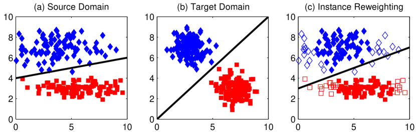
Instance weighting is one of the most popular strategies adopted by shallow DA methods for medical image analysis [40, 41, 42, 43, 44]. In this strategy, samples/instances in the source domain are assigned with different weights according to their relevance with target samples/instances. Generally, source instances that are more relevant to the target instances will be assigned larger weights. After instance weighting, a learning model (e.g., classifier or regressor) is trained on the re-weighted source samples, thus reducing domain shift between the source and target domains. Fig. 4 illustrates the effect of instance weighting strategy on the source and target domains. From Fig. 4(a)-(b), we can see that the domain difference between the source and target domain is large. After instance weighting, the domain difference is reduced based on those re-weighted source instances, as shown in Fig. 4(b)-(c).
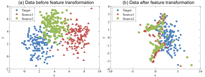
Feature transformation strategy focuses on transforming source and target samples from their original feature spaces to a new shared feature representation space [46, 47, 48]. As shown in Fig. 5, the goal of feature transformation for DA is to construct a common/shared feature space for the source and target domains to reduce their distribution gap, based on various techniques such as low-rank representation [48]. Then, a learning model can be trained on the new feature space, which is less affected by the domain shift in the original feature space between the two domains.
III-B Supervised Shallow DA
Wachinger et al. [40] propose an instance weighting-based DA method for Alzheimer’s disease (AD) classification. Specifically, labeled target samples are used to estimate the target distribution. Then source samples are re-weighted by calculating the probability of source samples in the target domain. A multi-class logistic regression classifier is finally trained on the re-weighted samples and labeled target data (with volume, thickness, and anatomical shape features), and applied to the target domain. Experiments on the ADNI [49], AIBL [50] and CADDementia challenge datasets [51] show that the DA model can achieve better results than methods that only use data from either the source or target domain. Goetz et al. [41] propose an instance weighting-based DA method for brain tumor segmentation. To compute the weights of source samples, a domain classifier (logistic regression) is trained with paired samples from source and target domains. The output of the domain classifier (in terms of probability) is then used to calculate the adjusted weights for source samples. A random forest classifier is trained to perform segmentation. Experiments on the BraTS dataset [52] demonstrate the effectiveness of this method. Van Opbroek et al. [42] propose an instance weighting-based DA method for brain MRI segmentation. In their approach, each training image in the source domain is assigned a weight that can minimize the difference between the weighted probability density function (PDF) of voxels in the source and target data. The re-weighted training data are then used to train a classifier for segmentation. Experiments on three segmentation tasks (i.e., brain-tissue segmentation, skull stripping, and white-matter-lesion segmentation) demonstrate its effectiveness.
Becker et al. [46] propose a feature transformation-based DA method for microscopy image analysis. Specifically, they propose to learn a nonlinear mapping that can project samples in both domains to a shared discriminative latent feature space. Then, they develop a boosting classifier trained on the data in the transformed common space. In [53, 54], the authors propose a domain transfer support vector machine (DTSVM) for mild cognitive impairment (MCI) classification. Based on the close relationship of Alzheimer’s disease (AD) and MCI, DTSVM is trained on MCI subjects in the target domain and fine-tuned with an auxiliary AD dataset to enhance its generalization ability. They validate their method on the ADNI database and achieve good performance in MCI conversion prediction.
III-C Semi-Supervised Shallow DA
Conjeti et al. [55] propose a two-step DA framework for ultrasound image classification. In the first step, they use principle component analysis (PCA) to transform the source and target domains to a common latent space, through which the global domain difference is minimized. In the second stage, a random forest classifier is trained on the transformed source domain, and then fine-tuned with a few labeled target data to further reduce the domain shift.
III-D Unsupervised Shallow DA
Cheplygina et al. [43] employ the instance weighting strategy for lung disease classification. Specifically, Gaussian texture features are first extracted from all CT images, based on which a weighted logistic classifier is trained. Source samples that are similar to the target data are assigned high weights to reduce domain shift. Experiments on four chest CT image datasets show that this method can significantly improve the classification performance. Heimann et al. [44] use instance weighting-based DA strategy for ultrasound transducer localization in X-ray images. The instance weights are computed through a domain classifier (i.e., logistic regression). Based on Haar-like features of X-ray images, a cascade of tree-based classifiers is trained for ultrasound transducer localization. Li et al. [56] propose a subspace alignment DA method for AD classification using functional MRI (fMRI) data. They first conduct feature extraction and selection for source and target samples, followed by a modified subspace alignment strategy to align samples from both domains into a shared subspace. Finally, the aligned samples in the shared subspace are used as an integrated dataset to train a discriminant analysis classifier. This method is validated on both ADNI and a private fMRI datasets. Kamphenkel et al. [57] propose an unsupervised DA method for breast cancer classification based on diffusion-weighted MR images (DWI). They use the Diffusion Kurtosis Imaging (DKI) algorithm to transform the target data to the source domain without any label information of target data.
III-E Multi-Source Shallow DA
Wang et al. [47] propose a multi-source DA framework for Autism Spectrum Disorder (ASD) classification with resting-state functional MRI (rs-fMRI) data. Each subject/sample is represented by both functional connectivity (FC) features in gray matter regions and functional correlation tensor features extracted from white matter regions. All subjects in each of the multiple source domains are transformed to the target domain via low-rank regularization and graph embedding. Then, a classifier is trained on the source samples (with transformed features). This method is validated on the ABIDE dataset [58]. Wang et al. [48] propose a low-rank representation based multi-source domain adaptation framework for ASD classification. They assume that multiple domains share an intrinsic latent data structure, and map multiple source data and target data into a common latent space via a low-rank representation to reduce the domain shift. Experimental results on both synthetic data and the ABIDE dataset suggest the good performance of their method. Cheng et al. [59] propose a Multi-Domain Transfer Learning (MDTL) framework for the early diagnosis of AD and MCI. They first select a subset of discriminative features (i.e., gray matter tissue volume) by using the training data from multiple auxiliary domains and target domain. Then, they construct a multi-domain transfer classifier based on the ensemble of the classifiers learned from auxiliary domains and an adaptive SVM [60] learned from the labeled target samples.
IV Deep Domain Adaptation Methods
Deep learning [61, 62] has greatly pushed forward the development of artificial intelligence and machine learning. As one of the most popular deep learning models, CNN has demonstrated its superiority over conventional human-engineered imaging features. Trained with large-scale labeled data in a full supervision manner, CNN has made breakthroughs in computer vision and medical image analysis [63, 64, 65]. It has been revealed that CNN is able to learn generic low-level features (e.g., textures and edges) that can be transferable to different image analysis tasks [66, 67]. A milestone is the AlexNet [68] which is trained with millions of natural images from ImageNet [6]. Following AlexNet, several representative CNN models have been proposed, including VGG [69], Inception [70], ResNet [71], and DenseNet [72]. These four types of CNNs have been widely used in various domain adaptation models for medical image analysis [23].
IV-A Supervised Deep DA
With deep features (e.g., extracted by CNNs), several studies focus on using shallow DA models for medical image analysis. Kumar et al. [73] use ResNet as the feature extractor of mammographic images. Based on the CNN features, they evaluate three shallow domain adaptation methods, i.e., Transfer Component Analysis (TCA) [74], Correlation Alignment (CORAL) [75], and Balanced Distribution Adaptation (BDA) [76] for breast cancer classification, and provide some empirical results on the DDSM [77] and InBreast [78] datasets. Huang et al. [79] propose to use LeNet-5 to extract features of histological images from different domains for epithelium-stroma classification, and then project them into a subspace (via PCA) to align them for adaptation. Experiments on the NKI, VGH [80] and IHC [81] datasets verify its effectiveness.
Another research direction is to transfer models learned on the source domain onto the target domain with fine-tuning. Ghafoorian et al. [31] evaluate the impact of fine-tuning strategy on brain lesion segmentation, based on CNN models pre-trained on brain MRI scans. Their experimental results reveal that using only a small number of target training examples for fine-tuning can improve the transferability of models. They further evaluate the influence of both the size of the target training set and different network architectures on the adaptation performance. Based on similar findings, numerous methods have been proposed to leverage CNNs that are well pre-trained on ImageNet to tackle medical image analysis problems. Samala et al. [82] propose to first pre-train an AlexNet-like network on ImageNet, and then fine-tune it with regions-of-interest (ROI) from 2,454 mass lesions for breast cancer classification. Khan et al. [83] propose to pre-train a VGG network on ImageNet, and then use labeled MRI data to fine-tune it for Alzheimer’s disease (AD) classification. They propose to use image entropy to select the most informative training samples in the target domain, and verify their method on the ADNI database [49]. Similarly, Swati et al. [84] pre-train a VGG network on ImageNet and re-train the higher layers of the network with labeled MR images for brain tumor classification. Experiments are conducted on a publicly available CE-MRI dataset111https://figshare.com/articles/brain_tumor_dataset/1512427. Abbas et al. [85] employ the ImageNet to pre-train CNNs for chest X-ray classification. To deal with the irregularities in the datasets, they introduce class decomposition into the network learning process, by partitioning each class within the image dataset into subsets and then assign new labels to the new set. Three different cohorts of chest X-ray images, histological images of human colorectal cancer, and digital mammograms are used for performance evaluation.
The above-mentioned methods employ the one-step DA strategy, i.e., directly transferring the pre-trained model to the target domain. When facing the problem setting where target samples are too few to fine-tune a model, several studies propose to use intermediate domains to facilitate multi-step DA. Gu et al. [86] develop a two-step adaptation method for skin cancer classification. First, they fine-tune a ResNet on a relatively large medical image dataset (for skin cancer). Then, the network is trained on the target domain which is a relatively small medical image dataset. Their experimental results on the MoleMap dataset222http://molemap.co.nz and the HAM10000 dataset333https://www.kaggle.com/kmader/skin-cancer-mnist-ham10000 show that the two-step adaptation method achieves better results than the directly transferring methods.
Although adopting pre-trained models on a dataset (e.g., ImageNet) is a popular way for supervised DA for medical image analysis, the 2D CNN structure may not able to fully explore the rich information conveyed in 3D medical images. To this end, some researchers deliberately design task-specific 3D CNNs that are trained with medical images as the backbone to facilitate the subsequent data adaptation tasks. Hosseini-Asl et al. [87] design a 3D CNN for brain MR images classification. The network is pre-trained with MR images in the source domain. Then, its upper fully-connected layers are fine-tuned with samples in the target domain. Experiments on ADNI and CADDementia demonstrate its effectiveness. Valverde et al. [88] propose a similar 3D CNN for brain MR images segmentation. Instead of using the whole brain MRI, their network takes 3D image patches as input, while only partial fully-connected layers are fine-tuned using the target data. This method is evaluated on the ISBI2015 dataset [89] for MRI segmentation. Kaur et al. [90] propose to first pre-train a 3D U-Net on a source domain that has relevant diseases with a large number of samples, and then use a few labeled target data to fine-tune the network. Experiments on the BraTS dataset [52] show this strategy achieves a better performance than the network trained from scratch. Similar strategy is also used in [91] where a network is pre-trained using a large number of X-ray computed tomography (CT) and synthesized radial MRI datasets and then fine-tuned with only a few labeled target MRI scans. Zhu et al. [92] propose a boundary-weighted domain adaptive neural network for prostate segmentation. A domain feature discriminator is co-trained with the segmentation networks in an adversarial learning manner to reduce domain shift. Aiming to tackle the difficulty caused by unclear boundaries, they design a boundary-weighted loss and add it into the training process for DA and segmentation. The boundary contour needs to be extracted from the ground truth label to generate a boundary-weighted map as the supervision information to minimize the loss. Experiments on the PROMISE12 challenge dataset444https://promise12.grand-challenge.org/ and BWH dataset [93] demonstrate its effectiveness. Bermúdez-Chacón et al. [94] design a two-stream U-Net for electron microscopy image segmentation. One stream uses source domain samples while the other uses target data. They utilize Maximum Mean Discrepancy (MMD) and correlation alignment as the domain regularization for DA. With a few labeled target data to fine-tune the model, their method achieves promising performance in comparison with several state-of-the-art ones on a private dataset. Laiz et al. [95] propose to use triplet loss for DA in endoscopy image classification. Each triplet consists of an anchor sample A from the source domain, a positive sample B from the target domain with the same label of A, and a negative sample C in the source domain. Through minimizing the triplet loss, their model can reduce the domain shift while keeping discrimination on different diseases.
IV-B Semi-Supervised Deep DA
Roels et al. [96] propose a semi-supervised DA method for electron microscopy image segmentation. They design a “Y-Net” with one feature encoder and two decoders. One decoder is used for segmentation, while a reconstruction decoder is designed to reconstruct images from both the source and target domains. The network is initially trained in an unsupervised manner. Then, the reconstruction decoder is discarded, and the whole network is fine-tuned with labeled target samples to make the model adapt to the target domain.
Madani et al. [97] propose a semi-supervised generative adversarial network (GAN) based DA framework for chest X-ray image classification. Different from conventional GAN, this model takes labeled source data, unlabeled target data and generated images as input. The discriminator performs three-category classification (i.e., normal, disease, or generated image). During training, unlabeled target data can be classified as any of those three classes, but can contribute to loss computation when they are classified as generated images. Through this way, both labeled and unlabeled data can be incorporated into a semi-supervised manner. Experiments on the NIH PLCO dataset [98] and NIH Chest X-Ray dataset [99] demonstrate its effectiveness.
IV-C Unsupervised Deep DA
Unsupervised deep domain adaptation has attracted increasing attention [11] in the field of medical image analysis, due to its advantage that does not require any labeled target data. We now introduce the existing unsupervised deep DA methods based on their specific strategies for knowledge transfer.
IV-C1 Feature Alignment
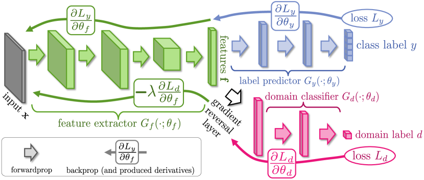
This line of research aims to learn domain-invariant features across domains through specifically designed CNN models. Most of the models adopt a siamese architecture similar to the Domain Adversarial Neural Network (DANN) structure [100] as shown in Fig. 6. Kamnitsas et al. [33] propose a DANN-based multi-connected adversarial network for brain lesion segmentation. In their model, the domain discriminator is trained simultaneously with a segmentation network. In addition, the authors argue that only adapting the last layers of the segmentor is not ideal, thus the domain discriminator is connected at multiple layers of the network to make it less susceptible to image-quality variations between different domains. Javanmardi et al. [101] propose a DANN-based model for eye vasculture segmentation. A U-Net and a domain discriminator are co-trained during the training process. Experiments on the DRIVE [102] and STARE [103] datasets demonstrate its effectiveness. Yang et al. [104] employ the DANN for lung texture classification, with experiments performed on the SPIROMICS dataset555https://www.spiromics.org/spiromics/. Panfilov et al. [105] develop an adversarial learning-based model for knee tissue segmentation. A U-Net-based segmentor and a domain discriminator with adversarial learning are co-trained for DA. Experiments on three knee MRI datasets verify its effectiveness. Zhang et al. [106] propose an adversarial learning-based DA for AD/MCI classification on ADNI. A classifier and a domain discriminator are co-trained to enhance the model’s transferability across different domains.
Different from the above-mentioned studies, Dou et al. [107] develop a cross-modality DA framework for cardiac MR and CT image segmentation, by only adapting low-level layers (with higher layers fixed) to reduce domain shift during training. They assume that the data shift between cross-modality domains mainly lies in low-level characteristics, and validate this assumption through experiments on the MM-WHS dataset [108]. Shen et al. [109] employ adversarial learning for fundus image quality assessment. Due to the demand for image assessment, they fix high-level weights during the adversarial training to focus on low-level feature adaptation. Yan et al. [110] propose an adversarial learning-based DA method for MR image segmentation. A domain discriminator is co-trained with the segmentor to learn domain-invariant features for the task of segmentation. To enhance the model’s attention to edges, Canny edge detector is introduced into the adversarial learning process. Experiments on images from three independent MR vendors (Philips, Siemens, and GE) demonstrate the effectiveness. Shen et al. [111] propose an adversarial learning-based method for mammogram detection which is an essential step in breast cancer diagnosis. A segmentor based on fully convolutional network (FCN) and a domain discriminator are co-trained for domain adaptation. Experiments on the public CBIS-DDSM [77], InBreast [78] and a self-collected dataset demonstrate its effectiveness. Yang et al. [112] propose an adversarial learning-based DA method for lesion detection within the Faster RCNN framework [113]. Besides global feature alignment, they also extract ROI-based region proposals to facilitate local feature alignment. Through combining these two level features, their method shows robustness in the experiments on two datasets from two independent medical centers.
Gao et al. [114] propose an unsupervised method for classification of brain activity based on fMRI data from the Human Connectome Project (HCP) dataset [115]. They use the central moment discrepancy (CMD) which matches the higher-order central moments of the data distributions as the domain regularization loss to facilitate adaptation. They assume that the high-level features extracted by fully-connected layers have large domain shift, thus CMD is imposed on these layers to perform adaptation. Bateson et al. [116] propose an unsupervised constrained DA framework for disc MR image segmentation. They propose to use some useful prior knowledge that is invariant across domains as an inequality constraint, and impose such constraints on the predicted results of unlabeled target samples as the domain adaptation regularization. In practice, they employ the size of target segmentation regions as a constraint for network optimization, with experiments performed on the MICCAI 2018 IVDM3Seg Challenge666https://ivdm3seg.weebly.com/. Mahapatra et al. [117] develop a deep DA approach for cross-modality image registration. To transfer knowledge across domains, they design a convolutional auto-encoder to learn latent feature representations of images, followed by a generator to synthesize the registered image. Trained on the latent feature space instead of on the original images, their model is less limited to any imaging modality. Experiments on chest X-ray, retinal and brain MR images show that their model (trained on one dataset) gives better registration performance for other datasets.
IV-C2 Image Alignment
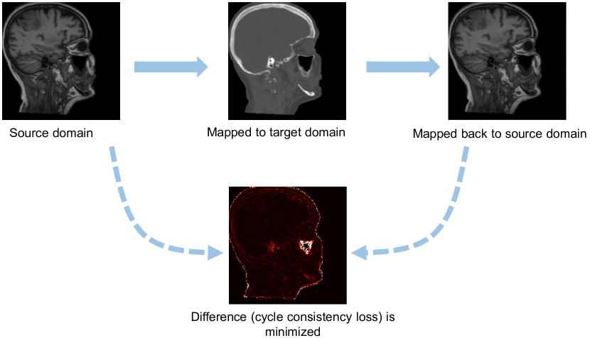
Image-level alignment is also used for domain adaptation based on deep generative models, such as Generative Adversarial Network (GAN) [120]. The original GAN takes random noise as input. As a significant improvement, Zhu et al. [118] propose a cycle-consistent GAN (CycleGAN) model which can translate one image domain into another without the demand for paired training samples. In medical image analysis, CycleGAN has been used for image synthesis (see Fig. 7), where an MR image is firstly mapped to the target domain (CT image) and then mapped back to the input domain. A cycle consistency loss is used to measure the difference between the input image and the reconstructed image. Through minimizing this loss, the CycleGAN can realize image-to-image translation without paired training samples.
Wollmann et al. [121] propose a CycleGAN-based DA method for breast cancer classification. They first use CycleGAN to transform whole-slide images (WSIs) of lymph nodes from a source domain (i.e., a medical center) to the target domain (i.e., another medical center). Then a densely connected deep neural network (DenseNet) is used for breast cancer classification. Image patches from region-of-interest (ROIs) are used as the input to facilitate two-level classification. Experiments on the CAMELYON17 challenge dataset777https: //camelyon17.grand-challenge.org/ demonstrate the effectiveness. Manakov et al. [122] propose to leverage unsupervised DA to tackle retinal optical coherence tomography (OCT) image denoising problem. They treat image noises as domain shift between high and low noise domains. A CycleGAN-based DA mode is designed to learn a mapping between the source (i.e., high noise) and the target domain (i.e., low noise) on unpaired OCT images, thus achieving the goal of image denoising. Experiments on numerous in-house OCT images demonstrate the effectiveness of this method. Gholami et al. [123] propose to use CycleGAN to generate more training data for brain tumor segmentation. They first generate synthetic tumor-bearing MR images using their in-house simulation model, and then transform them to real MRIs using CycleGAN to augment training samples. They conduct experiment on the BraST [52] dataset and achieve good segmentation results for brain tumors.
Zhang et al. [124] propose a Noise Adaptation Generative Adversarial Network (NAGAN) for eye blood vessel segmentation, by formulating DA as a noise style transfer task. They use an image-to-image translation generator to map the target image to the source domain. Besides a normal discriminator which enforces the content similarity between generated images and real images, they also design a style discriminator which enforces the generated images to have the same noise patterns as those from the target domain. Experiments on the SINA [125] and a local dataset demonstrate the effectiveness. Mahmood et al. [126] propose a Reverse Domain Adaptation method based on GAN for endoscopy image analysis. Different from traditional GAN, they reverse the flow and transform real images to synthetic images, based on the insight that subjects with optical endoscopy images have patient-specific details and these details do not generalize across patients. To remove these details from real images while maintaining useful diagnostic details, they transform real images in the target domain to synthetic ones, thus bridging the gap between the target and source domains.
IV-C3 Image+Feature Alignment
Chen et al. [36, 127] combine image alignment and feature alignment for cross-modality cardiac image segmentation. They firstly use CycleGAN to transform labeled source images into target-like images. Then, the synthesized target images and real target images are fed into a two-stream CNN with a domain discriminator which can further reduce the domain gap via adversarial learning. Experiments on the MM-WHS dataset [108] demonstrate its effectiveness. Yan et al. [37] propose a similar unsupervised framework for cross-vendor cardiac cine MRI segmentation. They first train a U-Net using labeled source samples, and then train a CycleGAN to facilitate image-level adaptation. In addition, they design a feature-level alignment mechanism by computing the Mean Square Error (MSE) of features between the original and translated images. Three major vendors including Philips, Siemens, and GE, are treated as three domains. For example, the Philips data are treated as the source domain, while the Siemens and GE data are defined as two target domains. The short-axis steady-state free precession (SSFP) cine MR images of 144 subjects acquired by those three vendors are used for performance evaluation.
IV-C4 Disentangled Representation
Yang et al. [35] propose a cross-modality (between CT and MRI) DA method via disentangled representation for liver segmentation. Through disentangle presentation learning, images from each domain are embedded into two spaces, i.e., a shared domain-invariant content space and a domain-specific style space. Then, domain adaptation is performed in the domain-invariant space. Experiments on the LiTS benchmark dataset [128] and a local dataset demonstrate the effectiveness of this method.
IV-C5 Ensemble Learning
Following [129], Perone et al. [24] propose a self-ensemble based DA method for medical image segmentation. A baseline CNN, called student network, takes labeled samples from the source domain, and makes its predictions after training with a task (segmentation) loss. Another network, called teacher network, gives its predictions using only unlabeled samples in the target domain. The teacher network is updated by the exponential moving average of the weights of the student network (a temporal ensemble strategy). During training, the domain shift is minimized by a consistency loss by comparing predictions from both the student and teacher networks. This method is evaluated on the SCGM challenge dataset [130], which is a multi-center multi-vendor dataset of spinal cord anatomical MR images from healthy subjects. Shanis et al. [131] employ a similar DA architecture for brain tumor segmentation. Besides a consistency loss that measures the difference between predictions of the teacher and student networks, they also utilize an adversarial loss to further improve adaptation performance. Effectiveness is validated with experiments on the BraTS dataset [52].
IV-C6 Soft Labels
Bermúdez-Chacón et al. [132] propose an unsupervised DA method to reduce domain shift in electron microscopy images. Based on the observation that some ROIs in medical images still resemble each other across domains, the authors propose to use Normalized Cross Correlation (NCC) which computes the similarity of two structures from two different images (visual correspondence) to construct a consensus heatmap. The high-scoring regions of the heatmap are used as soft labels for the target domain, which are then used to train the target classifier for segmentation.
IV-C7 Feature Learning
Ahn et al. [133, 134] introduce a convolutional auto-encoder to learn features for domain adaptation. Other than using labeled target data to fine-tune a pre-trained AlexNet, the authors propose to use a zero-bias convolutional auto-encoder as an adapter to transform the feature maps from the last convolution layer of AlexNet to relevant medical image features. The learned features can further be used for classification tasks. Experimental results on the ImageCLEF dataset888https://www.imageclef.org/ and ISIC dataset [135] validate the effectiveness of this method.
IV-D Multi-Target Deep DA
Orbes-Arteaga et al. [136] propose an unsupervised DA framework with two target domains for brain lesion segmentation. Their network is first trained with labeled source data. During the adaptation phase, labeled source data and the paired unlabeled data from the two target domains are fed into the DA model. An adversarial loss is imposed to reduce domain differences. To minimize the output probability distribution on the target data, a consistency loss is imposed on the paired target data. Experiments are performed on the MICCAI 2017 WMH Challenge [137] and two independent datasets. Karani et al. [29] propose a life-long multi-domain adaptation framework for brain structure segmentation. In their method, batch normalization plays the key role in adaptation, as suggested in [138]. They first train a network on source domains with shared convolutional filters but different batch normalization (BN) parameters, and each domain corresponds to a specific set of BN parameters. By fine-tuning the BN parameters with a few labeled data in each new target domain, the model can then be adapted to those multi-target domains. This method is evaluated on 4 publicly available datasets, including HCP [115], ADNI [49], ABIDE [58], and IXI999brain-development.org/ixi-dataset/.
| Reference | Task | Dataset | Image Type | Method |
| Brain | ||||
| Wachinger et al. [40] | AD classification | ADNI, AIBL, CADDementia | MRI | Instance weighting |
| Cheng et al. [54] | MCI conversion prediction | ADNI | MRI, PET | Adaptive SVM |
| Cheng et al. [59] | AD classification | ADNI | MRI | Ensemble, Adaptive SVM |
| Li et al. [56] | AD classification | ADNI | fMRI | Subspace alignment |
| Hofer et al. [139] | AD classification | ADNI, IXI | MRI | Instance alignment |
| Wang et al. [47] | ASD classification | ABIDE | fMRI | low-rank regularization |
| Wang et al. [48] | ASD classification | ABIDE | fMRI | low-rank regularization |
| Goetz et al. [41] | Tumor segmentation | BraTS | MRI | Instance weighting |
| Kaur et al. [90] | Tumor segmentation | BraTS | MRI | CNN fine-tuning |
| Gholami et al. [123] | Tumor segmentation | BraTS | MRI | GAN |
| Shanis et al. [131] | Tumor segmentation | BraTS | MRI | CNN, feature alignment |
| Swati et al. [84] | Tumor classification | CE-MRI | MRI | CNN fine-tuning |
| Karani et al. [29] | Structure segmentation | HCP, ADNI, ABIDE, IXI | MRI | CNN, feature alignment |
| Khan et al. [83] | AD classification | ADNI | MRI | CNN fine-tuning |
| Hosseini-Asl et al. [87] | AD classification | ADNI, CADDementia | MRI | CNN fine-tuning |
| Zhang et al. [106] | AD classification | ADNI | MRI | Adversarial learning |
| Guan et al. [140] | AD classification | ADNI, AIBL | MRI | CNN, feature alignment |
| Mahapatra et al. [117] | Registration | ADNI | MRI | CNN, feature alignment |
| Valverde et al. [88] | Lesion segmentation | ISBI2015 | MRI | CNN fine-tuning |
| Orbes-Arteaga et al. [136] | Lesion segmentation | MICCAI WMH Challenge | MRI | CNN, feature alignment |
| Gao et al. [114] | Brain activity classification | HCP | fMRI | CNN, feature alignment |
| Lung | ||||
| Cheplygina et al. [43] | COPD classification | DLCST, COPDGene | CT | Instance weighting |
| Yang et al. [104] | Lung texture classification | SPIROMICS | CT | CNN, feature alignment |
| Mahapatra et al. [117] | Registration | NIH ChestXray14 | X-ray | CNN, feature alignment |
| Heart | ||||
| Madani et al. [97] | Cardiac abnormalities identity | NIH PLCO, NIH Chest | X-ray | GAN |
| Dou et al. [107] | Cardiac segmentation | MM-WHS | CT, MRI | CNN, feature alignment |
| Chen et al. [36] | Cardiac segmentation | MM-WHS | CT, MRI | GAN, feature alignment |
| Breast | ||||
| Kumar et al. [73] | Cancer classification | CBIS-DDSM, InBreast, MIAS | X-ray | CNN feature, shallow |
| Shen et al. [111] | Mass detection | CBIS-DDSM, InBreast | X-ray | CNN, feature alignment |
| Wollmann et al. [121] | Cancer classification | CAMELYON17 | Histological image | GAN |
| Skin | ||||
| Gu et al. [86] | Skin disease classification | MoleMap, HAM10000 | dermoscopic, camera images | GAN |
| Ahn et al. [134] | Skin disease classification | ISIC | dermoscopic images | CNN, feature alignment |
| Eye | ||||
| Javanmardi et al. [101] | Retina segmentation | DRIVE, STARE | Color retinal images | CNN, feature alignment |
| Zhang et al. [124] | Vasculture segmentation | SINA, AROD | OCT | GAN |
| Abdomen | ||||
| Zhu et al. [92] | Prostate segmentation | PROMISE12 | MRI | CNN fine-tuning |
| Yang et al. [35] | Liver segmentation | LiTS | CT, MRI | GAN |
| Histology and Microscopy | ||||
| Huang et al. [79] | Epithelium-stroma classification | NKI, VGH and IHC | Histological image | CNN fine-tuning |
| Others | ||||
| Bateson et al. [116] | Disc segmentation | MICCAI2018 IVDM3Seg | MRI | CNN, feature alignment |
| Perone et al. [24] | Spinal cord gray matter segmentation | SCGM Challenge | MRI | CNN, feature alignment |
| Shanis et al. [131] | Spinal cord gray matter segmentation | SCGM Challenge | MRI | CNN, feature alignment |
V Benchmark Datasets
We now introduce benchmark datasets for domain adaptation in medical image analysis (see Table I) in terms of different research objects/organs.
V-A Brain Images
V-A1 ADNI
V-A2 AIBL
The Australian Imaging, Biomarkers and Lifestyle (AIBL) [50] dataset is a benchmark for AD research, including MRI and PET data. All the data are collected from two medical centers in Australia.
V-A3 CADDementia
The Computer-Aided Diagnosis of Dementia (CADDementia) dataset [51] consists of clinical-representative T1-weighted MRIs of patients from multiple centers with Alzheimer’s disease (AD), mild cognitive impairment (MCI) and healthy controls.
V-A4 IXI
The Information eXtraction from Images (IXI) datasetIXI101010brain-development.org/ixi-dataset/ contains 600 MR images from normal, healthy subjects, collected from three different hospitals in London with different scanners.
V-A5 ABIDE
Autism Brain Imaging Data Exchange (ABIDE) is a benchmark for the research of Autism [58]. It has aggregated numerous functional and structural brain imaging data from more than 24 imaging centers around the world.
V-A6 ISBI2015
The ISBI2015 MS lesion challenge [89] is a public dataset for lesion segmentation. It consists of 5 training and 14 testing subjects, and each subject has 4 or 5 different images at different time-points.
V-A7 BraTS
The multi-modal brain tumor image segmentation benchmark (BraTS) is a popular dataset for brain tumor segmentation tasks [52]. The MR images are collected from four different centers with different scanners.
V-A8 MICCAI WMH Challenge
The MICCAI White Matter Hyperintensities (WMH) Challenge dataset [137] is a collection of brain MR images, consisting of 60 training and 110 test images. All the imaging data are acquired from five different scanners in three different institutes.
V-A9 CE-MRI dataset
The T1-weighted contrast-enhanced magnetic resonance images (CE-MRI) benchmark dataset111111https://figshare.com/articles/brain_tumor_dataset/1512427 is a brain tumor dataset which consists of 3,064 T1 contrast-enhanced MRIs with three categories of brain tumor.
V-A10 HCP
The Human Connectome Project (HCP) [115]. is a large database of brain research data provided by the National Institutes of Health (NIH). The dataset consists of 1,200 subjects which are scanned in four modalities, i.e., structural MRI, resting-state fMRI, task fMRI and diffusion MRI, on a 3T scanner at Washington University (WashU). A subset with 200 subjects is scanned on a 7T scanner at the University of Minnesota (UMinn).
V-B Lung Images
V-B1 NIH ChestXray14
V-B2 DLCST
The Danish Lung Cancer Screening Trial (DLCST) [143] contains CT images for lung cancer research.
V-B3 COPDGene
The COPDGene dataset [144] contains chest CT images, and all images are acquired from 21 clinical study centers in the United States.
V-C Heart Images
V-C1 MM-WHS
The MICCAI 2017 Multi-Modality Whole Heart Segmentation challenge (MM-WHS) dataset [108] provides 20 MR and 20 CT cardiac images with pixel-wise segmentation annotations. The images are unpaired and come from different sites, which enables it suitable for cross-modality adaptation research.
V-C2 NIH PLCO
The National Institute of Health (NIH) Prostate, Lung, Colorectal, and Ovarian (PLCO) cancer dataset [98] consists of about 196,000 X-ray images acquired from 10 imaging centers throughout the United States.
V-C3 NIH Chest
The NIH Chest X-Ray dataset [99] is a collection of chest X-ray images. It consists of 8,121 images acquired from 2 large hospital systems within the Indiana Network by Indiana University.
V-D Eye Images
V-D1 DRIVE
The DRIVE dataset [102] is a popular eye blood vessel segmentation dataset. It contains 40 color images with corresponding pixel-level ground truth. All the images are divided into a training and test set, each containing 20 images.
V-D2 STARE
The STructured Analysis of the Retina (STARE) dataset121212https://cecas.clemson.edu/ ahoover/stare/ is a popular project for retinal imaging. It consists of 400 eye images, with partially labeled data for blood vessel segmentation.
V-D3 SINA
The SINA [125] is a publicly optical coherence tomography (OCT) dataset. It has 220 B-scans from 20 volumes of eyes, collected using OCT imaging systems.
V-E Breast Images
V-E1 CBIS-DDSM
The Curated Breast Imaging Subset of DDSM (CBISD-DSM) [77] is a publicly available dataset with 3,103 scanned mammogram images. All the images are labeled as Benign or Malignant with verified pathology information. Most of the mammogram images have cranial cardo (CC) and media later oblique (MLO) views.
V-E2 InBreast
The InBreast [78] is a public full-field digital mammography dataset with 10 mammogram images (labeled as benign or malignant) from 115 patients. It includes BI-RAIDS readings for images, but lacks biopsy confirmation.
V-E3 CAMELYON
The CAMELYON challenge dataset131313https: //camelyon17.grand-challenge.org contains histological images from 100 patients collected by five different medical centers in the Netherlands.
V-E4 MIAS
The MIAS dataset [145] has 322 mammogram images with MLO view. All the images are scanned copies of films with the size of .
V-F Skin Images
V-F1 MoleMap
The MoleMap dataset141414http://molemap.co.nz consists of 102,451 skin images. There are totally 25 skin conditions in these images from 3 cancerous categories and 22 benign categories.
V-F2 HAM10000
The Human Against Machine with 10,000 training images (HAM10000) dataset151515https://www.kaggle.com/kmader/skin-cancer-mnist-ham10000 is a large collection of multi-source dermatoscopic images of skin lesions. It consists of 10,015 dermatoscopic images with 7 skin categories, including 2 cancerous categories and 5 benign categories.
V-F3 ISIC
The International Skin Imaging Collaboration (ISIC) challenge dataset [135] is a public dermoscopic skin image dataset. It houses 20,000 images in 3 skin condition categories that are acquired from several centers.
V-G Abdomen Images
V-G1 PROMISE12
The MICCAI 2012 Prostate MR Image Segmentation challenge dataset (PROMISE12)161616https://promise12.grand-challenge.org/ is a publicly available dataset for evaluating prostate segmentation algorithms from MR images. Overall, it consists of 50 T2-weighted MRIs of the prostate and the corresponding segmentation ground truth acquired in different hospitals.
V-G2 BWH
The Brigham and Women’s Hospital (BWH) Multiparametric MR (mpMR) prostate image dataset [93] is a collection of prostate MRIs acquired from 15 subjects.
V-G3 LiTS
The Liver Tumor Segmentation Benchmark (LiTS) is a publicly available dataset for liver segmentation. It consists of 201 CT scans of patients, acquired by several hospitals and research institutions. Annotations are manually reviewed independently by three radiologists.
V-H Histology and Microscopy Images
V-H1 NKI, VGH
The Netherland Cancer Institute (NKI) and Vancouver General Hospital (VGH) datasets are collected independently by the Netherland Cancer Institute (248 patients, 778 images) and Vancouver General Hospital (328 patients, 666 images) [80]. Both NKI and VGH consist of histological images from breast cancer tissue microarrays, and each image has the size of .
V-H2 IHC
The Immunohistochemistry Dataset (IHC) [81] contains 1,377 rectangular tissue samples from 643 patients with colorectal cancer at Helsinki University, and each sample is labeled as epithelium or stroma.
V-I Others
V-I1 MICCAI2018 IVDM3Seg
This is a public dataset for disc localization and segmentation171717https://ivdm3seg.weebly.com, containing 16 multi-modality MRIs of lower spine with manual segmentations.
V-I2 SCGM
The Spinal Cord Gray Matter Challenge (SCGM) dataset [130] is a publicly-available MRI data collection. It contains 80 healthy subjects from 4 medical centers.
VI Discussion
VI-A Challenges of Data Adaptation for Medical Image Analysis
VI-A1 3D/4D Volumetric Representation
Medical images are generally high-dimensional and stored in 3D or 4D formats (e.g., with the temporal dimension). Especially for time-series data, e.g., function MRI, there are a series of 3D volumes for each subject, and each 3D volume is composed of hundreds of 2D image slices. These slices usually contain rich structural context information that is significant for representing medical images. It is challenging to design advanced domain adaptation models to effectively capture the 3D or 4D structural information convened in medical images.
VI-A2 Limited Training Data
The existing medical image datasets usually contain a limited number of samples, ranging from a few hundred [50] to several hundred thousand [142]. Also, labeled medical images are generally even fewer, since labeling medical images is a time-consuming and expensive task which demands for the participation of medical experts. Even though one can adapt/transfer models pre-trained on large-scale ImageNet, the existing off-the-shelf deep models may not be well adapted to medical images, since they are designed for 2D image analysis and have a huge number of parameters at the higher layers for classification of a large number of categories (e.g., ). Raghu et al. [32] conduct an empirical study on two large-scale medical imaging datasets, and find that transferring standard CNN models pre-trained on ImageNet to medical imaging analysis does not benefit performance a lot. The problem of limited data has posed a great challenge to effective training of domain adaptation models, especially for deep learning-based ones.
VI-A3 Inter-Modality Heterogeneity
Various digital imaging techniques have been developed to generate heterogeneous visual representations of each subject, such as CT, structural MRI, function MRI, and positron emission tomography (PET). While these multi-modality data provide complementary information, the inter-modality heterogeneity brings many challenges for domain adaptation [36]. For instance, a pair of structural MRI and PET scans from the same subject contains large inter-modality discrepancy [146]. The large inter-modality difference brings many difficulties actually for efficient knowledge transfer between different domains.
VI-B Future Research Trends
VI-B1 Task-Specific 3D/4D Models for Domain Adaptation
Compared with 2D CNNs pre-trained on ImageNet, 3D models are usually more powerful to explore medical image features and yield better learning performance [147]. For more high-dimensional medical images such as fMRI that involves temporal information, a 4D CNN has been introduced in the literature [148]. Besides, medical images usually contain redundant or noisy regions, while task-specific ROIs can help filter out these redundant/noisy regions. For instance, brain structural changes caused by AD usually locate in a limited number of ROIs in structural MRIs, such as hippocampus [149] and ventricles [150]. However, effectively defining these task-specific ROIs in medical images has always been an open issue. Currently, there are very few works on developing 3D/4D DA models with task-specific ROI definitions for medical image analysis, and we believe this is a promising future direction.
VI-B2 Unsupervised Domain Adaptation
The lack of labeled data is one of the most significant challenges for medical image research. To tackle this problem, many recent studies tend to avoid using labeled target data for model fine-tuning, by using various unsupervised domain adaptation methods [11, 12] for medical image analysis. Also, completely avoiding any target data (even those unlabeled ones) for model training is an interesting research topic. Thus, research on domain generalization [34, 151, 152, 153] and zero-shot learning [154, 155, 156] will be welcome in the future.
VI-B3 Multi-Modality Domain Adaptation
To make use of complementary but heterogeneous multi-modality neuroimaging data, it is desired to develop domain adaptation models that are trained on one modality and can be well generalized to another modality. For more challenging problems where each domain contains multiple modalities (e.g., MRI and CT), it is meaningful to consider both inter-modality heterogeneity and inter-domain difference when designing domain adaptation models. Several techniques including CycleGAN [36, 119] and disentangle learning [35, 146] have been introduced into this emerging area, while further exploration of multi-modality DA is required for medical image analysis.
VI-B4 Multi-Source/Multi-Target Domain Adaptation
Existing DA methods usually focus on single-source domain adaptation, i.e., training a model on one source domain, but there may be multiple source domains (e.g., multiple imaging centers) in real-world applications. Multi-source domain adaptation [14, 15, 157, 158], aiming to utilize training data from multi-source domains to improve models’ transferability on the target domain, is of great clinical significance. It is also practical to transfer a model to multiple target domains, i.e., multi-target DA. Currently, very limited works have been done on multi-source/multi-target DA in medical image analysis, so there is still a lot of room for future research.
VII Conclusion
In this paper, we provide a survey of recent advances in domain adaptation for medical image analysis. We categorize existing methods into shallow and deep models, and each category is further divided into supervised, semi-supervised and unsupervised methods. We also introduce the existing benchmark medical image datasets used for domain adaptation. We summarize potential challenges and discuss future research directions. We hope this survey can offer the readers a better understanding of the current DA-related studies and future directions in this promising research area.
References
- [1] L. G. Valiant, “A theory of the learnable,” Communications of the ACM, vol. 27, no. 11, pp. 1134–1142, 1984.
- [2] S. Ben-David, J. Blitzer, K. Crammer, and F. Pereira, “Analysis of representations for domain adaptation,” in Advances in Neural Information Processing Systems (NeurIPS), 2007, pp. 137–144.
- [3] A. Torralba and A. A. Efros, “Unbiased look at dataset bias,” in IEEE conference on Computer Vision and Pattern Recognition (CVPR), 2011, pp. 1521–1528.
- [4] J. Quionero-Candela, M. Sugiyama, A. Schwaighofer, and N. D. Lawrence, Dataset shift in machine learning. The MIT Press, 2009.
- [5] J. Donahue, Y. Jia, O. Vinyals, J. Hoffman, N. Zhang, E. Tzeng, and T. Darrell, “Decaf: A deep convolutional activation feature for generic visual recognition.” in International Conference on Machine Learning (ICML), 2014, pp. 647–655.
- [6] O. Russakovsky, J. Deng, H. Su, J. Krause et al., “ImageNet large scale visual recognition challenge,” International Journal of Computer Vision, vol. 115, no. 3, pp. 211–252, 2015.
- [7] D. Mahajan, R. Girshick, V. Ramanathan, K. He et al., “Exploring the limits of weakly supervised pretraining,” in European Conference on Computer Vision (ECCV), 2018, pp. 181–196.
- [8] O. Ronneberger, P. Fischer, and T. Brox, “U-Net: convolutional networks for biomedical image segmentation,” in International Conference on Medical Image Computing and Computer-Assisted Intervention (MICCAI), 2015, pp. 234–241.
- [9] G. Csurka, “A comprehensive survey on domain adaptation for visual applications,” in Domain Adaptation in Computer Vision Applications. Springer, 2017, pp. 1–35.
- [10] M. Wang and W. Deng, “Deep visual domain adaptation: A survey,” Neurocomputing, vol. 312, pp. 135–153, 2018.
- [11] G. Wilson and D. J. Cook, “A survey of unsupervised deep domain adaptation,” ACM Transactions on Intelligent Systems and Technology, vol. 11, no. 5, pp. 1–46, 2020.
- [12] W. M. Kouw and M. Loog, “A review of domain adaptation without target labels,” IEEE Transactions on Pattern Analysis and Machine Intelligence, 2019.
- [13] V. M. Patel, R. Gopalan, R. Li, and R. Chellappa, “Visual domain adaptation: A survey of recent advances,” IEEE Signal Processing Magazine, vol. 32, no. 3, pp. 53–69, 2015.
- [14] S. Sun, H. Shi, and Y. Wu, “A survey of multi-source domain adaptation,” Information Fusion, vol. 24, pp. 84–92, 2015.
- [15] S. Zhao, B. Li, C. Reed, P. Xu, and K. Keutzer, “Multi-source domain adaptation in the deep learning era: A systematic survey,” arXiv preprint arXiv:2002.12169, 2020.
- [16] S. J. Pan and Q. Yang, “A survey on transfer learning,” IEEE Transactions on Knowledge and Data Engineering, vol. 22, no. 10, pp. 1345–1359, 2010.
- [17] L. Shao, F. Zhu, and X. Li, “Transfer learning for visual categorization: A survey,” IEEE Transactions on Neural Networks and Learning Systems, vol. 26, no. 5, pp. 1019–1034, 2015.
- [18] J. Zhang, W. Li, P. Ogunbona, and D. Xu, “Recent advances in transfer learning for cross-dataset visual recognition: A problem-oriented perspective,” ACM Computing Surveys, vol. 52, no. 1, pp. 1–38, 2019.
- [19] C. Tan, F. Sun, T. Kong, W. Zhang, C. Yang, and C. Liu, “A survey on deep transfer learning,” in International Conference on Artificial Neural Networks. Springer, 2018, pp. 270–279.
- [20] F. Zhuang, Z. Qi, K. Duan, D. Xi, Y. Zhu, and H. Zhu, “A comprehensive survey on transfer learning,” arXiv preprint arXiv:1911.02685, 2019.
- [21] L. Zhang, “Transfer adaptation learning: A decade survey,” arXiv preprint arXiv:1903.04687, 2019.
- [22] V. Cheplygina, M. de Bruijne, and J. P. Pluim, “Not-so-supervised: A survey of semi-supervised, multi-instance, and transfer learning in medical image analysis,” Medical Image Analysis, vol. 54, pp. 280–296, 2019.
- [23] M. A. Morid, A. Borjali, and G. Del Fiol, “A scoping review of transfer learning research on medical image analysis using ImageNet,” arXiv preprint arXiv:2004.13175, 2020.
- [24] C. S. Perone, P. Ballester, R. C. Barros, and J. Cohen-Adad, “Unsupervised domain adaptation for medical imaging segmentation with self-ensembling,” NeuroImage, vol. 194, pp. 1–11, 2019.
- [25] Moreno-Torres, J. G., T. Raeder, R. Alaiz-RodríGuez, N. V. Chawla, and F. Herrera, “A unifying view on dataset shift in classification,” Pattern Recognition, vol. 45, no. 1, pp. 521–530, 2012.
- [26] E. H. Pooch, P. L. Ballester, and R. C. Barros, “Can we trust deep learning models diagnosis? The impact of domain shift in chest radiograph classification,” arXiv preprint arXiv:1909.01940, 2019.
- [27] K. Stacke, G. Eilertsen, J. Unger, and C. Lundström, “A closer look at domain shift for deep learning in histopathology,” arXiv preprint arXiv:1909.11575, 2019.
- [28] F. Prados, J. Ashburner, C. Blaiotta, T. Brosch et al., “Spinal cord grey matter segmentation challenge,” NeuroImage, vol. 152, pp. 312–329, 2017.
- [29] N. Karani, K. Chaitanya, C. Baumgartner, and E. Konukoglu, “A lifelong learning approach to brain MR segmentation across scanners and protocols,” in International Conference on Medical Image Computing and Computer-Assisted Intervention (MICCAI), 2018, pp. 476–484.
- [30] E. A. AlBadawy, A. Saha, and M. A. Mazurowski, “Deep learning for segmentation of brain tumors: Impact of cross‐institutional training and testing,” Medical Physics, vol. 45, no. 3, pp. 1150–1158, 2018.
- [31] M. Ghafoorian, A. Mehrtash, T. Kapur, N. Karssemeijer et al., “Transfer learning for domain adaptation in MRI: Application in brain lesion segmentation,” in International Conference on Medical Image Computing and Computer-Assisted Intervention (MICCAI), 2017, pp. 516–524.
- [32] M. Raghu, C. Zhang, J. Kleinberg, and S. Bengio, “Transfusion: Understanding transfer learning for medical imaging,” in Advances in Neural Information Processing Systems (NeurIPS), 2019, pp. 3347–3357.
- [33] K. Kamnitsas, C. Baumgartner, C. Ledig, V. Newcombe, J. Simpson et al., “Unsupervised domain adaptation in brain lesion segmentation with adversarial networks,” in International Conference on Information Processing in Medical Imaging, 2017, pp. 597–609.
- [34] L. Zhang, X. Wang, D. Yang, T. Sanford et al., “Generalizing deep learning for medical image segmentation to unseen domains via deep stacked transformation,” IEEE Transactions on Medical Imaging, 2020.
- [35] J. Yang, N. C. Dvornek, F. Zhang, J. Chapiro, M. Lin, and J. S. Duncan, “Unsupervised domain adaptation via disentangled representations: Application to cross-modality liver segmentation,” in International Conference on Medical Image Computing and Computer-Assisted Intervention (MICCAI), 2019, pp. 255–263.
- [36] C. Chen, Q. Dou, H. Chen, J. Qin, and P. A. Heng, “Unsupervised bidirectional cross-modality adaptation via deeply synergistic image and feature alignment for medical image segmentation,” IEEE Transactions on Medical Imaging, 2020.
- [37] W. Yan, Y. Wang, S. Gu, L. Huang, F. Yan, L. Xia, and Q. Tao, “The domain shift problem of medical image segmentation and vendor-adaptation by Unet-GAN,” in International Conference on Medical Image Computing and Computer-Assisted Intervention (MICCAI), 2019, pp. 623–631.
- [38] B. Tan, Y. Song, E. Zhong, and Q. Yang, “Transitive transfer learning,” in 21th ACM SIGKDD International Conference on Knowledge Discovery and Data Mining, 2015, pp. 1155–1164.
- [39] B. Tan, Y. Zhang, S. J. Pan, and Q. Yang, “Distant domain transfer learning,” in AAAI conference on Artificial Intelligence, 2017, pp. 2604–2610.
- [40] C. Wachinger and M. Reuter, “Domain adaptation for Alzheimer’s disease diagnostics,” NeuroImage, vol. 139, pp. 470–479, 2016.
- [41] M. Goetz, C. Weber, F. Binczyk, J. Polanska et al., “Dalsa: domain adaptation for supervised learning from sparsely annotated MR images,” IEEE Transactions on Medical Imaging, vol. 35, no. 1, pp. 184–196, 2016.
- [42] A. van Opbroek, M. W. Vernooij, M. A. Ikram, and M. de Bruijne, “Weighting training images by maximizing distribution similarity for supervised segmentation across scanners,” Medical Image Analysis, vol. 24, no. 1, pp. 245–254, 2015.
- [43] V. Cheplygina, I. P. Pena, J. H. Pedersen, D. A. Lynch, L. Sørensen, and M. de Bruijne, “Transfer learning for multicenter classification of chronic obstructive pulmonary disease,” IEEE Journal of Biomedical and Health Informatics, vol. 22, no. 5, pp. 1486–1496, 2018.
- [44] T. Heimann, P. Mountney, M. John, and R. Ionasec, “Learning without labeling: Domain adaptation for ultrasound transducer localization,” in International Conference on Medical Image Computing and Computer-Assisted Intervention (MICCAI), 2013, pp. 49–56.
- [45] M. Long, J. Wang, G. Ding, J. Sun, and P. S. Yu, “Transfer joint matching for unsupervised domain adaptation,” in IEEE conference on Computer Vision and Pattern Recognition (CVPR), 2014, pp. 1410–1417.
- [46] C. Becker, C. M. Christoudias, and P. Fua, “Domain adaptation for microscopy imaging,” IEEE Transactions on Medical Imaging, vol. 34, no. 5, pp. 1125–1139, 2015.
- [47] J. Wang, L. Zhang, Q. Wang, L. Chen et al., “Multi-class ASD classification based on functional connectivity and functional correlation tensor via multi-source domain adaptation and multi-view sparse representation,” IEEE Transactions on Medical Imaging, 2020.
- [48] M. Wang, D. Zhang, J. Huang, P. T. Yap, D. Shen, and M. Liu, “Identifying autism spectrum disorder with multi-site fMRI via low-rank domain adaptation,” IEEE Transactions on Medical Imaging, vol. 39, no. 3, pp. 644–655, 2019.
- [49] C. R. Jack Jr, M. A. Bernstein, N. C. Fox et al., “The Alzheimer’s Disease Neuroimaging Initiative (ADNI): MRI methods,” Journal of Magnetic Resonance Imaging, vol. 27, no. 4, pp. 685–691, 2008.
- [50] K. A. Ellis, A. I. Bush, D. Darby, D. De Fazio et al., “The Australian Imaging, Biomarkers and lifestyle (AIBL) study of aging: Methodology and baseline characteristics of 1112 individuals recruited for a longitudinal study of Alzheimer’s disease,” International Psychogeriatrics, vol. 21, no. 4, pp. 672–687, 2009.
- [51] E. E. Bron, M. Smits, W. M. Van Der Flier, H. Vrenken et al., “Standardized evaluation of algorithms for computer-aided diagnosis of dementia based on structural MRI: the CADDementia challenge,” NeuroImage, vol. 111, pp. 562–579, 2015.
- [52] B. H. Menze, A. Jakab, S. Bauer, J. Kalpathy-Cramer et al., “The multimodal brain tumor image segmentation benchmark (BRATS),” IEEE Transactions on Medical Imaging, vol. 34, no. 10, pp. 1993–2024, 2014.
- [53] B. Cheng, D. Zhang, and D. Shen, “Domain transfer learning for MCI conversion prediction,” in International Conference on Medical Image Computing and Computer-Assisted Intervention (MICCAI), 2012, pp. 82–90.
- [54] B. Cheng, M. Liu, D. Zhang, B. C. Munsell, and D. Shen, “Domain transfer learning for MCI conversion prediction,” IEEE Transactions on Biomedical Engineering, vol. 62, no. 7, pp. 1805–1817, 2015.
- [55] S. Conjeti, A. Katouzian, A. G. Roy, L. Peter et al., “Supervised domain adaptation of decision forests: transfer of models trained in vitro for in vivo intravascular ultrasound tissue characterization,” Medical Image Analysis, vol. 32, pp. 1–17, 2016.
- [56] W. Li, Y. Zhao, X. Chen, Y. Xiao, and Y. Qin, “Detecting Alzheimer’s disease on small dataset: A knowledge transfer perspective,” IEEE Journal of Biomedical and Health Informatics, vol. 23, no. 3, pp. 1234–1242, 2019.
- [57] J. Kamphenkel, P. F. Jäger, S. Bickelhaupt et al., “Domain adaptation for deviating acquisition protocols in CNN-based lesion classification on diffusion-weighted MR images,” in Image Analysis for Moving Organ, Breast, and Thoracic Images, 2018, pp. 73–80.
- [58] A. Di Martino, C. G. Yan, Q. Li, E. Denio et al., “The autism brain imaging data exchange: Towards a large-scale evaluation of the intrinsic brain architecture in autism,” Molecular Psychiatry, vol. 19, no. 6, pp. 659–667, 2014.
- [59] B. Cheng, M. Liu, D. Shen, Z. Li, and D. Zhang, “Multi-domain transfer learning for early diagnosis of Alzheimer’s disease,” Neuroinformatics, vol. 15, no. 2, pp. 115–132, 2017.
- [60] J. Yang, R. Yan, and A. G. Hauptmann, “Cross-domain video concept detection using adaptive SVMs,” in 15th ACM international conference on Multimedia, 2007, pp. 188–197.
- [61] Y. LeCun, Y. Bengio, and G. Hinton, “Deep learning,” Nature, vol. 521, no. 7553, pp. 436–444, 2015.
- [62] A. Khan, A. Sohail, U. Zahoora, and A. S. Qureshi, “A survey of the recent architectures of deep convolutional neural networks,” Artificial Intelligence Review, pp. 1–62, 2020.
- [63] D. Shen, G. Wu, and H. I. Suk, “Deep learning in medical image analysis,” Annual Review of Biomedical Engineering, vol. 19, pp. 221–248, 2017.
- [64] G. Litjens, T. Kooi, B. E. Bejnordi, A. A. A. Setio et al., “A survey on deep learning in medical image analysis,” Medical Image Analysis, vol. 42, pp. 60–88, 2017.
- [65] M. A. Mazurowski, M. Buda, A. Saha, and M. R. Bashir, “Deep learning in radiology: An overview of the concepts and a survey of the state of the art with focus on MRI,” Journal of Magnetic Resonance Imaging, vol. 49, pp. 939–954, 2019.
- [66] M. Oquab, L. Bottou, I. Laptev, and J. Sivic, “Learning and transferring mid-level image representations using convolutional neural networks,” in IEEE conference on Computer Vision and Pattern Recognition (CVPR), 2014, pp. 1717–1724.
- [67] M. D. Zeiler and R. Fergus, “Visualizing and understanding convolutional networks,” in European conference on Computer Vision (ECCV), 2014, pp. 818–833.
- [68] A. Krizhevsky, I. Sutskever, and G. E. Hinton, “ImageNet classification with deep convolutional neural networks,” in Advances in Neural Information Processing Systems (NeurIPS), 2012, pp. 1097–1105.
- [69] K. Simonyan and A. Zisserman, “Very deep convolutional networks for large-scale image recognition,” in International Conference on Learning Representations (ICLR), 2015.
- [70] C. Szegedy, S. Ioffe, V. Vanhoucke, and A. A. Alemi, “Inception-v4, inception-resnet and the impact of residual connections on learning,” in AAAI conference on Artificial Intelligence, 2017.
- [71] K. He, X. Zhang, S. Ren, and J. Sun, “Deep residual learning for image recognition,” in IEEE conference on Computer Vision and Pattern Recognition (CVPR), 2016, pp. 770–778.
- [72] G. Huang, Z. Liu, L. Van Der Maaten, and K. Weinberger, “Densely connected convolutional networks,” in IEEE conference on Computer Vision and Pattern Recognition (CVPR), 2017, pp. 4700–4708.
- [73] D. Kumar, C. Kumar, and M. Shao, “Cross-database mammographic image analysis through unsupervised domain adaptation,” in IEEE International Conference on Big Data, 2017, pp. 4035–4042.
- [74] S. J. Pan, I. W. Tsang, J. T. Kwok, and Q. Yang, “Domain adaptation via transfer component analysis,” IEEE Transactions on Neural Networks, vol. 22, no. 2, pp. 199–210, 2010.
- [75] B. Sun, J. Feng, and K. Saenko, “Return of frustratingly easy domain adaptation,” in AAAI Conference on Artificial Intelligence, 2016, pp. 2058–2065.
- [76] J. Wang, Y. Chen, S. Hao, W. Feng, and Z. Shen, “Balanced distribution adaptation for transfer learning,” in IEEE International Conference on Data Mining (ICDM), 2017, pp. 1129–1134.
- [77] R. S. Lee, F. Gimenez, A. Hoogi, and D. Rubin, “Curated breast imaging subset of DDSM,” The Cancer Imaging Archive, 2016.
- [78] I. C. Moreira, I. Amaral, I. Domingues, A. Cardoso, M. J. Cardoso, and J. S. Cardoso, “Inbreast: Toward a full-field digital mammographic database,” Academic Radiology, vol. 19, no. 2, pp. 236–248, 2012.
- [79] Y. Huang, H. Zheng, C. Liu, X. Ding, and G. K. Rohde, “Epithelium-stroma classification via convolutional neural networks and unsupervised domain adaptation in histopathological images,” IEEE Journal of Biomedical and Health Informatics, vol. 21, no. 6, pp. 1625–1632, 2017.
- [80] A. H. Beck, A. R. Sangoi, S. Leung, R. J. Marinelli et al., “Systematic analysis of breast cancer morphology uncovers stromal features associated with survival.” Science Translational Medicine, vol. 3, no. 108, pp. 108–113, 2011.
- [81] N. Linder, J. Konsti, R. Turkki, E. Rahtu et al., “Identification of tumor epithelium and stroma in tissue microarrays using texture analysis,” Diagnostic Pathology, vol. 7, no. 22, pp. 1–11, 2012.
- [82] R. K. Samala, H. P. Chan, L. Hadjiiski, M. A. Helvie, C. Richter, and K. Cha, “Cross-domain and multi-task transfer learning of deep convolutional neural network for breast cancer diagnosis in digital breast tomosynthesis,” in Medical Imaging 2018: Computer-Aided Diagnosis, 2018.
- [83] N. M. Khan, N. Abraham, and M. Hon, “Transfer learning with intelligent training data selection for prediction of Alzheimer’s disease,” IEEE Access, vol. 7, pp. 72 726–72 735, 2019.
- [84] Z. N. K. Swati, Q. Zhao, M. Kabir, F. Ali, Z. Ali, S. Ahmed, and J. Lu, “Brain tumor classification for MR images using transfer learning and fine-tuning,” Computerized Medical Imaging and Graphics, vol. 75, pp. 34–46, 2019.
- [85] A. Abbas, M. M. Abdelsamea, and M. M. Gaber, “Detrac: Transfer learning of class decomposed medical images in convolutional neural networks,” IEEE Access, vol. 8, pp. 74 901–74 913, 2020.
- [86] Y. Gu, Z. Ge, C. P. Bonnington, and J. Zhou, “Progressive transfer learning and adversarial domain adaptation for cross-domain skin disease classification,” IEEE Journal of Biomedical and Health Informatics, vol. 24, no. 5, pp. 1379–1393, 2019.
- [87] E. Hosseini-Asl, R. Keynton, and A. El-Baz, “Alzheimer’s disease diagnostics by adaptation of 3D convolutional network,” in IEEE International Conference on Image Processing (ICIP), 2016, pp. 126–130.
- [88] S. Valverde, M. Salem, M. Cabezas, D. Pareto et al., “One-shot domain adaptation in multiple sclerosis lesion segmentation using convolutional neural networks,” NeuroImage: Clinical, vol. 21, 2019.
- [89] A. Carass, S. Roy, A. Jog, J. L. Cuzzocreo et al., “Longitudinal multiple sclerosis lesion segmentation: Resource and challenge,” NeuroImage, vol. 148, pp. 77–102, 2017.
- [90] B. Kaur, P. Lemaître, R. Mehta, N. M. Sepahvand, D. Precup, D. Arnold, and T. Arbel, “Improving pathological structure segmentation via transfer learning across diseases,” in Domain Adaptation and Representation Transfer and Medical Image Learning with Less Labels and Imperfect Data, 2019, pp. 90–98.
- [91] Y. Han, J. Yoo, H. H. Kim, H. J. Shin, K. Sung, and J. C. Ye, “Deep learning with domain adaptation for accelerated projection-reconstruction MR,” in Magnetic Resonance in Medicine, vol. 80, no. 3, 2018, pp. 1189–1205.
- [92] Q. Zhu, B. Du, and P. Yan, “Boundary-weighted domain adaptive neural network for prostate MR image segmentation,” IEEE Transactions on Medical Imaging, vol. 39, no. 3, pp. 1149–1159, 2019.
- [93] A. Fedorov, M. Schwier, D. Clunie, C. Herz et al., “An annotated test-retest collection of prostate multiparametric MRI,” IEEE Transactions on Medical Imaging, vol. 5, no. 1, p. Scientific Data, 2018.
- [94] R. Bermúdez-Chacón, P. Márquez-Neila, M. Salzmann, and P. Fua, “A domain-adaptive two-stream U-Net for electron microscopy image segmentation,” in 15th IEEE International Symposium on Biomedical Imaging (ISBI), 2018, pp. 400–404.
- [95] P. Laiz, J. Vitria, and S. Seguí, “Using the triplet loss for domain adaptation in WCE,” in IEEE International Conference on Computer Vision Workshops, 2019.
- [96] J. Roels, J. Hennies, Y. Saeys, W. Philips, and A. Kreshuk, “Domain adaptive segmentation in volume electron microscopy imaging,” in 16th IEEE International Symposium on Biomedical Imaging (ISBI), 2019, pp. 1519–1522.
- [97] A. Madani, M. Moradi, A. Karargyris, and T. Syeda-Mahmood, “Semi-supervised learning with generative adversarial networks for chest X-ray classification with ability of data domain adaptation,” in 15th IEEE International Symposium on Biomedical Imaging (ISBI), 2018, pp. 1038–1042.
- [98] M. M. Oken, W. G. Hocking, P. A. Kvale, G. L. Andriole et al., “Screening by chest radiograph and lung cancer mortality: The prostate, lung, colorectal, and ovarian (PLCO) randomized trial,” Jama, vol. 306, no. 17, pp. 1865–1873, 2011.
- [99] D. Demner-Fushman, M. D. Kohli, M. B. Rosenman, S. E. Shooshan et al., “Preparing a collection of radiology examinations for distribution and retrieval,” Journal of the American Medical Informatics Association, vol. 23, no. 2, pp. 304–310, 2016.
- [100] Y. Ganin, E. Ustinova, H. Ajakan et al., “Domain-adversarial training of neural networks,” Journal of Machine Learning Research, vol. 17, no. 1, pp. 2096–2030, 2016.
- [101] M. Javanmardi and T. Tasdizen, “Domain adaptation for biomedical image segmentation using adversarial training,” in 15th IEEE International Symposium on Biomedical Imaging (ISBI), 2018, pp. 554–558.
- [102] J. Staal, M. D. Abràmoff, M. Niemeijer, M. A. Viergever, and B. Van Ginneken, “Ridge-based vessel segmentation in color images of the retina,” IEEE Transactions on Medical Imaging, vol. 23, no. 4, pp. 501–509, 2004.
- [103] A. D. Hoover, V. Kouznetsova, and M. Goldbaum, “Locating blood vessels in retinal images by piecewise threshold probing of a matched filter response,” IEEE Transactions on Medical Imaging, vol. 19, no. 3, pp. 203–210, 2004.
- [104] J. Yang, T. Vetterli, P. P. Balte, R. G. Barr, A. F. Laine, and E. D. Angelini, “Unsupervised domain adaption with adversarial learning (UDAA) for emphysema subtyping on cardiac CT scans: The mesa study,” in 16th IEEE International Symposium on Biomedical Imaging (ISBI), 2019, pp. 289–293.
- [105] E. Panfilov, A. Tiulpin, S. Klein, M. T. Nieminen, and S. Saarakkala, “Improving robustness of deep learning based knee MRI segmentation: Mixup and adversarial domain adaptation,” in IEEE International Conference on Computer Vision Workshops, 2019.
- [106] J. Zhang, M. Liu, Y. Pan, and D. Shen, “Unsupervised conditional consensus adversarial network for brain disease identification with structural MRI,” in International Workshop on Machine Learning in Medical Imaging, 2019, pp. 391–399.
- [107] Q. Dou, C. Ouyang, C. Chen, H. Chen, B. Glocker, X. Zhuang, and P. A. Heng, “Pnp-adanet: Plug-and-play adversarial domain adaptation network at unpaired cross-modality cardiac segmentation,” IEEE Access, vol. 7, pp. 99 065–99 076, 2019.
- [108] X. Zhuang and J. Shen, “Multi-scale patch and multi-modality atlases for whole heart segmentation of MRI preparing a collection of radiology examinations for distribution and retrieval,” Medical Image Analysis, vol. 31, pp. 77–87, 2016.
- [109] Y. Shen, B. Sheng, R. Fang, H. Li et al., “Domain-invariant interpretable fundus image quality assessment,” Medical Image Analysis, vol. 61, 2020.
- [110] W. Yan, Y. Wang, M. Xia, and Q. Tao, “Edge-guided output adaptor: Highly efficient adaptation module for cross-vendor medical image segmentation,” IEEE Signal Processing Letters, vol. 26, no. 11, pp. 1593–1597, 2019.
- [111] R. Shen, J. Yao, K. Yan, K. Tian, C. Jiang, and K. Zhou, “Unsupervised domain adaptation with adversarial learning for mass detection in mammogram,” Neurocomputing, vol. 393, pp. 27–37, 2020.
- [112] S. Yang, X. Zhou, J. Wang, G. Xie et al., “Unsupervised domain adaptation for cross-device OCT lesion detection via learning adaptive features,” in 17th IEEE International Symposium on Biomedical Imaging (ISBI), 2020, pp. 1570–1573.
- [113] S. Ren, K. He, R. Girshick, and J. Sun, “Faster R-CNN: Towards real-time object detection with region proposal networks,” in Advances in Neural Information Processing Systems (NeurIPS), 2015, pp. 91–99.
- [114] Y. Gao, Y. Zhang, Z. Cao, X. Guo, and J. Zhang, “Decoding brain states from fMRI signals by using unsupervised domain adaptation,” IEEE Journal of Biomedical and Health Informatics, vol. 24, no. 6, pp. 1677–1685, 2020.
- [115] B. H. Menze, A. Jakab, S. Bauer, Kalpathy-Cramer et al., “The WU-Minn human connectome project: An overview,” NeuroImage, vol. 80, pp. 62–79, 2013.
- [116] M. Bateson, H. Kervadec, J. Dolz, H. Lombaert, and I. B. Ayed, “Constrained domain adaptation for segmentation,” in International Conference on Medical Image Computing and Computer-Assisted Intervention (MICCAI), 2019, pp. 326–334.
- [117] D. Mahapatra and Z. Ge, “Training data independent image registration using generative adversarial networks and domain adaptation,” Pattern Recognition, vol. 100, 2020.
- [118] J. Y. Zhu, T. Park, P. Isola, and A. A. Efros, “Unpaired image-to-image translation using cycle-consistent adversarial networks,” in IEEE International Conference on Computer Vision (ICCV), 2017, pp. 2223–2232.
- [119] J. M. Wolterink, A. M. Dinkla, M. H. Savenije, P. R. Seevinck et al., “Deep MR to CT synthesis using unpaired data,” in International Workshop on Simulation and Synthesis in Medical Imaging, 2017, pp. 14–23.
- [120] I. Goodfellow, J. Pouget-Abadie, M. Mirza, B. Xu et al., “Generative adversarial nets,” in Advances in Neural Information Processing Systems (NeurIPS), 2014, pp. 2672–2680.
- [121] T. Wollmann, C. S. Eijkman, and K. Rohr, “Adversarial domain adaptation to improve automatic breast cancer grading in lymph nodes,” in 15th IEEE International Symposium on Biomedical Imaging (ISBI), 2018, pp. 582–585.
- [122] I. Manakov, M. Rohm, C. Kern, B. Schworm, K. Kortuem, and V. Tresp, “Noise as domain shift: denoising medical images by unpaired image translation,” in Domain Adaptation and Representation Transfer and Medical Image Learning with Less Labels and Imperfect Data, 2019, pp. 3–10.
- [123] A. Gholami, Subramanian, V. S., Shenoy, N. Himthani et al., “A novel domain adaptation framework for medical image segmentation,” in International MICCAI Brainlesion Workshop, 2018, pp. 289–298.
- [124] T. Zhang, J. Cheng, H. Fu, Z. Gu et al., “Noise adaptation generative adversarial network for medical image analysis,” IEEE Transactions on Medical Imaging, vol. 39, no. 4, pp. 1149–1159, 2019.
- [125] A. D. Hoover, V. Kouznetsova, and M. Goldbaum, “Validated automatic segmentation of AMD pathology including drusen and geographic atrophy in SD-OCT images,” Investigative ophthalmology & Visual science, vol. 53, no. 1, pp. 53–61, 2012.
- [126] F. Mahmood, R. Chen, and N. J. Durr., “Unsupervised reverse domain adaptation for synthetic medical images via adversarial training,” IEEE Transactions on Medical Imaging, vol. 37, no. 12, pp. 2572–2581, 2018.
- [127] C. Chen, Q. Dou, H. Chen, J. Qin, and P. A. Heng, “Synergistic image and feature adaptation: Towards cross-modality domain adaptation for medical image segmentation,” in AAAI Conference on Artificial Intelligence, vol. 33, 2019, pp. 865–872.
- [128] P. Bilic, P. F. Christ, E. Vorontsov, G. Chlebus et al., “The liver tumor segmentation benchmark (LiTS),” arXiv preprint arXiv:1901.04056, 2019.
- [129] G. French, M. Mackiewicz, and M. Fisher, “Self-ensembling for visual domain adaptation,” in International Conference on Learning Representations (ICLR), 2018.
- [130] F. Prados, J. Ashburner, C. Blaiotta, T. Brosch et al., “Spinal cord grey matter segmentation challenge,” IEEE Transactions on Medical Imaging, vol. 152, pp. 312–329, 2017.
- [131] Z. Shanis, S. Gerber, M. Gao, and A. Enquobahrie, “Intramodality domain adaptation using self ensembling and adversarial training,” in Domain Adaptation and Representation Transfer and Medical Image Learning with Less Labels and Imperfect Data, 2019, pp. 28–36.
- [132] R. Bermúdez-Chacón, O. Altingövde, C. Becker, M. Salzmann, and P. Fua, “Visual correspondences for unsupervised domain adaptation on electron microscopy images,” IEEE Transactions on Medical Imaging, vol. 39, no. 4, pp. 1256–1267, 2020.
- [133] E. Ahn, A. Kumar, D. Feng, M. Fulham, and J. Kim, “Unsupervised deep transfer feature learning for medical image classification,” in 16th IEEE International Symposium on Biomedical Imaging (ISBI), 2019, pp. 1915–1918.
- [134] E. Ahn, A. Kumar, M. Fulham, D. Feng, and J. Kim, “Unsupervised domain adaptation to classify medical images using zero-bias convolutional auto-encoders and context-based feature augmentation,” IEEE Transactions on Medical Imaging, 2020.
- [135] N. C. Codella, D. Gutman, M. E. Celebi, B. Helba et al., “Skin lesion analysis toward melanoma detection: A challenge at the 2017 international symposium on biomedical imaging (ISBI), hosted by the international skin imaging collaboration (ISIC),” in 15th IEEE International Symposium on Biomedical Imaging (ISBI), 2018, pp. 168–172.
- [136] M. Orbes-Arteaga, T. Varsavsky, C. H. Sudre, Z. Eaton-Rosen et al., “Multi-domain adaptation in brain MRI through paired consistency and adversarial learning,” in Domain Adaptation and Representation Transfer and Medical Image Learning with Less Labels and Imperfect Data, 2019, pp. 54–62.
- [137] H. J. Kuijf, J. M. Biesbroek, J. De Bresser, R. Heinen et al., “Standardized assessment of automatic segmentation of white matter hyperintensities and results of the WMH segmentation challenge,” IEEE Transactions on Medical Imaging, vol. 38, no. 11, pp. 2556–2568, 2019.
- [138] Y. Li, N. Wang, J. Shi, J. Liu, and X. Hou, “Revisiting batch normalization for practical domain adaptation,” in International Conference on Learning Representations (ICLR) Workshop, 2017.
- [139] C. Hofer, R. Kwitt, Y. Höller, E. Trinka, and A. Uhl, “Simple domain adaptation for cross-dataset analyses of brain MRI data,” in 14th IEEE International Symposium on Biomedical Imaging (ISBI), 2017, pp. 441–445.
- [140] H. Guan, E. Yang, P. T. Yap, D. Shen, and M. Liu, “Attention-guided deep domain adaptation for brain dementia identification with multi-site neuroimaging data,” in 2nd MICCAI Workshop on Domain Adaptation and Representation Transfer, 2020.
- [141] M. W. Weiner, D. P. Veitch, P. S. Aisen, L. A. Beckett et al., “The Alzheimer’s Disease Neuroimaging Initiative 3: continued innovation for clinical trial improvement,” Alzheimer’s & Dementia, vol. 13, no. 5, pp. 561–571, 2017.
- [142] X. Wang, Y. Peng, L. Lu, Z. Lu, M. Bagheri, and R. M. Summers, “Chestx-ray8: Hospital-scale chest X-ray database and benchmarks on weakly-supervised classification and localization of common thorax diseases,” in IEEE conference on Computer Vision and Pattern Recognition (CVPR), 2017, pp. 2097–2106.
- [143] J. H. Pedersen, H. Ashraf, A. Dirksen, K. Bach et al., “The danish randomized lung cancer CT screening trial—overall design and results of the prevalence round,” Journal of Thoracic Oncology, vol. 4, no. 5, pp. 608–614, 2009.
- [144] E. A. Regan, J. E. Hokanson, J. R. Murphy, B. Make et al., “Genetic epidemiology of COPD (COPDGene) study design,” COPD: Journal of Chronic Obstructive Pulmonary Disease, vol. 7, no. 1, pp. 32–43, 2011.
- [145] J. Suckling, J. Parker, D. Dance, S. Astley et al., “The mammographic image analysis society digital mammogram database exerpta medica,” Exerpta Medica. International Congress Series, pp. 375–378, 1994.
- [146] E. Yang, D. Yao, B. Cao, H. Guan, P.-T. Yap, D. Shen, and M. Liu, “Deep disentangled hashing with momentum triplets for neuroimage search,” in International Conference on Medical Image Computing and Computer-Assisted Intervention (MICCAI), 2020.
- [147] S. P. Singh, L. Wang, S. Gupta, H. Goli, P. Padmanabhan, and B. Gulyás, “3D deep learning on medical images: A review,” arXiv preprint arXiv:2004.00218, 2020.
- [148] Y. Zhao, X. Li, W. Zhang, S. Zhao, M. Makkie, M. Zhang, Q. Li, and T. Liu, “Modeling 4D fMRI data via spatio-temporal convolutional neural networks,” in International Conference on Medical Image Computing and Computer-Assisted Intervention (MICCAI), 2018, pp. 181–189.
- [149] Y. Mu and F. H. Gage, “Adult hippocampal neurogenesis and its role in Alzheimer’s disease,” Molecular Neurodegeneration, vol. 6, no. 1, p. 85, 2011.
- [150] B. R. Ott, R. A. Cohen, A. Gongvatana et al., “Brain ventricular volume and cerebrospinal fluid biomarkers of Alzheimer’s disease,” Journal of Alzheimer’s Disease, vol. 20, no. 2, pp. 647–657, 2010.
- [151] C. Gan, T. Yang, and B. Gong, “Learning attributes equals multi-source domain generalization,” in IEEE conference on Computer Vision and Pattern Recognition (CVPR), 2016, pp. 87–97.
- [152] M. Ghifary, D. Balduzzi, W. B. Kleijn, and M. Zhang, “Scatter component analysis: A unified framework for domain adaptation and domain generalization,” IEEE Transactions on Pattern Analysis and Machine Intelligence, vol. 39, no. 7, pp. 1414–1430, 2016.
- [153] H. Li, S. Jialin Pan, S. Wang, and A. C. Kot, “Domain generalization with adversarial feature learning,” in IEEE conference on Computer Vision and Pattern Recognition (CVPR), 2018, pp. 5400–5409.
- [154] W. Wang, V. W. Zheng, H. Yu, and C. Miao, “A survey of zero-shot learning: Settings, methods, and applications,” ACM Transactions on Intelligent Systems and Technology (TIST), vol. 10, no. 2, pp. 1–37, 2019.
- [155] E. Kodirov, T. Xiang, Z. Fu, and S. Gong, “Unsupervised domain adaptation for zero-shot learning,” in IEEE international Conference on Computer Vision (ICCV), 2015, pp. 2452–2460.
- [156] M. Palatucci, D. Pomerleau, G. E. Hinton, and T. M. Mitchell, “Zero-shot learning with semantic output codes,” in Advances in Neural Information Processing Systems (NeurIPS), 2009, pp. 1410–1418.
- [157] S. Zhao, G. Wang, S. Zhang, Y. Gu et al., “Multi-source distilling domain adaptation,” in AAAI Conference on Artificial Intelligence, 2020, pp. 12 975–12 983.
- [158] H. Zhao, S. Zhang, G. Wu, J. M. Moura, J. P. Costeira, and G. J. Gordon, “Adversarial multiple source domain adaptation,” in Advances in Neural Information Processing Systems (NeurIPS), 2018, pp. 8559–8570.