DeepRA: Predicting Joint Damage from Radiographs Using CNN with Attention
Abstract
Joint damage in Rheumatoid Arthritis (RA) is assessed by manually inspecting and grading radiographs of hands and feet. This is a tedious task which requires trained experts whose subjective assessment leads to low inter-rater agreement. An algorithm which can automatically predict the joint level damage in hands and feet can help optimize this process, which will eventually aid the doctors in better patient care and research. In this paper, we propose a two-staged approach which amalgamates object detection and convolution neural networks with attention which can efficiently and accurately predict the overall and joint level narrowing and erosion from patients radiographs. This approach has been evaluated on hands and feet radiographs of patients suffering from RA and has achieved a weighted root mean squared error (RMSE) of 1.358 and 1.404 in predicting joint level narrowing and erosion Sharp/van der Heijde (SvH) scores which is 31% and 19% improvement with respect to the baseline SvH scores, respectively. The proposed approach achieved a weighted absolute error of 1.456 in predicting the overall damage in hands and feet radiographs for the patients which is a 79% improvement as compared to the baseline. Our method also provides an inherent capability to provide explanations for model predictions using attention weights, which is essential given the black box nature of deep learning models. The proposed approach was developed during the RA2 Dream Challenge hosted by Dream Challenges 111http://dreamchallenges.org/challenges/ and secured 4th and 8th position in predicting overall and joint level narrowing and erosion SvH scores from radiographs.
Keywords Attention CNNs Rheumatoid Artheritis Object Detection
1 Introduction
Rheumatoid arthritis (RA) is an inflammatory rheumatic disease with progressive course affecting articular and extra-articular structures resulting in pain, disability, and mortality Birch and Bhattacharya (2010). Persistent inflammation leads to erosive joint damage and functional impairment in the vast majority of patients Combe (2009). The extent of joint damage can be assessed from radiographs using the established and validated Sharp/van der Heijde (SvH) method Van der Heijde et al. (1995). However, this requires experienced radiologists to manually inspect and grade the images. This is not only very time consuming, but also highly subjective, ultimately leading to low inter-rater reliability, where even trained experts often disagree on the final score Renshaw and Gould (2005). An automated scoring algorithm which can efficiently and accurately predict the narrowing and erosion SvH scores is an important and unmet need for better patient care for a number of reasons. First, RA-associated joint damage is not quantitative in clinical practice, as radiology reports typically state mild, moderate, or severe damage, which is highly subjective. Second, existing radiographs are a very objective measure of patient outcomes, but are not optimally used in patient care because of the lack of a method to quickly quantify damage. Third, there is often a disagreement between patient symptoms and the degree of damage in radiographs RA2 (2019 (accessed January, 2021). This discordance makes early identification and treatment of damage critical, as many biologics have clinical indications to prevent structural radiographic damage. Convolution Neural Networks (CNNs) have delivered outstanding performance in solving problems related to the medical imaging domain in the past, such as Classification of skin cancer Fujisawa et al. (2019), prediction of lung cancer mutations Coudray et al. (2018), prediction of covid 19 from chest X-Rays Boudrioua (2020) where the authors leverage deep neural networks to extract the hidden representation from the images and accurately predict the outcome and have achieved outstanding results. With recent developments in computer vision multitude of works have incorporated attention mechanism within CNNs which helps in boosting their representation learning capacity, such as CBAM, Dynamic convolution, Attention augmented CNNs Woo et al. (2018); Bello et al. (2019); Chen et al. (2020). Attention mechanisms have also been implemented in the domain of medical imaging such as CABNet He et al. (2020) where the authors proposed an attention module which can be leveraged within CNNs for imbalanced diabetic retinopathy grading. Integrating attention blocks enhances the CNNs to focus on salient regions in the image but also acts as an inherent ability to explain the predictions made by the models.
In this paper, we propose a two staged approach for predicting joint narrowing and erosion SvH scores from hands and feet radiographs for patients using object detection and CNN with an attention mechanism. First stage being object detection, RetinaNet object detection models are trained on hands and feet radiographs to detect fingers/wrist joints from the radiographs. The second stage deals with developing joint level models which comprise of CNNs with attention to predict the joint-wise narrowing and erosion SvH scores on top of joints extracted from the first stage. Incorporating an attention mechanism assists the joint level models to focus on the salient regions in the joint radiographs to make damage predictions, additionally visualizing these attention maps helps in explaining the model predictions.
To summarize the contribution of this work
-
We deal with the problem of non-availability of annotations for detecting fingers and wrist in hands and feet radiographs by training object detection models which can accurately detect these joints for further downstream tasks
-
We tackle the time consuming nature of grading radiographs by training joint level attention based CNN models which provide accurate predictions for narrowing and erosion SvH scores which also provides the overall RA damage for the patients
-
Given the black-box nature of deep learning models, our method provides inherent explanability using attention maps, which establishes confidence in the model predictions
Rest of the paper is organized as follows. In Section 2, we briefly describe the related works in this domain. We describe the overall methodology in detail i.e., object detection, CNN with attention in Section 3. In Section 4, we discuss the experiments and results and present the attention maps generated from the joint level models, we finally conclude the paper in Section 5.
2 Related Work
Earlier works on the radiographic scoring of patients with rheumatoid arthritis (RA) such as Snekhalatha and Anburajan (2017); Langs et al. (2008) focuses on solving the problem related to joint space narrowing, these methods lack extensive validation using the recently modified Sharp/van der Heijde (SvH) scores and these approaches also involve some degree of manual effort in generating hand crafted features. Other traditional methods to score radiographs are Hoving et al. (2004); Døhn et al. (2008) which rely on magnetic resonance imaging and sonography for damage predictions in patients with Rheumatoid arthritis. Some recent work which deals with scoring radiographs also exists for the degenerative, non-inflammatory osteoarthritis such as in Tiulpin et al. (2018) the authors proposed a deep siamese CNN for scoring the severity of osteoarthritis in knee radiographs and in Xue et al. (2017) authors propose a method to detect osteoarthritis in hip radiographs using pretrained VGG Simonyan and Zisserman (2014) models. More recently, the representation learning capabilities of CNNs have also been leveraged to categorize the level of joint damage from radiographs in limited works such as Rohrbach et al. (2019) where the author developed a VGG Simonyan and Zisserman (2014) inspired CNN model on hand radiographs at joint level to categorize the joint damage, another work which classifies the degree of joint damage from hand radiographs has been proposed in Hirano et al. (2019) where the authors use haar cascade features for joint detection and further leverage a CNN model to categorize the joint level damage, classifying the degree of joint damage can be higly subjective, additionally these approaches lack the inherent explainability of the model predictions. Other approaches developed during the challenge extracted joint patches from hands and feet radiographs using object detection and employed CNN models to predict the overall and joint level damage of the patients, although the performance of these approaches is promising however these approaches lacked inherent explanability for the model predictions. In this work, we propose a novel solution where we detect finger rather than finger joints from hands and feet radiographs and develop a attention-based CNN architecture which provides accurate predictions at par with the joint level approaches developed during the challenge and in prior works, additionally our approach has an inherent explainability for the model predictions.
3 Methodology
In this section, we discuss object detection using RetinaNet and Convolution Neural Networks with attention, which are leveraged in the proposed approach.
3.1 Object Localization
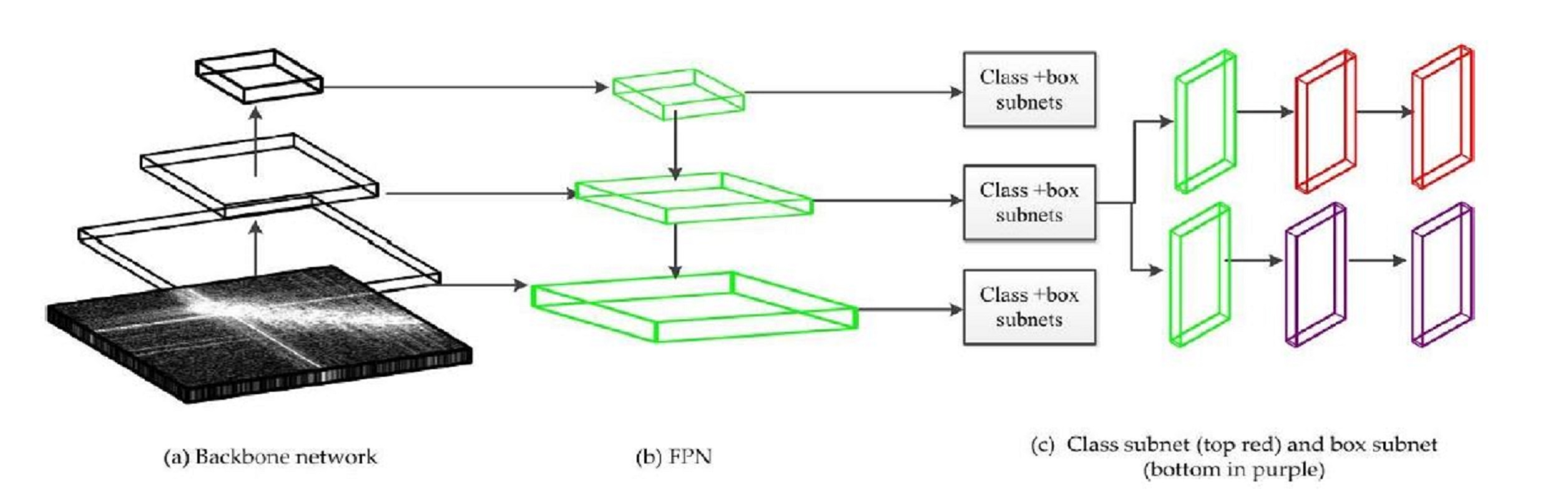
We address the challenge of localizing joints from radiographs using object detection, which exploits RetinaNet Lin et al. (2017a) a one-stage detector model which is able to accurately detect fingers/wrists from the radiographs. Object detectors are generally classified into two major categories i.e., two-stage detectors and one-stage detectors, where the former performs better in terms of accuracy in identifying the objects whereas the latter outperforms in terms of speed and simplicity. RetinaNet being a one-stage detector outperforms two-stage detectors in terms of accuracy, speed, and simplicity by utilizing a modified loss function known as focal loss Lin et al. (2017a) which tends to give higher weightage to hard examples and give a lower weight to easily classified examples, focal loss also reduces the contribution from easy examples and increases the importance of correcting misclassified examples. The architecture of RetinaNet constitutes of three major components, i.e., a backbone network for deep feature extraction, Feature Pyramid Network (FPN) to extract multi-scale features, and two subnetworks, i.e., one which predicts the class of the object and the second which predicts the coordinates of the bounding boxes in the input images. As shown In Fig. 1. RetinaNet employs backbone networks, such as residual networks (ResNet) He et al. (2016), convolutional neural networks named by Visual Geometry Group (VGG) Simonyan and Zisserman (2014), or densely connected convolutional networks (DenseNet) Huang et al. (2017) to extract higher semantic feature maps and then applies FPNs Lin et al. (2017b) to extract the multi-scale features from the backbone network, it adopts a pyramidal top-down architecture with lateral connections to capture the high-level feature maps at all scales and helps the model to detect objects of smaller size Lin et al. (2017b). FPN extracts the features from the images at different scales and merges them with the features of consecutive layers of the backbone network, which effectively propagate these at different levels and scales to the succeeding layer Li and Ren (2019). The pyramidal features are then sent to the classification and regression subnets to classify and locate the desired objects.
3.2 Convolution with Attention
Convolutional Neural Network (CNN) has been recently employed to solve problems from both the computer vision and medical image analysis domains. Various research groups have developed innovative CNN architectures such as DenseNets Huang et al. (2017), ResNets He et al. (2016), VGG Simonyan and Zisserman (2014) which tend to be computationally expensive, Inception network utilizes a unique architecture which lowers the computational cost compared to the previous CNN architectures and achieves state-of-the-art performance in the ImageNet Large-Scale Visual Recognition Challenge 2014 (ILSVRC14) Szegedy et al. (2015). In recent years, many attempts has been made to improve CNN performance by expanding them in different dimensions i.e width (W), depth (D) and resolution (R) with limited computational budget, although it is possible to scale two or three dimensions arbitrarily, this involves tedious manual tuning and generally yields substandard accuracy and efficiency. Increasing the width of the network helps the model to learn more complex features which causes a problem of vanishing gradients which has been mitigated with the use of skip connections as mentioned in He et al. (2016). Wider network corresponds to the use of a higher number of channels which aim to capture more fine-grained patterns in the images. Using images with higher resolutions, Huang et al. (2019) have also achieved state-of-the-art ImageNet accuracy. However, as we increase the W, D and R of the network, the accuracy quickly saturates after a certain point Tan and Le (2019). Inspired by these observations more recently, a family of models named EfficientNets Tan and Le (2019) has been proposed, which tend to improve the performance of CNNs by scaling them in three dimensions, i.e., W, D, and R using a set of fixed scaling coefficients that meet a specific computational constraint. The family of EfficientNet models ranges from B0-B7 where B0 is the baseline network which was developed by neural architecture search (NAS) which tends to arrive at the optimal values for W, D, and R for a CNN given a computational constraint. Rescaling the architecture of the B0 model along three dimensions, i.e., W, D, and R constitute of 7 other models denoted by B1-B7. The EfficientNet-B1 is 7.6x smaller and 5.7x faster than ResNet-152 and EfficientNet-B7 achieves new state-of-the-art 84.3% top-1 accuracy on ImageNet task but is 8.4x smaller and 6.1x faster than GPipe Huang et al. (2019) as mentioned in Tan and Le (2019). The proposed approach leveraged the EfficientNet B3-B5 models as backbones in the joint level models to grade radiographs.
3.2.1 Attention
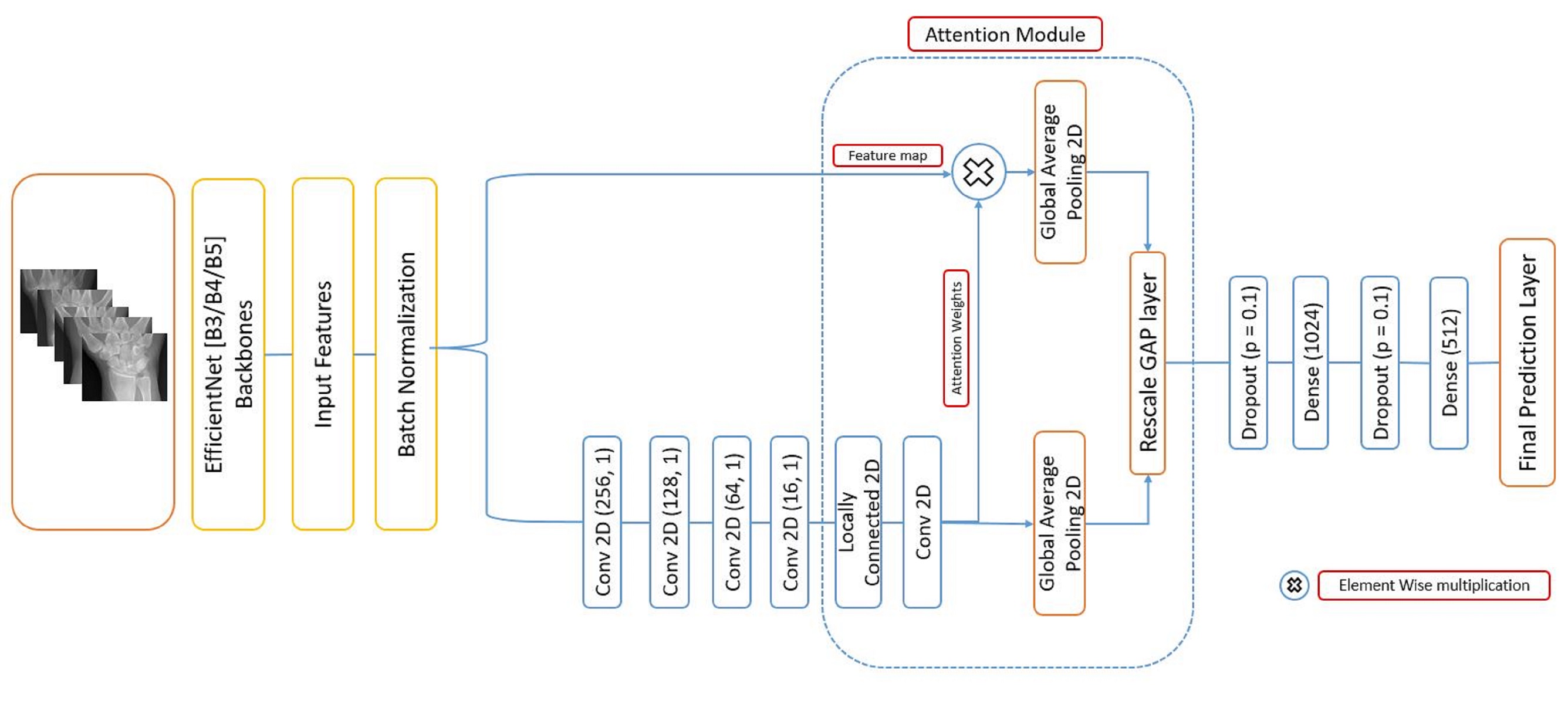
Attention mechanism has revolutionized the Natural Language Processing (NLP) domain. More recently numerous efforts have been made to incorporate attention mechanisms in CNNs such as Xu et al. (2015) where authors introduce the concept of soft and hard attention in images to generate image captions. We incorporate an attention mechanism in the EfficientNet architecture which enhances the representation learning capacity of the EfficientNet architecture. This proposed approach is inspired by Schlemper et al. (2019) in which the authors apply attention gated CNN network for object localization in a 3D CT abdominal dataset. Attention module not only helps the model to eventually learn the salient regions in the radiographs while making predictions but also adds a layer of explainability by extraction of attention weights. These weights are obtained by passing the input feature maps from the EfficientNet backbone through various convolution layers and finally utilizing a locally connected 2D layer with sigmoid activation which computes the weights at a pixel level ranging from 0-1. A element-wise multiplication of these attention weights with the feature maps helps the model to turn off the pixel values where the weights are zero. Fig. 2. depicts the final model architecture which is trained on the extracted joints from the first stage, i.e., object detection. We employ task-specific EfficientNet backbones and train an end-to-end network which predicts the joint damage from the radiographs. Code for this approach is publicly available at https://github.com/NC717/DeepRA.
4 Experimentation and Results
4.1 Dataset used
The Dataset used, for the analyses in this paper was contributed by University of Alabama at Birmingham. This dataset was obtained as part of the RA2-DREAM Challenge: Automated Scoring of Radiographic Damage in Rheumatoid Arthritis through Synapse ID [syn20545111] Bridges Jr et al. (2010); Ormseth et al. (2016). The training dataset consisted of 1,468 radiographs corresponding to 367 patients with manually annotated narrowing and erosion SvH scores at a patient level. Leaderboard performance was evaluated on hands and feet radiographs for 300 patients which along-with the final evaluation test set was not available to the participants during the course of the RA2 Dream Challenge. SvH scoring system considers joints in the hands/wrist and feet that are typical targets of inflammation in rheumatoid Arthritis (RA), which includes joints in the wrists, metacarpal phalangeal (MCP) and proximal interphalangeal (PIP) joints of the hands, and the metatarsal phalangeal (MTP) and the PIP joints in feet. The challenge was to predict narrowing and erosion scores corresponding to these joints at a patient level, which can further be used to get the overall damage in the radiographs Sun et al. (2021).
4.2 Joint localization
First stage of the proposed approach, i.e., object detection, was performed to extract the fingers and wrist from the radiographs for both hands and feet.
4.2.1 Implementation Details
We trained RetinaNet model with ResNet50 He et al. (2016) as the backbone network, which was pretrained on COCO image dataset Lin et al. (2014). All weights of the backbone network were trainable owing to the inherent differences in the data distributions in COCO dataset and the hands and feet radiographs. Fingers in hands and feet and wrist in hand were manually annotated for 914 radiographs using an open source tool labellmg222https://github.com/tzutalin/labelImg, which were finally used to train and validate two separate detector models to detect fingers/wrist from the radiographs. First model was developed to detect fingers and wrist in hand and the second model detected fingers in feet. We had 341 and 57 radiographs in the training and validation sets, respectively, to develop the hand finger/wrist detection model, whereas the feet finger detection model consisted of 355 and 61 radiographs in the training and validation sets, respectively. During training, we limit the dimensions of the input radiographs with image Min/Max dimension bounded at 1,000 and 1,400 respectively. We employ Adam Kingma and Ba (2014) optimizer with a learning rate of 0.001 which is reduced during training by a factor of 0.1 if the mean Average Precision (mAP) has not improved after 4 consecutive epochs. Batch size of 8 is used during training and to reduce the overfitting, early stopping is used if the mAP has not improved for 5 consecutive epochs on the validation set. We use Tesla K80 GPU on Google Colab to train and test the models.
4.2.2 Results
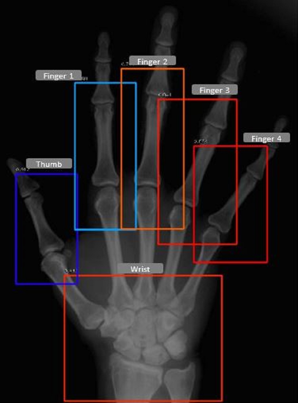
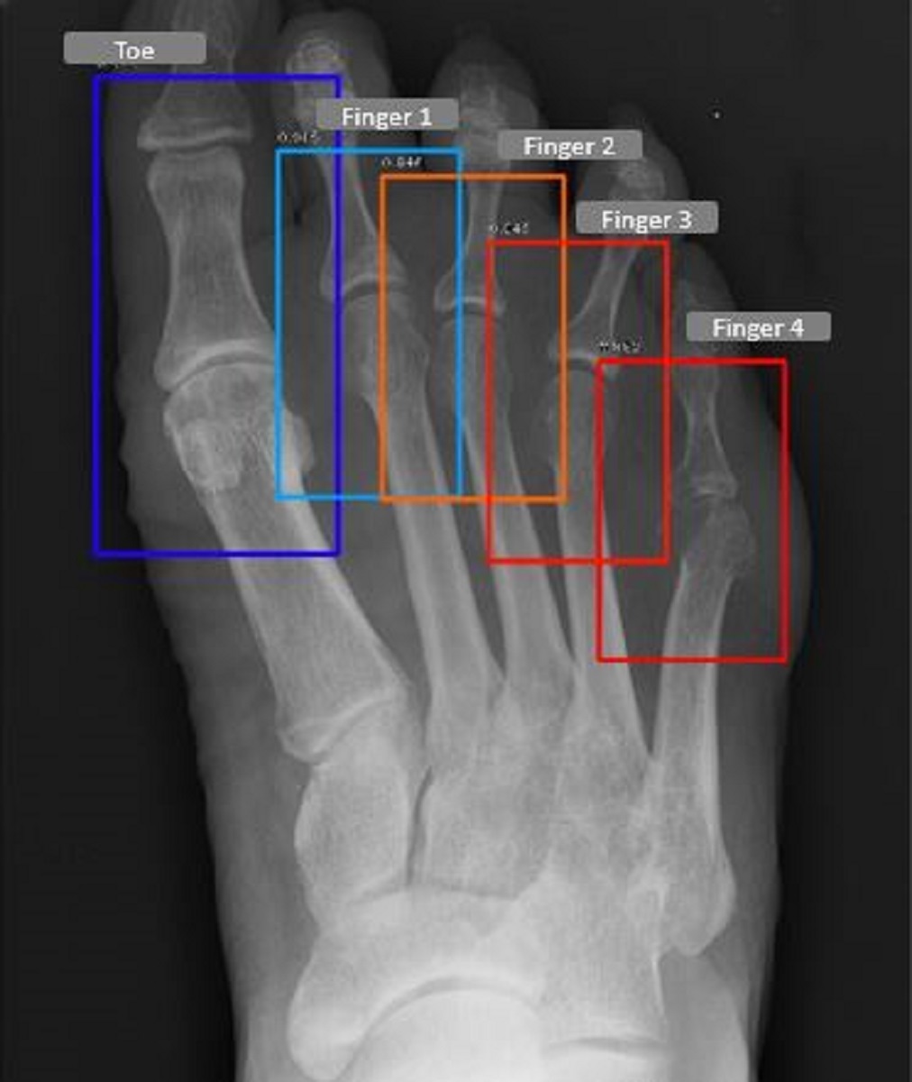
We evaluate the detector models on the test dataset which comprised of 50 Hands/Feet radiographs respectively, these radiographs were not used during the training and validation. The detector model, for the feet was able to achieve a mAP of 0.965, whereas the hand model achieved a mAP of 0.981 at an intersection over union threshold (IOU) of 0.6. In Table 1, we report the joint wise average precision (AP), we observe that the wrist joint being the easiest to detect, achieves the highest AP, whereas all other joints have a relatively similar AP in the case of both detector models. Fig. 4 represents the joints detected for a patient in the test set.
|
|
|
|||||
|---|---|---|---|---|---|---|---|
| Thumb/Toe | 0.986 | 0.983 | |||||
| Finger 1 | 0.979 | 0.973 | |||||
| Finger 2 | 0.983 | 0.955 | |||||
| Finger 3 | 0.971 | 0.947 | |||||
| Finger 4 | 0.977 | 0.965 | |||||
| Wrist | 0.993 | - | |||||
| Overall mAP | 0.981 | 0.965 |
4.3 Joint Level Models
Second stage of the proposed approach consists of training joint level models, to predict the joint narrowing and erosion SvH scores from the radiographs. We describe the implementation details and the results in the following sections.
4.3.1 Implementation Details
We train 6 joint level models which are based on the EfficientNet [B3-B4-B5] backbones pretrained on ImageNet dataset along-with the attention mechanism. Finger joints extracted from object detection stage were resized to whereas wrist joints were resized to while training. We use a 80:20 train test split to train all joint level models. During training, we use Adam optimizer Kingma and Ba (2014) and mean squared error (MSE) as the loss function with an initial learning rate of 0.001 which is decayed by a factor of 0.2 if the validation RMSE does not improve within 5 epochs. We train the models for 200 epochs and finally save the model which has the least RMSE on the validation set. To make the model robust, we employ horizontal flipping of the joint images in every joint model. During training 70% weights of the EfficientNet backbones were unfreezed since the distribution of the medical radiographs is entirely different from the ImageNet dataset which was originally used to train these models. Approximately 70% of the joints had a Narrowing/Erosion SvH score of zero in the training data which caused the issue of imbalance while training. We tackled this by training the model by randomly sampling the equivalent number of joints with non-zero Narrowing/Erosion scores from zero damage joint radiographs, so that there is no class imbalance while training the model. We use a batch-size of 32 and all the models are implemented with Keras using Tensorflow backend. All the experiments are performed on Tesla K80 GPUs on Google Colab. In Table 2, we describe the training specifications for the joint level models which describe the EfficientNet backbones utilized, the number of joint images extracted from the radiographs using the detector models, and the number of predictions to be made in every model.
| Model type |
|
|
|
|
|||||||
|---|---|---|---|---|---|---|---|---|---|---|---|
| Erosion | Hand Fingers & Thumb/Toe | B3 | 4,404 | 2 | |||||||
| Narrowing | Hand Fingers & Toe | B3 | 3,670 | 2 | |||||||
| Narrowing | Feet Fingers & Thumb | B4 | 3,670 | 1 | |||||||
| Erosion | Feet Fingers | B4 | 2,936 | 1 | |||||||
| Erosion & Narrowing | Wrist | B5 | 734 | 6 |
4.3.2 Evaluation Metrics
The RA2 Dream challenge utilized the weighted (Wt.) root mean squared error (RMSE) to evaluate the joint level narrowing and erosion SvH scores, whereas weighted (Wt.) absolute error was used to evaluate the overall RA damage at a patient level. The weights used for calculating the joint wise RMSE were not released during the RA2 Dream Challenge.
4.3.3 Results
The proposed approach was evaluated on leaderboard and the final test dataset which comprised on 1200 radiographs corresponding to 300 patients each, these datasets were not available to the participants during the challenge. The RA2 Dream baseline uses the distribution of scores in the training data to create a random prediction. In Table 3, we describe multiple experiments and the associated Weighted AE for overall RA damage prediction and Weighted RMSE for joint narrowing and erosion SvH score prediction on the Leaderboard/Final test datasets. Initial experiments were performed with a DenseNet-169 model pretrained on ImageNet dataset which is fine tuned further on hands and feet radiographs without joint detection and attention to predict the joint wise and overall SvH scores, which showed a minor improvement w.r.t baseline score, whereas when we include joint detection and attention into the same model architecture we were able to improve the model performance significantly. We further went on and incorporated various pretrained EfficientNet backbones into the joint level model architecture and observed that the EfficientNet models with joint detection and attention outperformed all the other experiments which were executed during the challenge. Our EfficientNet based joint models with Joint detection and attention achieved 4th place in predicting the overall damage and 8th place in predicting the joint Narrowing and Erosion SvH scores on the final test dataset.
| Experiments |
|
|
|
||||||
|---|---|---|---|---|---|---|---|---|---|
| Baseline | 7.023 | 1.96 | 1.728 | ||||||
|
2.979 | 1.659 | 1.487 | ||||||
|
1.465 | 1.449 | 1.394 | ||||||
|
1.371 | 1.433 | 1.397 | ||||||
|
1.456 | 1.358 | 1.404 |
4.4 Attention Map Analysis
While dealing with healthcare related problems we need to be sure of what we are predicting for a patient. In this case since we are predicting joint level narrowing and erosion SvH scores we need to have some evidence in our model predictions, otherwise it is tough to acknowledge that the predictions are accurate. Here we demonstrate that the attention mechanism we introduced within the model architecture is not only capable of boosting the model performance but also provides a layer of explanability for the model predictions. We extracted the attention weights and overlayed them onto the joint images. In Fig. 5, we demonstrate the attention maps for narrowing SvH score prediction problem for hand/feet fingers, and wrist.
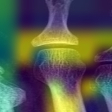
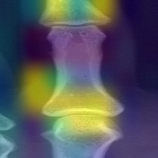
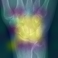
In Fig. 5(a), and 5(b), the joint region in the feet and hand fingers is activated to provide the final predictions, similarly, we see that in Fig. 5(c), all wrist joints are given a higher weightage by the model while making the predictions. Our attention map provides an inbuilt explanability component which can aid the radiologist to believe in the model predictions.
5 Conclusion
In this paper, we propose an automated method for detecting joints from radiographs and predicting the associated damage in the joints by leveraging attention based CNN framework. Due to the lack of availability of annotations for the the joints we propose an object detection model which is capable of correctly identifying and localizing the joints in the radiographs which can be leveraged for downstream tasks. Since manual grading of radiographs requires experienced radiologists, we tackle this with an attention-based CNN method which can accurately grade the radiographs and aid radiologists in better patient care and research. Not only the model is able to give better predictions but it also provides a sense of the salient regions in the joint radiographs where the model latches on to make those predictions. While the achieved performance is encouraging, further evaluation of this approach on broader dataset is required to have a more reliable estimation of the model predictions. In the future, we aim to develop joint level detector models instead of a finger level models and employ the similar model architecture which could provide a great deal of understanding while predicting the scores and assist the radiologists in making their decisions, also directly comparing the explainability of inherent attention maps and other offline explainability methods may instill confidence in the predictions.
References
- Birch and Bhattacharya [2010] James T Birch and Shelley Bhattacharya. Emerging trends in diagnosis and treatment of rheumatoid arthritis. Primary Care: Clinics in Office Practice, 37(4):779–792, 2010.
- Combe [2009] Bernard Combe. Progression in early rheumatoid arthritis. Best Practice & Research Clinical Rheumatology, 23(1):59–69, 2009.
- Van der Heijde et al. [1995] DMFM Van der Heijde, MA Van Leeuwen, PLCM Van Riel, and LBA Van de Putte. Radiographic progression on radiographs of hands and feet during the first 3 years of rheumatoid arthritis measured according to sharp’s method (van der heijde modification). 1995.
- Renshaw and Gould [2005] Andrew A Renshaw and Edwin W Gould. Comparison of disagreement and error rates for three types of interdepartmental consultations. American journal of clinical pathology, 124(6):878–882, 2005.
- RA2 [2019 (accessed January, 2021] Ra2 dream challenge. https://www.synapse.org/#!Synapse:syn20545111/wiki/597242, 2019 (accessed January, 2021).
- Fujisawa et al. [2019] Y Fujisawa, Y Otomo, Y Ogata, Y Nakamura, R Fujita, Y Ishitsuka, R Watanabe, N Okiyama, K Ohara, and M Fujimoto. Deep-learning-based, computer-aided classifier developed with a small dataset of clinical images surpasses board-certified dermatologists in skin tumour diagnosis. British Journal of Dermatology, 180(2):373–381, 2019.
- Coudray et al. [2018] Nicolas Coudray, Paolo Santiago Ocampo, Theodore Sakellaropoulos, Navneet Narula, Matija Snuderl, David Fenyö, Andre L Moreira, Narges Razavian, and Aristotelis Tsirigos. Classification and mutation prediction from non–small cell lung cancer histopathology images using deep learning. Nature medicine, 24(10):1559–1567, 2018.
- Boudrioua [2020] Mohamed Samir Boudrioua. Covid-19 detection from chest x-ray images using cnns models: Further evidence from deep transfer learning. Available at SSRN 3630150, 2020.
- Woo et al. [2018] Sanghyun Woo, Jongchan Park, Joon-Young Lee, and In So Kweon. Cbam: Convolutional block attention module. In Proceedings of the European conference on computer vision (ECCV), pages 3–19, 2018.
- Bello et al. [2019] Irwan Bello, Barret Zoph, Ashish Vaswani, Jonathon Shlens, and Quoc V Le. Attention augmented convolutional networks. In Proceedings of the IEEE International Conference on Computer Vision, pages 3286–3295, 2019.
- Chen et al. [2020] Yinpeng Chen, Xiyang Dai, Mengchen Liu, Dongdong Chen, Lu Yuan, and Zicheng Liu. Dynamic convolution: Attention over convolution kernels. In Proceedings of the IEEE/CVF Conference on Computer Vision and Pattern Recognition, pages 11030–11039, 2020.
- He et al. [2020] Along He, Tao Li, Ning Li, Kai Wang, and Huazhu Fu. Cabnet: Category attention block for imbalanced diabetic retinopathy grading. IEEE Transactions on Medical Imaging, 2020.
- Snekhalatha and Anburajan [2017] U Snekhalatha and M Anburajan. Computer-based measurements of joint space analysis using metacarpal morphometry in hand radiograph for evaluation of rheumatoid arthritis. International journal of rheumatic diseases, 20(9):1120–1131, 2017.
- Langs et al. [2008] Georg Langs, Philipp Peloschek, Horst Bischof, and Franz Kainberger. Automatic quantification of joint space narrowing and erosions in rheumatoid arthritis. IEEE transactions on medical imaging, 28(1):151–164, 2008.
- Hoving et al. [2004] Jan Lucas Hoving, Rachelle Buchbinder, Stephen Hall, Gary Lawler, Peter Coombs, Stephen McNealy, Paul Bird, and David Connell. A comparison of magnetic resonance imaging, sonography, and radiography of the hand in patients with early rheumatoid arthritis. The Journal of Rheumatology, 31(4):663–675, 2004.
- Døhn et al. [2008] Uffe Møller Døhn, Bo J Ejbjerg, Maria Hasselquist, Eva Narvestad, Jakob Møller, Henrik S Thomsen, and Mikkel Østergaard. Detection of bone erosions in rheumatoid arthritis wrist joints with magnetic resonance imaging, computed tomography and radiography. Arthritis research & therapy, 10(1):R25, 2008.
- Tiulpin et al. [2018] Aleksei Tiulpin, Jérôme Thevenot, Esa Rahtu, Petri Lehenkari, and Simo Saarakkala. Automatic knee osteoarthritis diagnosis from plain radiographs: a deep learning-based approach. Scientific reports, 8(1):1–10, 2018.
- Xue et al. [2017] Yanping Xue, Rongguo Zhang, Yufeng Deng, Kuan Chen, and Tao Jiang. A preliminary examination of the diagnostic value of deep learning in hip osteoarthritis. PLoS One, 12(6):e0178992, 2017.
- Simonyan and Zisserman [2014] Karen Simonyan and Andrew Zisserman. Very deep convolutional networks for large-scale image recognition. arXiv preprint arXiv:1409.1556, 2014.
- Rohrbach et al. [2019] Janick Rohrbach, Tobias Reinhard, Beate Sick, and Oliver Dürr. Bone erosion scoring for rheumatoid arthritis with deep convolutional neural networks. Computers & Electrical Engineering, 78:472–481, 2019.
- Hirano et al. [2019] Toru Hirano, Masayuki Nishide, Naoki Nonaka, Jun Seita, Kosuke Ebina, Kazuhiro Sakurada, and Atsushi Kumanogoh. Development and validation of a deep-learning model for scoring of radiographic finger joint destruction in rheumatoid arthritis. Rheumatology advances in practice, 3(2):rkz047, 2019.
- Lin et al. [2017a] Tsung-Yi Lin, Priya Goyal, Ross Girshick, Kaiming He, and Piotr Dollár. Focal loss for dense object detection. In Proceedings of the IEEE international conference on computer vision, pages 2980–2988, 2017a.
- He et al. [2016] Kaiming He, Xiangyu Zhang, Shaoqing Ren, and Jian Sun. Deep residual learning for image recognition. In Proceedings of the IEEE conference on computer vision and pattern recognition, pages 770–778, 2016.
- Huang et al. [2017] Gao Huang, Zhuang Liu, Laurens Van Der Maaten, and Kilian Q Weinberger. Densely connected convolutional networks. In Proceedings of the IEEE conference on computer vision and pattern recognition, pages 4700–4708, 2017.
- Lin et al. [2017b] Tsung-Yi Lin, Piotr Dollár, Ross Girshick, Kaiming He, Bharath Hariharan, and Serge Belongie. Feature pyramid networks for object detection. In Proceedings of the IEEE conference on computer vision and pattern recognition, pages 2117–2125, 2017b.
- Li and Ren [2019] Yixing Li and Fengbo Ren. Light-weight retinanet for object detection. arXiv preprint arXiv:1905.10011, 2019.
- Lin et al. [2014] Tsung-Yi Lin, Michael Maire, Serge Belongie, James Hays, Pietro Perona, Deva Ramanan, Piotr Dollár, and C Lawrence Zitnick. Microsoft coco: Common objects in context. In European conference on computer vision, pages 740–755. Springer, 2014.
- Wang et al. [2019] Yuanyuan Wang, Chao Wang, Hong Zhang, Yingbo Dong, and Sisi Wei. Automatic ship detection based on retinanet using multi-resolution gaofen-3 imagery. Remote Sensing, 11(5):531, 2019.
- Tan and Le [2019] Mingxing Tan and Quoc V Le. Efficientnet: Rethinking model scaling for convolutional neural networks. arXiv preprint arXiv:1905.11946, 2019.
- Szegedy et al. [2015] Christian Szegedy, Wei Liu, Yangqing Jia, Pierre Sermanet, Scott Reed, Dragomir Anguelov, Dumitru Erhan, Vincent Vanhoucke, and Andrew Rabinovich. Going deeper with convolutions. In Proceedings of the IEEE conference on computer vision and pattern recognition, pages 1–9, 2015.
- Huang et al. [2019] Yanping Huang, Youlong Cheng, Ankur Bapna, Orhan Firat, Dehao Chen, Mia Chen, HyoukJoong Lee, Jiquan Ngiam, Quoc V Le, Yonghui Wu, et al. Gpipe: Efficient training of giant neural networks using pipeline parallelism. In Advances in neural information processing systems, pages 103–112, 2019.
- Xu et al. [2015] Kelvin Xu, Jimmy Ba, Ryan Kiros, Kyunghyun Cho, Aaron Courville, Ruslan Salakhudinov, Rich Zemel, and Yoshua Bengio. Show, attend and tell: Neural image caption generation with visual attention. In International conference on machine learning, pages 2048–2057, 2015.
- Schlemper et al. [2019] Jo Schlemper, Ozan Oktay, Michiel Schaap, Mattias Heinrich, Bernhard Kainz, Ben Glocker, and Daniel Rueckert. Attention gated networks: Learning to leverage salient regions in medical images. Medical image analysis, 53:197–207, 2019.
- Bridges Jr et al. [2010] S Louis Bridges Jr, Zenoria L Causey, Paula I Burgos, B Quynh N Huynh, Laura B Hughes, Maria I Danila, Amalia Van Everdingen, Stephanie Ledbetter, Doyt L Conn, Ashutosh Tamhane, et al. Radiographic severity of rheumatoid arthritis in african americans: results from a multicenter observational study. Arthritis care & research, 62(5):624–631, 2010.
- Ormseth et al. [2016] Michelle J Ormseth, Patricia G Yancey, Joseph F Solus, S Louis Bridges Jr, Jeffrey R Curtis, MacRae F Linton, Sergio Fazio, Sean S Davies, L Jackson Roberts, Kasey C Vickers, et al. Effect of drug therapy on net cholesterol efflux capacity of high-density lipoprotein–enriched serum in rheumatoid arthritis. Arthritis & rheumatology, 68(9):2099–2105, 2016.
- Sun et al. [2021] Dongmei Sun, Thanh M. Nguyen, Robert J. Allaway, Jelai Wang, Verena Chung, Thomas V Yu, Michael Mason, Isaac Dimitrovsky, Lars Ericson, Hongyang Li, Yuanfang Guan, Ariel Israel, Alex Olar, Balint Armin Pataki, RA2 DREAM Challenge Community, Gustavo Stolovitzky, Justin Guinney, Percio S. Gulko, Mason B. Frazier, James C. Costello, Jake Y. Chen, and S. Louis Bridges. A crowdsourcing approach to develop machine learning models to quantify radiographic joint damage in rheumatoid arthritis. medRxiv, 2021. doi: 10.1101/2021.10.25.21265495. URL https://www.medrxiv.org/content/early/2021/10/26/2021.10.25.21265495.
- Kingma and Ba [2014] Diederik P Kingma and Jimmy Ba. Adam: A method for stochastic optimization. arXiv preprint arXiv:1412.6980, 2014.