Analyzing Host-Viral Interactome of SARS-CoV-2 for Identifying Vulnerable Host Proteins during COVID-19 Pathogenesis
Abstract
The development of therapeutic targets for COVID-19 treatment is based on the understanding of the molecular mechanism of pathogenesis. The identification of genes and proteins involved in the infection mechanism is the key to shed out light into the complex molecular mechanisms. The combined effort of many laboratories distributed throughout the world has produced the accumulation of both protein and genetic interactions. In this work we integrate these available results and we obtain an host protein-protein interaction network composed by 1432 human proteins. We calculate network centrality measures to identify key proteins. Then we perform functional enrichment of central proteins. We observed that the identified proteins are mostly associated with several crucial pathways, including cellular process, signalling transduction, neurodegenerative disease. Finally, we focused on proteins involved in causing disease in the human respiratory tract. We conclude that COVID-19 is a complex disease, and we highlighted many potential therapeutic targets including RBX1, HSPA5, ITCH, RAB7A, RAB5A, RAB8A, PSMC5, CAPZB, CANX, IGF2R, HSPA1A, which are central and also associated with multiple diseases.
keywords:
SARS-CoV-2 , COVID-19 , Protein-protein interaction , Centrality , Pathways , Disease1 Introduction
The world is experiencing an unprecedented pandemic due to a massive outbreak of Severe Acute Respiratory Syndrome Corona Virus 2 (SARS-CoV-2 ) infected viral disease, COVID-19. SARS-CoV-2 , is a large enveloped coronavirus (family-Coronaviridae, subfamily-Coronavirinae) with non-segmented, single-stranded, and positive-sense RNA genomes [1], transmits rapidly through human to human contacts. Although SARS-CoV-2 is similar to other known coronaviruses, i.e. SARS-CoV and MERS-CoV [2, 3], it has demonstrated high rates of infection [4, 5]. Therefore there is the need to understand the disease pathogenesis of SAR-CoV-2 to develop effective therapies and vaccines.
The SARS-CoV-2 virus is responsible COVID-19 disease that causes damages in multiple organs as the disease progresses from an asymptomatic phase to a life-threatening disease [6]. Therefore, accurate molecular diagnosis of COVID-19 disease is essential by collecting the proper respiratory tract specimen [7]. In this context, the integrated analysis [8] of various data-sets, including clinical and imaging data, may explain, and hopefully predict, the longitudinal effects of SARS-CoV-2 infection [9, 10]. In particular, many independent projects throughout the world have focused on genomics and proteomics level [10], and then they integrated these data with clinical ones. These works have produced data about the infection’s effect at a molecular scale, evidencing genes and proteins’ role, such as the interactions among viral and human proteins. Interactions between a host and its pathogen, are primarily driven by interactions among the host proteins and pathogen proteins; also referred to as host-pathogen protein-protein interaction (PPI) network. The SARS-CoV-2 virus-host interactome have been studied focusing various virulence factors influencing SARS-CoV-2 pathogenesis and interacting mechanism [11, 12, 13, 14, 15, 16]. Further, many recent works also used host-viral protein-protein interaction network as an input to elucidate potential drug targets or repurposed drug molecules [17, 18, 19]. Host-pathogen protein interactions provide important insights into the molecular mechanisms of pathogenecity [20] and for understanding virulence factors influencing SARS-CoV-2 pathogenesis [21, 22]. SARS-CoV-2 is a newly found virus whose interacting human host proteins play a major disease progression role that needs to be investigated.
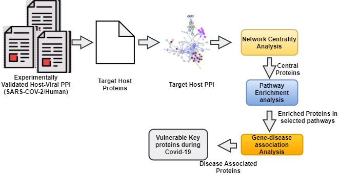
Protein-Protein Interactions (PPI) are usually modelled and analysed with graph theory [23]. In this formalism, the interactions are modelled as a graph whose nodes are proteins (or genes), and the edges are the interaction among them. Several studies have found that specific candidate proteins might play a crucial role [24, 25, 26, 27]. Protein-protein interaction networks are an essential ingredient for any systems-level understanding of cellular processes and modelling, and even drug discovery [28, 29, 30, 31, 32]. The key genes/proteins involved in the different biological pathways can give valuable insight for in-depth characterisation of disease progression [33, 34, 35]. It is well accepted that all the viruses have evolved to target proteins that are central and have strong control over the human interactome [36, 37, 38, 39, 40]. Exploring the predicted interaction networks can suggest new directions for future experimental research and provide cross-species predictions for efficient interaction mapping [41, 34]. The complete workflow of the current study can be seen from Figure 1.
This study aims to identify essential human host proteins based on topology analysis of the protein-protein interaction network of SARS-CoV-2 interacting human host proteins. We performed functional enrichment of the identified proteins to shed out light on cellular, signalling, and disease pathways.
2 Materials and Method
2.1 Dataset: Curated SARS-CoV-2 interacting human host proteins
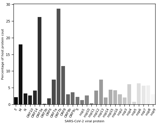
We use recently reported host proteins that are physically verified using Affinity purification mass spectrometry for their interactions with SARS-CoV-2 [17, 21, 42]. The used host-viral protein interactions are also available in BioGRID [43]. A total of 2489 host-viral interactions (consisting of 1432 unique host proteins interacting with 37 SARS-CoV-2 viral proteins) are obtained. In Figure 2, we provided the number of interacting host protein count. It is noted that the majority of the host proteins are targeted to the specific viral protein.
2.2 Construction of Host PPI network
Starting with the human proteins that are interacting with the virus, we build a host PPI by querying the Search Tool for the Retrieval of Interacting Genes/Proteins (STRING, Version 10.0; http://string-db.org/) [44].
The topology analysis of the PPI network is performed by using Cytoscape (http://apps.cytoscape.org), a general platform for complex network analysis and visualization [45].
2.3 Centrality analysis of host PPI network
In network analysis, indicators of centrality identify the most critical nodes in the network [46]. The centrality measure uses to characterise each node and edge in the PPI network. The degree measure is the most intuitive for topology analysis of the PPI network. Several other crucial factors that can influence network links are betweenness centrality, closeness centrality, clustering coefficient, topological coefficient, and neighbourhood connectivity.
-
(i)
Degree centrality: The degree centrality (simply degree) of a node in a network is defined as (), which indicates number of directly connected nodes to . The densely connected nodes in PPI network is considered hub nodes [47].
-
(ii)
Betweenness centrality: Betweenness centrality quantifies the number of times a node acts as a bridge along the shortest path between two other nodes [48]. The betweenness centrality of a node is represented as:
(1) where is the total number of shortest paths from node to node and is the number of those paths that pass through .
-
(iii)
Closeness centrality: Closeness centrality is a way of detecting nodes that are able to spread information very efficiently through the network [49]. It can be calculated as :
(2) where is the length of shortest path between node and , and denotes any other nodes that are reachable to node .
-
(iv)
Average shortest-path length: Shortest-path length between two nodes (say and ) in network topology is defined as the number of minimum steps that required to traverse between node and ![50]. The average shortest path length () of node is the average value of all pair of nodes shortest path from the node .
-
(v)
Clustering coefficient: Clustering coefficient is a measure of the degree to which nodes in a graph tend to cluster together [51]. In undirected networks, the clustering coefficient of a node is defined as:
(3) where is the number of neighbors of and is the number of connected pairs between all neighbors of .
-
(vi)
Topological coefficient: Topological coefficient is a relative measure for the extent to which a node shares neighbors with other nodes [52]. The topological coefficient of a node with neighbors is computed as follows:
(4) Where is defined for all nodes that share at least one neighbour with , and the value is the number of neighbours shared between the nodes and , plus one if there is a direct link between and .
-
(vii)
Neighborhood connectivity: Neighborhood connectivity () of a node is defined as the average connectivity of all neighbors of [53]. The neighborhood connectivity distribution gives the average of the neighborhood connectivities of all nodes with neighbors for .
We used NetworkAnalyzer [45] to calculate above centrality score. In NetworkAnalyzer, (Closeness centrality) is calculated as the reciprocal of the average shortest path length. So, high means highly central, and thus low .
2.4 Gene ontology and pathway enrichment analysis
We performed enrichment analysis to find out set of significantly enriched genes/proteins in different functional and biological pathways. We used KEGG (Kyoto Encyclopedia of Genes and Genomes) [54] for elucidating pathway enrichment of a host protein and Gene Ontology (GO) for the assessment of protein functions [55]. KEGG is a database resource for understanding high-level functions and utilities of the biological system [56].
2.5 Gene-disease association network
Complex diseases are caused by a group of genes known as disease genes. More often, a gene can participate in various disease conditions [57, 58]. It helps unravel the disease pathogenesis, which in turn help disease diagnosis, treatment, and disease prevention. We obtained gene-disease association network from DisGeNET (v7.0) database (https://www.disgenet.org/), which contains 1,134,942 gene-disease associations (GDAs), between 21,671 genes and 30,170 disease [59]. From this database, we considered curated gene-disease associations only.
3 Results and Discussion
Here, we report the outcomes of intermediate steps to reach to our objective of isolating key host proteins followed by their significance analysis.
3.1 Deriving PPI network for candidate host proteins
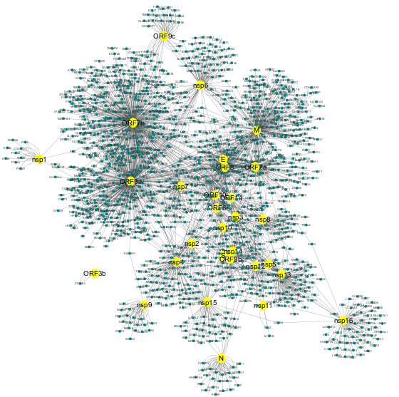
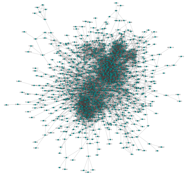
Our candidate host proteins list, collected from the reported host-viral networks (Section 2), consists of total of 1432 distinct proteins that are targeted by SARS-CoV-2 during COVID-19 . We rebuilt the PPI network centered around our candidate proteins using STRING DB. There are 7076 edges in the derived PPI network. We curated derived PPI by keeping only the interactions whose confidence scores are at least (high confidence). The derived PPI network is then analysed using Cytoscape. We identified the big connected component (also called gain/main component) of the PPI network. After discarding all disconnected components in the PPI network, we considered gain component of PPI network with 1111 nodes (Approx. ) and 7043 edges (Figure 3).
3.2 Network topology analysis of gain component
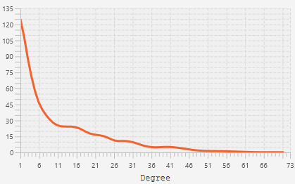
We performed topological analysis of the gain component using NetworkAnalyzer [45]. The degree distribution of all the candidate proteins in the gain component showed that the majority of the proteins in the gain component exhibit a higher degree of connectivity (Figure 4). Few proteins with degree (shown within parentheses) more than 50 are CDK1(73), PPP2R1A(65), NOP56(60), POLR2B(60), RAB1A(59), RBX1(58), SKIV2L2(57), NAPA(57), RPS14(56), STX5(54), TGOLN2(54), TCEB1(53), DCTN2(53), TCEB2(52), HSPA9(51), GNB2L1(50).
The histogram analysis of all the centrality measures (discussed in section 2.3) showing non-random distribution (Figure S1). We performed correlation (Pearson) analysis among all centrality scores (Table 1). The correlation score between degree centrality () scores and closeness centrality () scores observed to be the highest () in comparison to other measures. Although, we observed correlation between and neighbourhood centrality () is the third-highest (), but and showed less correlative (). Overall, we observed correlation score of three centrality measures () are quite closer. Therefore, we selected them in subsequent analysis. We identified 373 proteins in these criteria, which are considered highly central proteins (above the median score for all three selected parameters). When we considered all measures, we find only six common proteins (GEMIN4, DDX20, GOLGA3, FKBP15, PMPCA, AK4) above the median score in each category of centrality measurement, and that is the reason why we selected three centrality measures for our downstream analysis.
| 0.603 | 1 | ||||
| 0.209 | -0.168 | 1 | |||
| -0.32 | -0.283 | 0.451 | 1 | ||
| 0.557 | 0.1101 | 0.346 | -0.139 | 1 | |
| 0.759 | 0.5146 | 0.213 | -0.329 | 0.684 |
3.3 Pathway enrichment analysis of highly central proteins
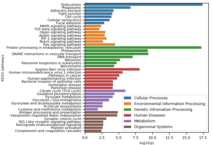
We performed KEGG pathway analysis of selected 373 highly central proteins. We obtained a total of 84 enriched KEGG pathways within the significant level (). The enriched pathways were involved in Cellular Processes (9), Environmental Information Processing (9), Genetic Information Processing (13), Human Disease (31), Organismal Systems (15), Metabolism (7). The top seven pathways in each category are shown in Figure 5.
Our current study mainly focused on the proteins that are involved in cellular process, signalling transduction, and human disease (viral and neurodegenerative) pathways, the most affected pathways in the context of COVID-19 disease [60, 61, 62, 63]. There are nine enriched pathways in cellular process (Endocytosis, Phagosome, Adherens junction, Tight junction, Cell cycle, Cellular senescence, Focal adhesion, Regulation of actin cytoskeleton, Lysosome), nine pathways in Environmental Information Processing-signalling transduction (Ras signalling pathway, HIF-1 signalling pathway, Hippo signalling pathway, Apelin signalling pathway, MAPK signalling pathway, TGF-beta signalling pathway, AMPK signalling pathway, NF-kappa B signalling pathway), nine pathways from human disease viral sub-category (Human immunodeficiency virus 1 infection, Human papillomavirus infection, Human cytomegalovirus infection, Hepatitis B, Human T-cell leukaemia virus 1 infection, Influenza A, Hepatitis C, Measles) and four pathways from neurodegenerative disease with sub-category (Huntington disease, Parkinson disease, Alzheimer disease, Prion diseases). A total of 141 distinct proteins (out of 373) were obtained from these pathways, which are then ranked based on presence in selected enriched pathways, and we found that 79 proteins are associated in our candidate pathways. All these proteins were then further studied for disease-gene association in the next.
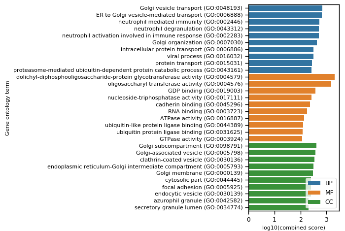
We also performed gene set enrichment analysis (Gene ontology). It is observed that out of selected genes mostly involved in Biological process (Supplementary-B). The top ten terms in each category of gene ontology (BP, MF, CC) are shown in Figure 6 that includes neutrophil mediated immunity (GO:0002446), neutrophil activation involved in immune response (GO:0002283) and viral process (GO:0016032) from BP category; dolichyl-diphosphooligosaccharide-protein glycotransferase activity (GO:0004579), GDP binding (GO:0019003), cadherin binding (GO:0045296) and ATPase activity (GO:0016887) from MF category; and focal adhesion (GO:0005925) from CC category.
3.4 Analysis of Disease-gene associations
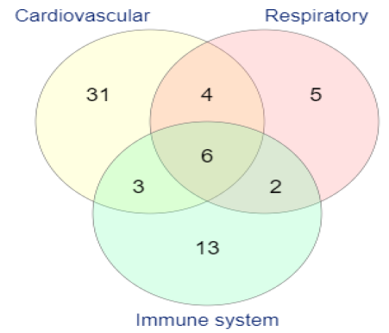
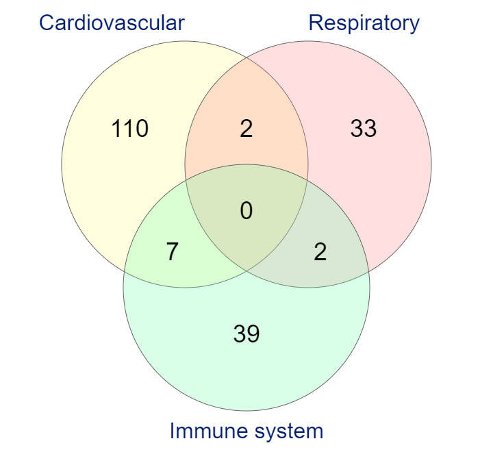
The identified 141 genes involved in four significant pathways (cellular process, signalling transduction, viral and neurodegenerative) are further screened by looking into their association with COVID-19 related disease. We particularly focused on three highly influential diseases during COVID-19 , namely cardiovascular, respiratory tract [64, 65, 66, 67] and immune system disease [68, 69]. To obtain disease-gene association, we used DisGeNET database [59] and selected source only. We found a total of 64 proteins (out of 141) playing roles in various diseases such as Asthma, Pneumonitis, Pneumonia, Influenza, Lung diseases, Cardiomyopathies, Coronary, Arteriosclerosis, Coronary Artery Disease, Heart failure, HIV Infections etc.(Supplementary-C). We compared proteins involved in all three disease categories and individual disease in each category (Figure 7). A total of 119, 37, and 48 unique diseases, and 44, 17, and 24 distinct proteins are associated with the Cardiovascular, Respiratory, and Immune system disease category, respectively. Interestingly, we found a few proteins that are associated with all three disease categories ( AREG, CAV1, IFIH1, PARP1, PLAU, TGFB1, ATM, B2M, DDX58, ENO1, HSPA5, PRKDC, STAT6, TGFBR1, TGFBR2). The top few proteins with ten or more disease associations are PLAU(59), TGFB1(29), CAV1(17), PARP1(17), TGFBR2(13), ATP2A2(11), AREG(10), FASN(10), IFIH1(10), ITGB1(10). The list of all 64 proteins and their associated quantitative parameters (degree, disease count (out of 204), disease type count (out of 3), and pathway count(out of 31) are presented in Table 2.
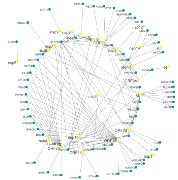
3.5 Viral proteins targeting key proteins
We then looked into source network (Figure 2) to identify the viral proteins that are targeting our selected 65 disease associated proteins. We found 25 SARS-CoV-2 proteins that are interacting with 65 proteins. Among 25 SARS-CoV-2 viral proteins, eight are accessory proteins (Orf3a, Orf7b, Orf6, Orf7a, Orf7b, Orf8, Orf9b, Orf10), four structural proteins (E,M,N,S) and thirteen non-structural poly-proteins (nsp1, nsp10, nsp12, nsp13, nsp14, nsp2, nsp3, nsp4, nsp5, nsp6, nsp7, nsp8, nsp9). It is observed that several host proteins are interacting with single viral protein. Very few host proteins are interacting with more than one viral proteins. The viral protein Orf7b exhibits the maximum number of target host proteins followed by Orf3a and M protein. Further, five host proteins are found to be common both in Orf3a and Orf7b.
We look further for any other viruses that are targeting our 65 host proteins. We mine VirusMINT [70], a virus-host association database, to find the other related viral diseases. We found that the majority of the highlighted host proteins are also targeted by Hepatitis C virus genotype 1b, Poliovirus Type 1, Human herpesvirus 1, Human papillomavirus type 16 & 31, Simian virus 40, Sendai virus, Human adenovirus 5 & 12, Epstein-Barr virus, Human SARS coronavirus Bovine papillomavirus type 1, and Epstein-Barr virus (Table 2). These proteins might be highly essential and need to put uttermost importance on developing host-directed antiviral therapies for COVID-19 .
| Gene | Degree | #Pathway | Disease category | #Disease count | Known target virus |
|---|---|---|---|---|---|
| ADAM17 | 10 | 2 | Immune | 1 | |
| ALDOA | 26 | 1 | Cardiovascular | 1 | |
| AP3B1 | 17 | 1 | Respiratory | 2 | |
| AREG | 20 | 2 | Respiratory, Cardiovascular, Immune | 10 | |
| ATM | 29 | 6 | Cardiovascular, Immune | 8 | |
| ATP2A2 | 10 | 1 | Cardiovascular | 11 | |
| ATP6 | 19 | 5 | Cardiovascular | 1 | Human SARS coronavirus, Bovine papillomavirus type 1, Human papillomavirus type 16 |
| ATR | 17 | 5 | Respiratory | 2 | Human adenovirus 5 |
| B2M | 25 | 4 | Cardiovascular, Immune | 4 | Hepatitis C virus genotype 1b (isolate Con1) |
| CANX | 31 | 2 | Cardiovascular | 1 | |
| CAPZB | 34 | 1 | Cardiovascular | 1 | |
| CAV1 | 22 | 2 | Respiratory, Cardiovascular, Immune | 17 | Poliovirus type 1 (strain Sabin) |
| CD44 | 19 | 1 | Immune | 1 | |
| COX2 | 9 | 3 | Cardiovascular | 1 | |
| CRKL | 11 | 5 | Cardiovascular | 4 | |
| DDX58 | 10 | 6 | Respiratory, Cardiovascular | 2 | |
| ENO1 | 13 | 1 | Cardiovascular, Immune | 2 | |
| EPHA2 | 9 | 2 | Cardiovascular | 5 | |
| FASN | 9 | 1 | Cardiovascular | 10 | |
| GAPDH | 23 | 2 | Cardiovascular | 1 | Hepatitis C virus genotype 1b (isolate Con1), Epstein-Barr virus (strain GD1) |
| GLA | 16 | 1 | Cardiovascular | 5 | |
| GNAQ | 14 | 5 | Cardiovascular | 1 | |
| GUSB | 16 | 1 | Immune | 1 | |
| HDAC2 | 12 | 5 | Respiratory | 2 | Human herpesvirus 1 (strain 17), Human papillomavirus type 16, Human papillomavirus type 31 |
| HLA-A | 15 | 8 | Immune | 5 | Epstein-Barr virus (strain GD1), Human papillomavirus type 16 |
| HLA-C | 14 | 8 | Immune | 7 | |
| HMGCR | 11 | 1 | Immune | 4 | |
| HSPA1A | 30 | 5 | Cardiovascular | 1 | Epstein-Barr virus (strain GD1) |
| HSPA5 | 46 | 1 | Respiratory, Cardiovascular | 3 | Epstein-Barr virus (strain GD1) |
| IFIH1 | 10 | 3 | Respiratory, Cardiovascular, Immune | 10 | Sendai virus (strain Fushimi) |
| IGF2R | 31 | 2 | Respiratory | 1 | |
| ITCH | 46 | 1 | Immune | 1 | Epstein-Barr virus (strain B95-8) |
| ITGA6 | 10 | 3 | Immune | 1 | |
| ITGB1 | 29 | 5 | Cardiovascular | 10 | Hepatitis C virus genotype 1b (isolate Con1) |
| JAK2 | 22 | 2 | Cardiovascular | 6 | |
| LDHA | 14 | 1 | Cardiovascular | 4 | |
| LDLR | 20 | 2 | Cardiovascular | 4 | |
| MET | 18 | 4 | Respiratory | 1 | |
| NDUFS2 | 20 | 3 | Cardiovascular | 3 | |
| NF1 | 16 | 2 | Cardiovascular | 3 | |
| NOTCH1 | 23 | 3 | Cardiovascular | 4 | Hepatitis C virus genotype 1b (isolate Con1) |
| NOTCH2 | 11 | 2 | Cardiovascular | 1 | |
| NOTCH3 | 15 | 3 | Cardiovascular | 2 | |
| PARP1 | 14 | 1 | Respiratory, Cardiovascular, Immune | 17 | Human herpesvirus 1 (strain 17) |
| PCNA | 25 | 3 | Immune | 1 | Human herpesvirus 1 (strain 17) |
| PDIA3 | 14 | 3 | Cardiovascular | 1 | |
| PLAU | 24 | 1 | Respiratory, Cardiovascular, Immune | 59 | |
| PPP1CB | 14 | 4 | Cardiovascular | 2 | |
| PRKDC | 15 | 1 | Respiratory, Immune | 3 | Human herpesvirus 1 (strain 17) |
| PSMC5 | 34 | 1 | Immune | 7 | Human adenovirus 5, Human adenovirus 12, Simian virus 40 |
| PSMD6 | 24 | 1 | Immune | 2 | |
| PTPN11 | 22 | 1 | Cardiovascular | 6 | |
| RAB5A | 40 | 3 | Cardiovascular | 1 | |
| RAB7A | 41 | 2 | Cardiovascular | 1 | |
| RAB8A | 40 | 3 | Immune | 1 | |
| RBX1 | 58 | 4 | Immune | 5 | |
| SERPINE1 | 16 | 4 | Cardiovascular | 8 | |
| SLC9A1 | 12 | 2 | Cardiovascular | 7 | |
| SORT1 | 14 | 1 | Cardiovascular | 3 | |
| STAT6 | 11 | 1 | Respiratory, Immune | 4 | |
| TGFB1 | 29 | 7 | Respiratory, Cardiovascular, Immune | 29 | Hepatitis C virus genotype 1b (isolate Con1) |
| TGFBR1 | 17 | 9 | Respiratory, Cardiovascular | 6 | |
| TGFBR2 | 16 | 8 | Respiratory, Cardiovascular | 13 | |
| XPO1 | 25 | 2 | Cardiovascular | 2 |
4 Conclusion
In this study, we have analysed human host protein-protein interaction network during the SARS-CoV-2 infection. We identified a set of proteins, including RBX1, HSPA5, ITCH, RAB7A, RAB5A, RAB8A, PSMC5, CAPZB, CANX, IGF2R, HSPA1A, which might influence the whole PPI network. These proteins were enriched for the following processes: cellular process, signalling, and neurodegenerative disease pathways as these pathways are known to be highly infectious for disease pathogenesis during COVID-19 . Finally, we have found 64 potential/key SARS-CoV-2 interacting human host proteins connected with respiratory, cardiovascular, and immune system disease. Many of them are known to target different other viruses and may be highly important for therapeutics treatment of COVID-19 viral disease. We strongly believe that the highlighted key proteins are an extremely promising target, which might play a crucial role during COVID-19 disease progression.
References
- Wrapp et al. [2020] D. Wrapp, N. Wang, K. S. Corbett, J. A. Goldsmith, C.-L. Hsieh, O. Abiona, B. S. Graham, J. S. McLellan, Cryo-em structure of the 2019-ncov spike in the prefusion conformation, Science 367 (2020) 1260–1263.
- Perlman and Netland [2009] S. Perlman, J. Netland, Coronaviruses post-sars: update on replication and pathogenesis, Nature reviews microbiology 7 (2009) 439–450.
- de Groot et al. [2013] R. J. de Groot, S. C. Baker, R. S. Baric, C. S. Brown, C. Drosten, L. Enjuanes, R. A. Fouchier, M. Galiano, A. E. Gorbalenya, Z. A. Memish, et al., Commentary: Middle east respiratory syndrome coronavirus (mers-cov): announcement of the coronavirus study group, Journal of virology 87 (2013) 7790–7792.
- Liu et al. [2020] Y. Liu, A. A. Gayle, A. Wilder-Smith, J. Rocklöv, The reproductive number of covid-19 is higher compared to sars coronavirus, Journal of travel medicine (2020).
- Surveillances [2020] V. Surveillances, The epidemiological characteristics of an outbreak of 2019 novel coronavirus diseases (covid-19)—china, 2020, China CDC Weekly 2 (2020) 113–122.
- Servick [2020] K. Servick, For survivors of severe covid-19, beating the virus is just the beginning, Science (2020).
- Whetton et al. [2020] A. D. Whetton, G. W. Preston, S. Abubeker, N. Geifman, Proteomics and informatics for understanding phases and identifying biomarkers in covid-19 disease, Journal of proteome research 19 (2020) 4219–4232.
- Antonelli et al. [2019] L. Antonelli, M. R. Guarracino, L. Maddalena, M. Sangiovanni, Integrating imaging and omics data: A review, Biomedical Signal Processing and Control 52 (2019) 264–280.
- Tang et al. [2020] Y.-W. Tang, J. E. Schmitz, D. H. Persing, C. W. Stratton, Laboratory diagnosis of covid-19: current issues and challenges, Journal of clinical microbiology 58 (2020).
- Das et al. [2020] J. K. Das, G. Tradigo, P. Veltri, P. H. Guzzi, S. Roy, Data science in unveiling covid-19 pathogenesis and diagnosis: Evolutionary origin to drug repurposing, Briefings in Bioinformatics (2020). URL: https://doi.org/10.1093/bib/bbaa420. doi:10.1093/bib/bbaa420.
- Guzzi et al. [2020] P. H. Guzzi, D. Mercatelli, C. Ceraolo, F. M. Giorgi, Master regulator analysis of the sars-cov-2/human interactome, Journal of clinical medicine 9 (2020) 982.
- Hoffmann et al. [2020] H.-H. Hoffmann, F. J. Sánchez-Rivera, W. M. Schneider, J. M. Luna, Y. M. Soto-Feliciano, A. W. Ashbrook, J. Le Pen, A. A. Leal, I. Ricardo-Lax, E. Michailidis, et al., Functional interrogation of a sars-cov-2 host protein interactome identifies unique and shared coronavirus host factors, Cell Host & Microbe (2020).
- Messina et al. [2020] F. Messina, E. Giombini, C. Agrati, F. Vairo, T. A. Bartoli, S. Al Moghazi, M. Piacentini, F. Locatelli, G. Kobinger, M. Maeurer, et al., Covid-19: viral–host interactome analyzed by network based-approach model to study pathogenesis of sars-cov-2 infection, Journal of Translational Medicine 18 (2020) 1–10.
- Li et al. [2020] J. Li, M. Guo, X. Tian, X. Wang, X. Yang, P. Wu, C. Liu, Z. Xiao, Y. Qu, Y. Yin, et al., Virus-host interactome and proteomic survey reveal potential virulence factors influencing sars-cov-2 pathogenesis, Med (2020).
- Cannataro et al. [2010] M. Cannataro, P. H. Guzzi, P. Veltri, Impreco: Distributed prediction of protein complexes, Future Generation Computer Systems 26 (2010) 434–440.
- Agapito et al. [2013] G. Agapito, M. Cannataro, P. H. Guzzi, F. Marozzo, D. Talia, P. Trunfio, Cloud4snp: distributed analysis of snp microarray data on the cloud, in: Proceedings of the International Conference on Bioinformatics, Computational Biology and Biomedical Informatics, 2013, pp. 468–475.
- Gordon et al. [2020] D. E. Gordon, G. M. Jang, M. Bouhaddou, J. Xu, K. Obernier, K. M. White, M. J. O’Meara, V. V. Rezelj, J. Z. Guo, D. L. Swaney, et al., A sars-cov-2 protein interaction map reveals targets for drug repurposing, Nature (2020) 1–13.
- Zhou et al. [2020] Y. Zhou, Y. Hou, J. Shen, Y. Huang, W. Martin, F. Cheng, Network-based drug repurposing for novel coronavirus 2019-ncov/sars-cov-2, Cell discovery 6 (2020) 1–18.
- Beck et al. [2020] B. R. Beck, B. Shin, Y. Choi, S. Park, K. Kang, Predicting commercially available antiviral drugs that may act on the novel coronavirus (sars-cov-2) through a drug-target interaction deep learning model, Computational and structural biotechnology journal (2020).
- Memišević et al. [2015] V. Memišević, N. Zavaljevski, S. V. Rajagopala, K. Kwon, R. Pieper, D. DeShazer, J. Reifman, A. Wallqvist, Mining host-pathogen protein interactions to characterize burkholderia mallei infectivity mechanisms, PLoS Comput Biol 11 (2015) e1004088.
- Liang et al. [2020] Q. Liang, J. Li, M. Guo, X. Tian, C. Liu, X. Wang, X. Yang, P. Wu, Z. Xiao, Y. Qu, et al., Virus-host interactome and proteomic survey of pmbcs from covid-19 patients reveal potential virulence factors influencing sars-cov-2 pathogenesis, bioRxiv (2020).
- Thiel et al. [2003] V. Thiel, K. A. Ivanov, A. Putics, T. Hertzig, B. Schelle, S. Bayer, B. Weißbrich, E. J. Snijder, H. Rabenau, H. W. Doerr, et al., Mechanisms and enzymes involved in sars coronavirus genome expression, Journal of General Virology 84 (2003) 2305–2315.
- Guzzi and Roy [2020] P. H. Guzzi, S. Roy, Biological Network Analysis: Trends, Approaches, Graph Theory, and Algorithms, Academic Press, 2020.
- Li et al. [2013] W. Li, L. Chen, W. He, W. Li, X. Qu, B. Liang, Q. Gao, C. Feng, X. Jia, Y. Lv, et al., Prioritizing disease candidate proteins in cardiomyopathy-specific protein-protein interaction networks based on “guilt by association” analysis, PloS one 8 (2013) e71191.
- Ferrari et al. [2018] R. Ferrari, D. A. Kia, J. E. Tomkins, J. Hardy, N. W. Wood, R. C. Lovering, P. A. Lewis, C. Manzoni, Stratification of candidate genes for parkinson’s disease using weighted protein-protein interaction network analysis, BMC genomics 19 (2018) 1–8.
- Galicia et al. [2020] J. C. Galicia, P. H. Guzzi, F. M. Giorgi, A. A. Khan, Predicting the response of the dental pulp to sars-cov2 infection: a transcriptome-wide effect cross-analysis, Genes & Immunity 21 (2020) 360–363.
- Lim et al. [2011] D. Lim, N.-K. Kim, H.-S. Park, S.-H. Lee, Y.-M. Cho, S. J. Oh, T.-H. Kim, H. Kim, Identification of candidate genes related to bovine marbling using protein-protein interaction networks, International journal of biological sciences 7 (2011) 992.
- Tucker et al. [2001] C. L. Tucker, J. F. Gera, P. Uetz, Towards an understanding of complex protein networks, Trends in cell biology 11 (2001) 102–106.
- Thakur et al. [2015] S. Thakur, M. Dhiman, G. Tell, A. K. Mantha, A review on protein–protein interaction network of ape1/ref-1 and its associated biological functions, Cell Biochemistry and Function 33 (2015) 101–112.
- Athanasios et al. [2017] A. Athanasios, V. Charalampos, T. Vasileios, et al., Protein-protein interaction (ppi) network: recent advances in drug discovery, Current drug metabolism 18 (2017) 5–10.
- Chautard et al. [2009] E. Chautard, N. Thierry-Mieg, S. Ricard-Blum, Interaction networks: from protein functions to drug discovery. a review, Pathologie Biologie 57 (2009) 324–333.
- Nietzsche et al. [2016] M. Nietzsche, R. Landgraf, T. Tohge, F. Börnke, A protein–protein interaction network linking the energy-sensor kinase snrk1 to multiple signaling pathways in arabidopsis thaliana, Current Plant Biology 5 (2016) 36–44.
- Lan et al. [2015] W. Lan, J. Wang, M. Li, W. Peng, F. Wu, Computational approaches for prioritizing candidate disease genes based on ppi networks, Tsinghua Science and Technology 20 (2015) 500–512.
- Safari-Alighiarloo et al. [2014] N. Safari-Alighiarloo, M. Taghizadeh, M. Rezaei-Tavirani, B. Goliaei, A. A. Peyvandi, Protein-protein interaction networks (ppi) and complex diseases, Gastroenterology and Hepatology from bed to bench 7 (2014) 17.
- Wang et al. [2018] Y. Wang, Y. Zhang, Q. Huang, C. Li, Integrated bioinformatics analysis reveals key candidate genes and pathways in breast cancer, Molecular medicine reports 17 (2018) 8091–8100.
- Jeong et al. [2001] H. Jeong, S. P. Mason, A.-L. Barabási, Z. N. Oltvai, Lethality and centrality in protein networks, Nature 411 (2001) 41–42.
- Bösl et al. [2019] K. Bösl, A. Ianevski, T. T. Than, P. I. Andersen, S. Kuivanen, M. Teppor, E. Zusinaite, U. Dumpis, A. Vitkauskiene, R. J. Cox, et al., Common nodes of virus–host interaction revealed through an integrated network analysis, Frontiers in immunology 10 (2019) 2186.
- Albert et al. [2000] R. Albert, H. Jeong, A.-L. Barabási, Error and attack tolerance of complex networks, nature 406 (2000) 378–382.
- Navratil et al. [2011] V. Navratil, B. de Chassey, C. R. Combe, V. Lotteau, When the human viral infectome and diseasome networks collide: towards a systems biology platform for the aetiology of human diseases, BMC systems biology 5 (2011) 13.
- Halehalli and Nagarajaram [2015] R. R. Halehalli, H. A. Nagarajaram, Molecular principles of human virus protein–protein interactions, Bioinformatics 31 (2015) 1025–1033.
- Xu and Li [2006] J. Xu, Y. Li, Discovering disease-genes by topological features in human protein–protein interaction network, Bioinformatics 22 (2006) 2800–2805.
- Stukalov et al. [2020] A. Stukalov, V. Girault, V. Grass, V. Bergant, O. Karayel, C. Urban, D. A. Haas, Y. Huang, L. Oubraham, A. Wang, et al., Multi-level proteomics reveals host-perturbation strategies of sars-cov-2 and sars-cov, Biorxiv (2020).
- Stark et al. [2006] C. Stark, B.-J. Breitkreutz, T. Reguly, L. Boucher, A. Breitkreutz, M. Tyers, Biogrid: a general repository for interaction datasets, Nucleic acids research 34 (2006) D535–D539.
- Szklarczyk et al. [2010] D. Szklarczyk, A. Franceschini, M. Kuhn, M. Simonovic, A. Roth, P. Minguez, T. Doerks, M. Stark, J. Muller, P. Bork, et al., The string database in 2011: functional interaction networks of proteins, globally integrated and scored, Nucleic acids research 39 (2010) D561–D568.
- Shannon et al. [2003] P. Shannon, A. Markiel, O. Ozier, N. S. Baliga, J. T. Wang, D. Ramage, N. Amin, B. Schwikowski, T. Ideker, Cytoscape: a software environment for integrated models of biomolecular interaction networks, Genome research 13 (2003) 2498–2504.
- Bonacich [1987] P. Bonacich, Power and centrality: A family of measures, American journal of sociology 92 (1987) 1170–1182.
- Han et al. [2004] J.-D. J. Han, N. Bertin, T. Hao, D. S. Goldberg, G. F. Berriz, L. V. Zhang, D. Dupuy, A. J. Walhout, M. E. Cusick, F. P. Roth, et al., Evidence for dynamically organized modularity in the yeast protein–protein interaction network, Nature 430 (2004) 88–93.
- Yoon et al. [2006] J. Yoon, A. Blumer, K. Lee, An algorithm for modularity analysis of directed and weighted biological networks based on edge-betweenness centrality, Bioinformatics 22 (2006) 3106–3108.
- Newman [2005] M. E. Newman, A measure of betweenness centrality based on random walks, Social networks 27 (2005) 39–54.
- Mao and Zhang [2013] G. Mao, N. Zhang, Analysis of average shortest-path length of scale-free network, Journal of Applied Mathematics 2013 (2013).
- Barabasi and Oltvai [2004] A.-L. Barabasi, Z. N. Oltvai, Network biology: understanding the cell’s functional organization, Nature reviews genetics 5 (2004) 101–113.
- Goldberg and Roth [2003] D. S. Goldberg, F. P. Roth, Assessing experimentally derived interactions in a small world, Proceedings of the National Academy of Sciences 100 (2003) 4372–4376.
- Maslov and Sneppen [2002] S. Maslov, K. Sneppen, Specificity and stability in topology of protein networks, Science 296 (2002) 910–913.
- Kanehisa and Goto [2000] M. Kanehisa, S. Goto, Kegg: kyoto encyclopedia of genes and genomes, Nucleic acids research 28 (2000) 27–30.
- Ashburner et al. [2000] M. Ashburner, C. A. Ball, J. A. Blake, D. Botstein, H. Butler, J. M. Cherry, A. P. Davis, K. Dolinski, S. S. Dwight, J. T. Eppig, et al., Gene ontology: tool for the unification of biology, Nature genetics 25 (2000) 25–29.
- Kuleshov et al. [2016] M. V. Kuleshov, M. R. Jones, A. D. Rouillard, N. F. Fernandez, Q. Duan, Z. Wang, S. Koplev, S. L. Jenkins, K. M. Jagodnik, A. Lachmann, et al., Enrichr: a comprehensive gene set enrichment analysis web server 2016 update, Nucleic acids research 44 (2016) W90–W97.
- Goh et al. [2007] K.-I. Goh, M. E. Cusick, D. Valle, B. Childs, M. Vidal, A.-L. Barabási, The human disease network, Proceedings of the National Academy of Sciences 104 (2007) 8685–8690.
- Consortium et al. [2007] W. T. C. C. Consortium, et al., Genome-wide association study of 14,000 cases of seven common diseases and 3,000 shared controls, Nature 447 (2007) 661.
- Piñero et al. [2020] J. Piñero, J. M. Ramírez-Anguita, J. Saüch-Pitarch, F. Ronzano, E. Centeno, F. Sanz, L. I. Furlong, The disgenet knowledge platform for disease genomics: 2019 update, Nucleic acids research 48 (2020) D845–D855.
- Seif et al. [2020] F. Seif, H. Aazami, M. Khoshmirsafa, M. Kamali, M. Mohsenzadegan, M. Pornour, D. Mansouri, Jak inhibition as a new treatment strategy for patients with covid-19, International Archives of Allergy and Immunology 181 (2020) 467–475.
- Ganesan et al. [2019] H. Ganesan, V. Balasubramanian, M. Iyer, A. Venugopal, M. D. Subramaniam, S.-G. Cho, B. Vellingiri, mtor signalling pathway-a root cause for idiopathic autism?, BMB reports 52 (2019) 424.
- Luo et al. [2020] W. Luo, Y.-X. Li, L.-J. Jiang, Q. Chen, T. Wang, D.-W. Ye, Targeting jak-stat signaling to control cytokine release syndrome in covid-19, Trends in pharmacological sciences (2020).
- Grimes and Grimes [2020] J. M. Grimes, K. V. Grimes, p38 mapk inhibition: A promising therapeutic approach for covid-19, Journal of Molecular and Cellular Cardiology (2020).
- Wu et al. [2020] F. Wu, S. Zhao, B. Yu, Y.-M. Chen, W. Wang, Z.-G. Song, Y. Hu, Z.-W. Tao, J.-H. Tian, Y.-Y. Pei, et al., A new coronavirus associated with human respiratory disease in china, Nature 579 (2020) 265–269.
- Clerkin et al. [2020] K. J. Clerkin, J. A. Fried, J. Raikhelkar, G. Sayer, J. M. Griffin, A. Masoumi, S. S. Jain, D. Burkhoff, D. Kumaraiah, L. Rabbani, et al., Covid-19 and cardiovascular disease, Circulation 141 (2020) 1648–1655.
- Konturek et al. [2020] P. C. Konturek, I. Harsch, M. Neurath, Y. Zopf, Covid-19-more than respiratory disease: a gastroenterologist’s perspective, J Physiol Pharmacol 71 (2020) 765–767.
- Agapito et al. [2018] G. Agapito, M. Simeoni, B. Calabrese, I. Caré, T. Lamprinoudi, P. H. Guzzi, A. Pujia, G. Fuiano, M. Cannataro, Dietos: A dietary recommender system for chronic diseases monitoring and management, Computer methods and programs in biomedicine 153 (2018) 93–104.
- Melenotte et al. [2020] C. Melenotte, A. Silvin, A.-G. Goubet, I. Lahmar, A. Dubuisson, A. Zumla, D. Raoult, M. Merad, B. Gachot, C. Hénon, et al., Immune responses during covid-19 infection, OncoImmunology 9 (2020) 1807836.
- Chowdhury et al. [2020] M. A. Chowdhury, N. Hossain, M. A. Kashem, M. A. Shahid, A. Alam, Immune response in covid-19: A review, Journal of Infection and Public Health (2020).
- Chatr-Aryamontri et al. [2009] A. Chatr-Aryamontri, A. Ceol, D. Peluso, A. Nardozza, S. Panni, F. Sacco, M. Tinti, A. Smolyar, L. Castagnoli, M. Vidal, et al., Virusmint: a viral protein interaction database, Nucleic acids research 37 (2009) D669–D673.