Determining the Proximity Effect Induced Magnetic Moment in Graphene by Polarized Neutron Reflectivity and X-ray Magnetic Circular Dichroism
Abstract
We report the magnitude of the induced magnetic moment in CVD-grown epitaxial and rotated-domain graphene in proximity with a ferromagnetic Ni film, using polarized neutron reflectivity (PNR) and X-ray magnetic circular dichroism (XMCD). The XMCD spectra at the C K-edge confirms the presence of a magnetic signal in the graphene layer and the sum rules give a magnetic moment of up to B/C atom induced in the graphene layer. For a more precise estimation, we conducted PNR measurements. The PNR results indicate an induced magnetic moment of 0.53 B/C atom at 10 K for rotated graphene and 0.38 B/C atom at 10 K for epitaxial graphene. Additional PNR measurements on graphene grown on a non-magnetic Ni9Mo1 substrate, where no magnetic moment in graphene is measured, suggest that the origin of the induced magnetic moment is due to the opening of the graphene’s Dirac cone as a result of the strong C pz-3d hybridization.
pacs:
I Introduction
Graphene is a promising material for many technological and future spintronic device applications such as spin-filters,1, 2, 3, 4, 5, 6, 7 spin-valves and spin field-effect transistors due to its excellent transport properties.8, 9 Graphene can have an intrinsic charge carrier mobility of more than 200,000 cm2V-1s-1 at room temperature (RT),10 and a large spin relaxation time as a result of its long electron mean free path and its negligible spin-orbit and hyperfine couplings.2, 11
Manipulating spins directly in the graphene layer has attracted great attention as it opens new ways for using this 2D material in spintronics applications.2, 12 This has been realized via various approaches such as through the proximity-induced effect,13, 14, 15, 16, 2 chemical doping of the graphene surface 11 or through a chemically-induced sublattice.17 Here, we report the feasibility of the first method in utilizing the exchange coupling of local moments between graphene and a ferromagnetic (FM) material to induce a magnetic moment in graphene.
Graphene is a zero-gap semiconductor because the and bands meet at the Fermi energy (F), at the corner of the graphene’s Brillouin zone ( points), i.e. at degenerate points forming the Dirac point (D), where the electronic structure of these bands can be described using the tight-binding model.18, 19 However, the adsorption of graphene on a strongly interacting metal distorts its intrinsic band structure around D. This is a result of the overlap of the graphene’s valence band with that of the metal substrate due to the breaking of degeneracy around D in a partially-filled d-metal, as discussed in the universal model proposed by Voloshina and Dedkov.20 Their model was supported by density functional theory calculations and proven experimentally using angle-resolved photoemission spectroscopy.18, 20, 21, 22, 23, 24, 25 Furthermore, a small magnetic signal was detected in the X-ray magnetic circular dichroism (XMCD) spectra of the graphene layer in proximity with a FM transition metal (TM) film, suggesting that a magnetic moment is induced in the graphene.13, 2, 26, 27 However, no direct quantitative analysis of the total induced magnetic moment has been reported.
It is widely accepted that graphene’s C atoms are assembled in what is known as the top-fcc configuration on top of close-packed (111) surfaces, where the C atoms are placed on top of the atoms in the first and third layers of the TM substrate.20, 22, 23 The strength of the graphene-TM interaction is influenced by the lattice mismatch, the graphene-TM bond length and the position of the d orbital of the TM relative to EF. Therefore, a Ni(111) substrate was used as the FM since it has a small lattice mismatch of -1.2%, a bond length of Å and their d orbitals are positioned eV below EF (i.e. forming d hybrid states around the points).20, 21, 22, 28 Epitaxial and rotated-domain graphene structures were investigated since rotated graphene is expected to interact more weakly with the TM film underneath. This is a result of the loss of epitaxial relationship and a lower charge transfer from the TM due to missing direct NitopC interaction and the smaller region covered by extended graphene layer as a result of the 3∘ rotation between the graphene and Ni .29, 22, 30, 31 Therefore, a smaller magnetic moment is expected to be induced in rotated-domain graphene.
We have studied the structural, magnetic and electronic properties of epitaxial- and rotated-domain graphene grown on Ni films, confirmed the presence of a magnetic moment in graphene by element-specific XMCD and measured the induced magnetic moment at 10 K and 300 K for rotated and epitaxial graphene using polarized neutron reflectivity (PNR).
To our knowledge, our attempt is the first reported approach in using PNR and XMCD sum rules combined to estimate the total induced magnetic moment in graphene. This is due to the thinness of graphene which is close to the resolution of the PNR technique, and the difficulty in processing the XMCD C K-edge signal due to the contribution of the carbon contamination in the beamline optics. We attribute the presence of an induced magnetic moment in graphene to the hybridization of the C pz orbital with the 3d bands of the TM, which is supported by additional PNR measurements on graphene grown on a non-magnetic Ni9Mo1(111) substrate, where no magnetic moment is detected in the graphene layer.
II Results and Discussion
II.1 Raman Spectroscopy Measurements
In order to evaluate the quality, number of graphene layers, the doping and defect density in the grown graphene samples we used Raman spectroscopy, which is a non-destructive technique known to be particularly sensitive to the structural and electronic properties of graphene.32, 33 For these measurements, the graphene layer was first transferred by a chemical etching process from the metallic film onto a Si substrate with a thermally oxidized SiO2 layer similar to that reported in Ref [34]. This was done to avoid loss in the resonance conditions due to the strong chemical interaction between the graphene orbital and the d-states of Ni and Ni9Mo1 which also alters the graphene’s pz orbitals (see Experimental section for further details).
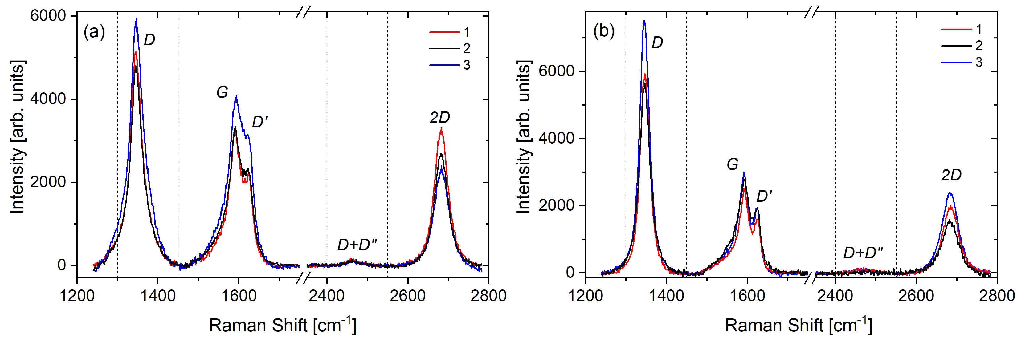
Figure 1 shows the Raman scans taken at three different regions of the graphene after being transferred from the Ni films (see Section IV.2 of the Experimental Section). All the spectra possess the D, G, D’, D+D” and 2D peaks.35, 36 Although all the 2D peaks shown in Figure 1 were fitted with single Lorentzians, they have relatively broad full-width at half-maximum (FWHM). The average FWHM of the 2D peak of epitaxial and rotated-domain graphene transferred from the Ni film are 40.8 cm-1 and 46.2 cm-1, respectively. Furthermore, the spectra of both samples show a high ID/IG ratio (average of 1.49 for epitaxial graphene and 2.35 for rotated graphene). The variation in the spectra of each sample, presence of second order and defect-induced peaks, the large FWHM of the 2D peak and the high ID/IG ratio could be a result of the chemical etching and transfer process (see the Sample Preparation section) or the chemical doping from the HNO3 used to etch the metallic films. Therefore, it is difficult to estimate the number of graphene layers based on the position of the G and 2D peaks and I2D/IG ratio. However, the SEM scan and LEED diffraction pattern in Figure 7 (a) and (c), respectively, show a single epitaxial graphene layer grown on Ni(111). The broader FWHM of the 2D and the higher average I2D/IG ratio in the rotated graphene compared to the epitaxial structure could be attributed to the formation of more defective or turbostratic (multilayer graphene with relative rotation between the layers) graphene as a result of the occasional overlap of the graphene domains.37 The Raman spectra for the graphene/Ni9Mo1 sample, as well as the full list of the peak positions and the 2D average FWHM of all the measured samples is provided in the supplementary material (SI).
II.2 X-ray Magnetic Circular Dichroism (XMCD)
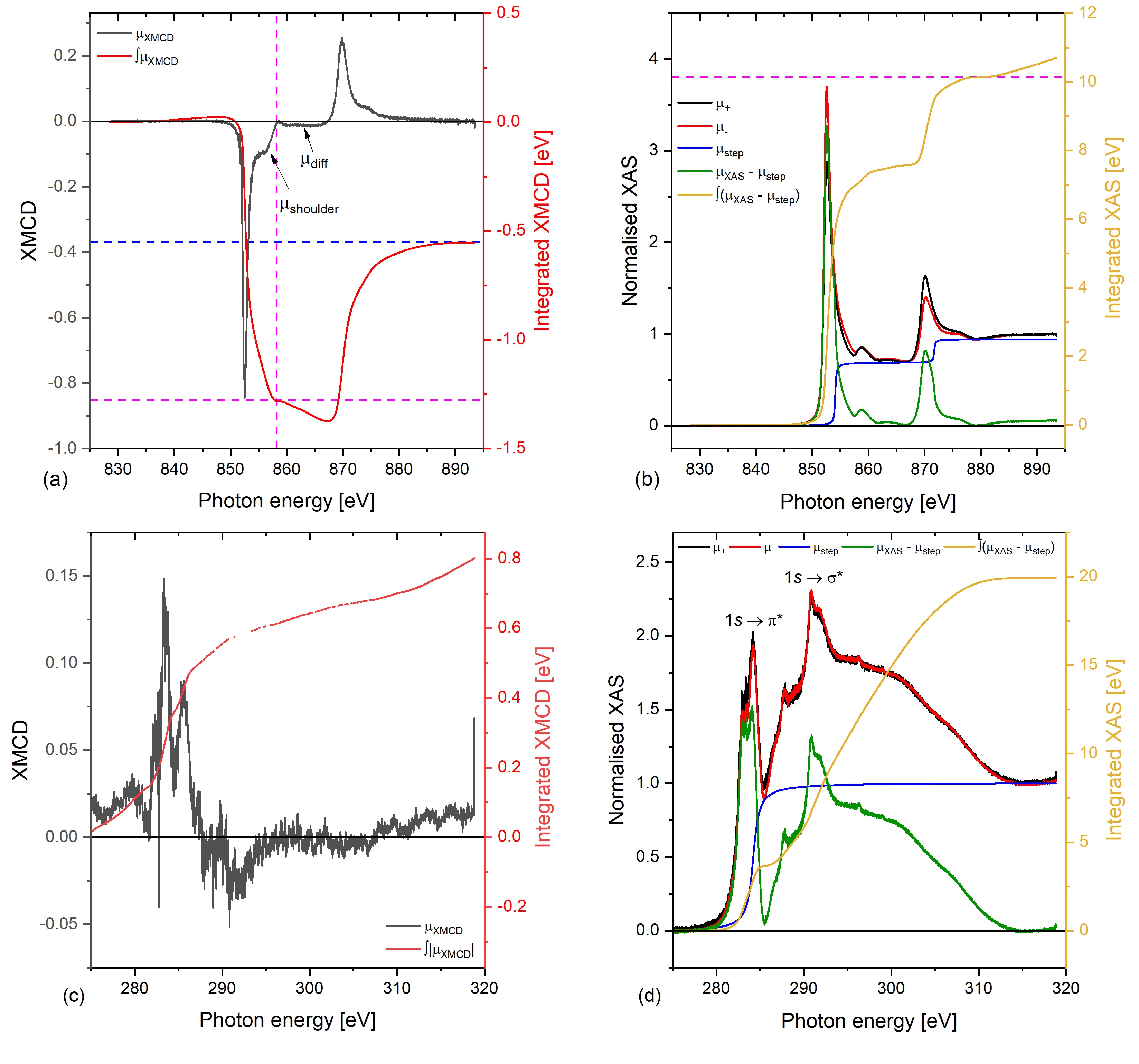
The X-ray absorption spectra (XAS) and the XMCD response (XMCD) at the Ni L2,3-edge in the rotated graphene/Ni sample are shown in Figure 2. The spectra show no sign of oxidation, proving that graphene acts as a good passivation layer against oxidation.38, 39 The region between the L3 and L2 edges with a constant negative intensity is known as the diffuse magnetism region, diff, and it has been observed and reported for Co, Ni and Fe XMCD spectra.40, 41 diff is expected to arise as a result of the opposite spin directions for the 4s and 3d electrons, interstitial and sp-projected magnetic moments, and the fact that it couples antiferromagnetically to the sample’s total magnetic moment in 3d elements, except for Mn.40 Although diff has been reported to contribute to about to the total magnetic moment in Ni,42, 40 since the sum rule does not account for diff, the integration range over the L3 was stopped just before diff for the calculation of the orbital magnetic moment, , and the spin magnetic moment, (858.7 eV). On the other hand, the main L3 peak and the shoulder, shoulder, are due to multiple initial-states configuration, and , respectively, and therefore they were accounted for in the sum rule calculations.40
Although the non-resonant contribution was subtracted from the and spectra, a higher background is measured at the post-edge ( eV). This tail has been excluded from the sum rules as it is considered part of the non-resonant contribution. The calculated and are 0.084 B/Ni atom and 0.625 B/Ni atom, respectively, and thus is 0.709 B/Ni atom (see the SI for the expressions for , and at the L-edge). Considering the 20 accuracy of the XMCD technique in estimating the magnetic moments of materials, the results obtained for Ni is consistent with the values reported in the literature. 43, 44
The C K-edge spectra for the rotated graphene/Ni sample are shown in Figure 2 (c) and (d). For the C K-edge, higher sources of errors are expected in the XMCD estimation attributed to the difficulty of applying the sum rules to the C K-edge spectra in comparison with that for Ni L2,3-edge. For instance, various studies have been reported for Ni 13, 42, 45, 23, 43, 46, 47 which can be used as references for our measurements, but the application of the sum rules has not been reported for graphene before. Also, the number of holes, , has not been measured for C previously. Moreover, the gyromagnetic factor () of the graphene was found to be different depending on the underlying substrate,48, 49, 50 and it has not been reported for graphene on Ni. It is also noteworthy to mention the difficulty associated with measuring the C K-edge due to the C contamination of the optical elements which appear as a significant reduction in the incoming intensity at this particular energy.
Nonetheless, we can obtain an upper limit to the orbital moment of the graphene layer by integrating the modulus of the dichroic signal, , which is shown in Figure 2 (c), red curve. Although the magnetic dichroism response is expected mainly at the peak corresponding to the 1s transition as a result of the C pzNi 3d hybridization,13 a small magnetic signal is observed at the 1s transition peak as well; a similar behaviour was reported for graphene/2 ML Co/Ir(111).24 The calculated upper bound for graphene is 0.062 B/C atom using ; which corresponds to an of 0.412 /C atom, using , which is the value reported for graphene grown on SiC.48 Therefore, of the rotated-domain graphene grown on Ni is 0.474 /C atom (see the SI for the expressions of , and at the K-edge).
Despite the large uncertainties expected for the estimated graphene moments, the XMCD results demonstrate the presence of magnetic polarization in graphene. For more precise, quantified and independent estimates, we turn to PNR.
II.3 Polarized Neutron Reflectivity
PNR experiments were carried out to measure the magnetic properties of each layer of the samples individually and to determine the value of the induced magnetic moment in graphene quantitatively.
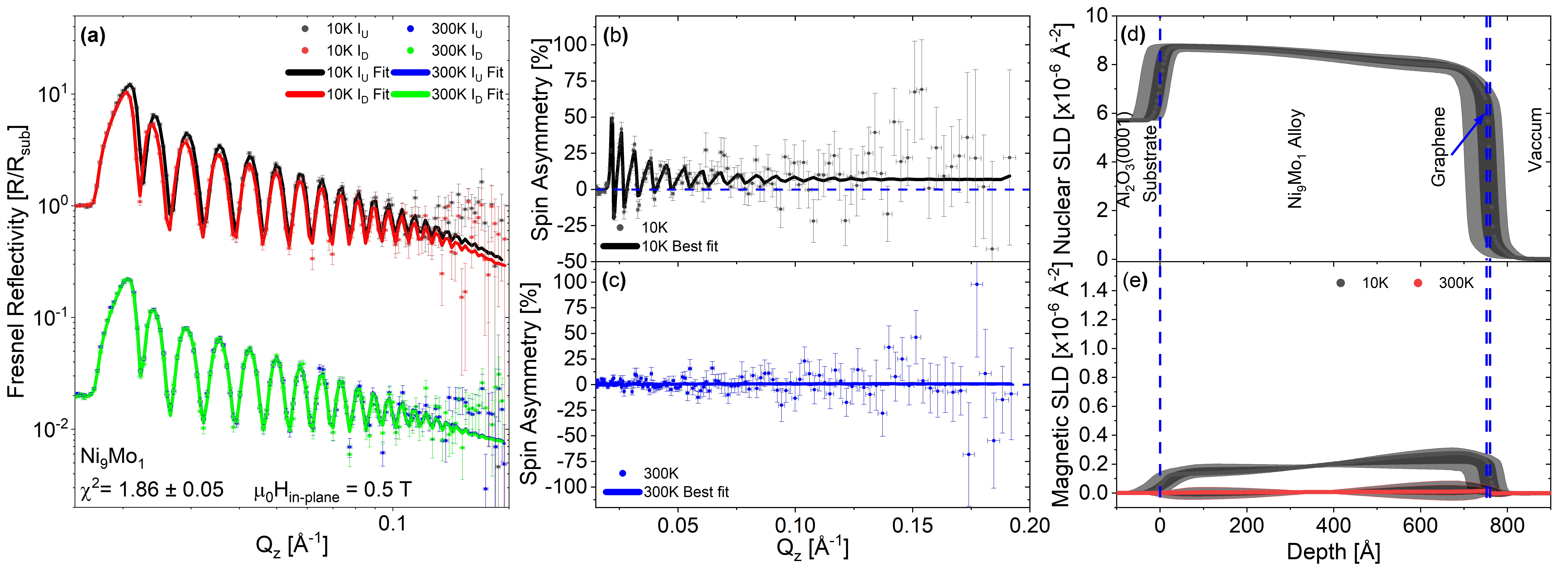
The PNR results for the rotated-domain graphene/Ni(111) and epitaxial graphene/Ni(111) samples, measured at 10 K and 300 K, are displayed in Figures 3 and 4, respectively. Panel (a) for each figure shows the Fresnel reflectivity profiles and panels (b) and (c) the spin asymmetry (, where and are the spin-up and spin-down neutron specular reflectivities, respectively). scales with the magnetic signal. A flat line at zero, shown as a blue dashed line, represents no net magnetic induction present in the system. Panel (d) displays the nuclear scattering length density (nSLD), for the sample structure. This structure is shared at both temperatures in the co-refinement, and panel (e) shows the magnetic scattering length density (mSLD) for each temperature.
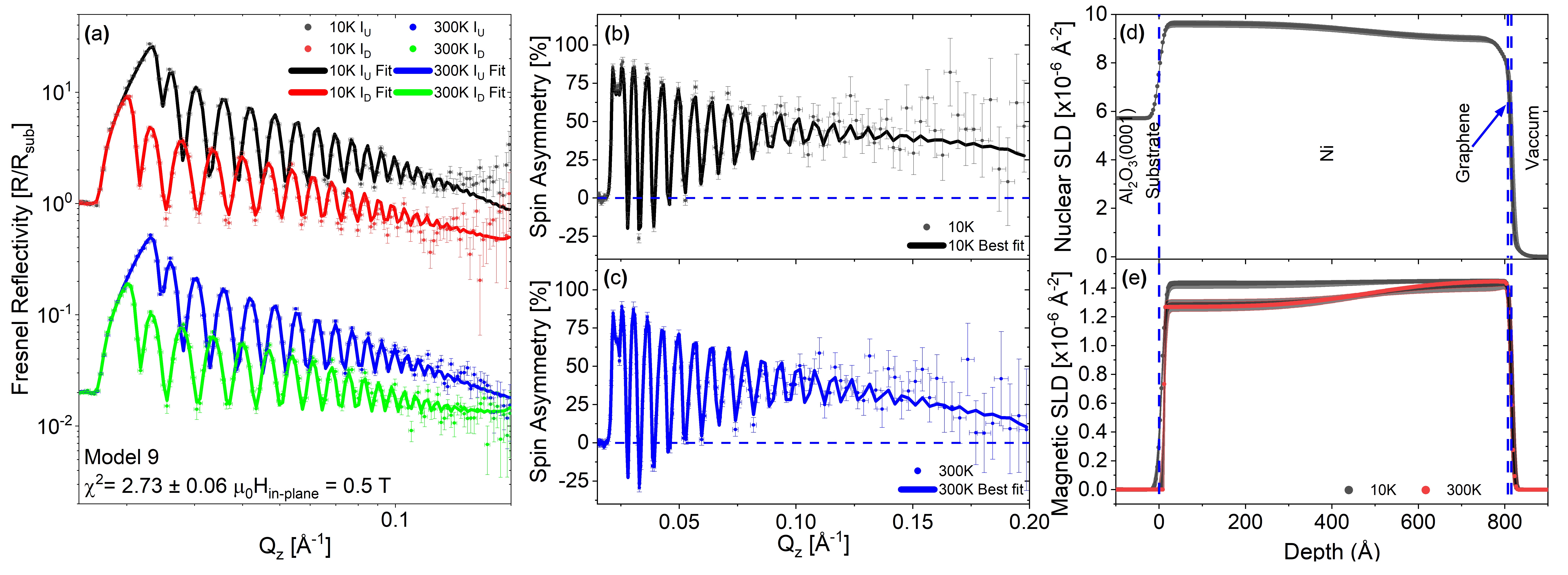
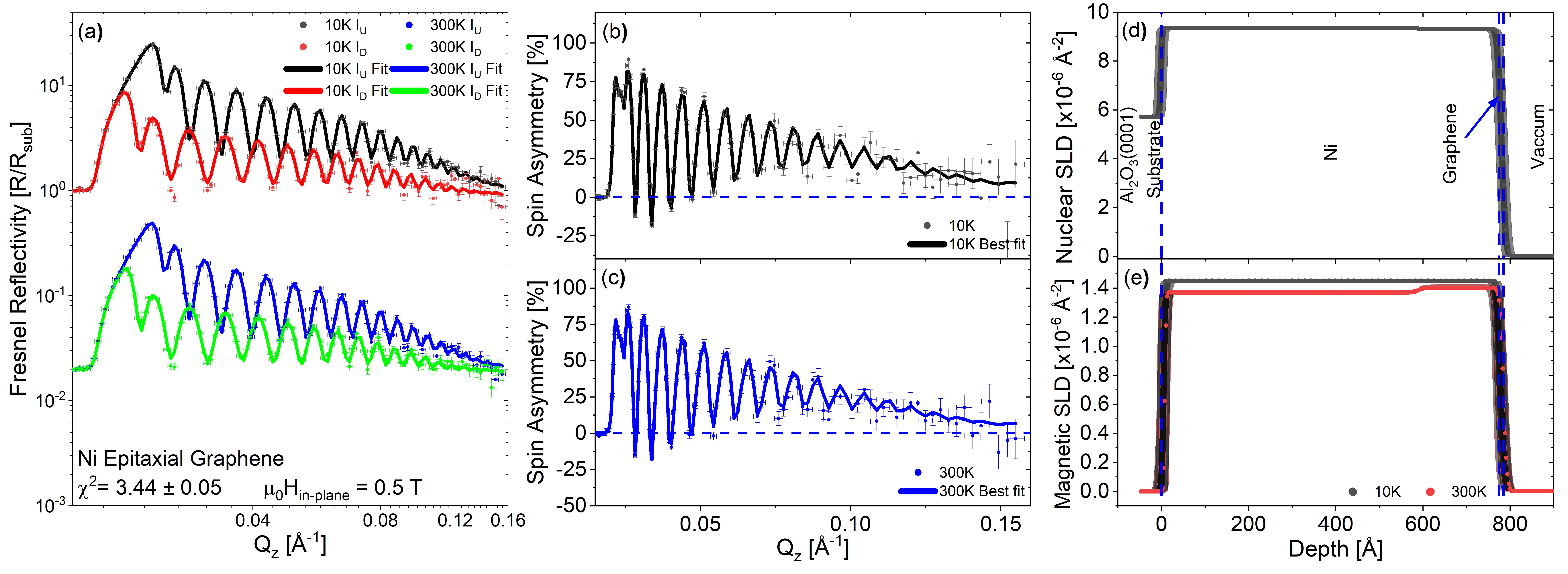
The fitting procedure is fully described in the SI, which contains the various models used to fit the rotated graphene/Ni sample. The 10 K and 300 K data shown in Figures 3 and 4 were fitted simultaneously with a shared nSLD and independent mSLD. The bulk of the fitting model selection was done on the rotated graphene/Ni sample. The study presented in the SI show the importance of prior knowledge of the sample properties to obtain the best PNR fit to estimate the induced magnetic moment in graphene. We used the information obtained from the structural characterizations (SEM and Raman spectroscopy) to obtain a lower bound on the graphene layer thickness. The fit tends to shrink the graphene thickness to less than one monolayer, if unrestrained, which does not agree with the SEM and Raman scans (refer to SI). This could be due to the limited (wavevector transfer) range measured in the time available for the experiment, as the fit is found to rely strongly on the high statistics. It should be noted that in model 9 shown in Figure 3 the magnetic moment of the graphene layer was allowed to fit to zero, but the analysis always required a non-zero value for the magnetic moment to obtain a good fit with low uncertainty, which is consistent with the XMCD results. The unrestrained graphene thickness models are shown in the SI. It is noteworthy that the improvement in the figure of merit () for the unconstrained to the constrained graphene thickness are within the error bar of each other. However, the results show that the graphene thickness has only a subtle influence, if any, on the amount of the induced magnetization as can be deduced from the values of the measured magnetic moments. To get greater certainty on the model selection given the data we used a nested sampler51, 52 to calculate the Bayesian evidence taking into account the structural information, providing a high degree of confidence in the final fit model as shown in the SI.
Additional scenarios were also tested. For example, oxidation of the Ni layer and the formation of Ni-carbide were examined by embedding an intermediate NiO and Ni2C layer, respectively, at the interface between the Ni and graphene, but this lead to poorer fits and the best results, shown in Figures 3 and 4, were achieved using a simpler model: Substrate/a FM layer split into two regions/graphene (see the modelling methodology in the SI).
The results of the fits to the PNR data are summarised in Table 1. Interestingly, it is the rotated-domain graphene rather than the epitaxially grown graphene that has the largest induced magnetic moment, counter to the initial hypothesis that the coupling between the graphene and Ni(111) would be weaker in the rotated-domain case. The greater uncertainty in the values of the moment obtained for the graphene in the epitaxial case can be attributed to the Bayesian analysis being sensitive to the reduced count time and short range that the epitaxial Ni was measured with, due to finite available beam time. Both Ni(111) samples have the full Ni moment at 10 K, which is only slightly reduced at 300 K. This reduction is associated with a magnetic gradient across the Ni(111) film. In the rotated-domain sample, this gradient, as shown in Figure 3 (d), starts with a magnetic moment close to the bulk nSLD of Ni (9.414 x 10Å-2) and reduces in size towards the surface, and is required in order to fit the lower features and allow the higher features to converge, paramount to getting certainty on the thin graphene layers. The mSLD consequently also has a gradient that oppositely mirrors the nSLD at 300 K and reaches the full moment for Ni (0.6 /Ni atom equivalent to an mSLD = 1.4514 x 10Å-2) near the surface and being slightly reduced near the substrate. The Ni(111) used for the epitaxial graphene, displays a much weaker gradient, being almost uniform across the Ni thickness but has the same general trends. We attribute the origin of the difference in the nSLD profiles to the fact that the samples were deposited at different times, following the growth recipe described in the Sample Preparation section.
The Ni9Mo1 sample was used to clarify whether the induced magnetic moments in graphene are due to the C pz-3d hybridization which result in opening the Dirac cone, as postulated in Refs. [23, 2, 28, 18, 20], or because of electron transfer (spin doping) and surface reconstruction, which distorts the d band of the TM as for fullerene/non-magnetic TM, as proposed in Refs. [53, 54, 55]. For this purpose, a Ni9Mo1 film was used with the aim of preserving the fcc crystal structure of Ni while suppressing its magnetization, as suggested in Ref. [56]. Since the Ni is doped by 10 only, the lattice mismatch and bond length to graphene are expected to be similar to that for graphene/Ni(111) sample, but the d-orbital position is considerably downshifted with respect to EF. The growth procedure and the structural properties of the sample is discussed in detail in the Experimental Section.
| FM Layer1 + FM layer2 | Graphene | ||||
|---|---|---|---|---|---|
| Sample | Temperature | Thickness | Magnetic Moment | Thickness | Magnetic Moment |
| [K] | [nm] | [/atom] | [nm] | [/atom] | |
| Rotated Gr/Ni | 10 K | ||||
| 300 K | |||||
| Epitaxial Gr/Ni | 10 K | ||||
| 300 K | |||||
| Gr/Ni9Mo1 | 10 K | ||||
| 300 K | |||||
The results of the PNR measurements of the graphene/Ni9Mo1 sample are shown in Figure 5. Again there is a gradient across the Ni9Mo1 film. At 10 K a small, but detectable spin splitting and a minute variation in the are observed. Surprisingly, a higher magnetization is detected in graphene () B/C atom) than in the Ni9Mo1 film () B/C atom), but the 95% confidence intervals indicate that we are not sensitive to this to a degree to say for sure given the Q range and counting statistics in the data. All we can ascertain is that there is a small moment in the Ni9Mo1 and a non zero moment in the graphene. At 300 K, both the alloy and the graphene have effectively zero moment within the 95% confidence intervals. The origin of the small residual moment at 10 K could arise from clusters of unalloyed Ni throughout the layer that become ferromagnetic at low temperature, which then polarize the graphene as per the Ni(111) samples. At 300 K, no magnetic moment in the Ni9Mo1 and graphene are detected. Therefore, the results support the hypothesis of the universal model, whereby the measured induced magnetic moment in graphene is due to the opening of the ED rather than the distortion of the d band. This is because no magnetic moment is detected in the Ni9Mo1 nor in the graphene layer.
The PNR fits have shown that at 10 K a magnetic moment of B/C atom (B/C atom) was induced in the rotated-domain (epitaxial) graphene grown on Ni films. These results indicate larger moments than previously reported.26, 13 In Ref. [26], Mertins et al. estimated the magnetic moment of graphene to be B/C atom when its grown on a hcp Co(0001) film. They obtained this value by comparing the XMCD reflectivity signal of graphene to that of the underlayer Co film.26 Weser et al. suggested a similar value (B/C atom) for the magnetic moment induced in a monolayer of graphene grown on a Ni(111) film.13 However, their assumption was based on comparing the graphene/Ni system with other C/3d TM structures, such as a C/Fe multilayer with 0.55 nm of C,58 and carbon nanotubes (CNTs) on a Co film,59 where a magnetic moment of 0.05 B/C atom and 0.1 B/C atom was estimated, respectively. Nonetheless, it is difficult to compare graphene-based heterostructures with other C allotropes/TM systems. This is because the Dirac cone is a characteristic feature of graphene and CNTs of the carbon allotropes. Therefore, one cannot exclude that a different mechanism other than the break of degeneracy around ED may be responsible for the magnetic moment detected in the C layer of a C/Fe multilayer system. On the other hand, for the CNTs/Co heterostructure, although Dirac cones exist in CNTs, a direct quantitative analysis of the induced magnetic moment was not possible from the MFM images reported in Ref. [59]. In contrast, PNR provides a direct estimation of the induced magnetic moment in graphene.
III Conclusion
In summary, we have successfully grown graphene by chemical vapor deposition (CVD) on different TM substrates. Induced magnetic moment in rotated-domain graphene as a result of the proximity effect in the vicinity of a FM substrate was detected by element-specific XMCD measurements at the C K-edge. PNR experiments were carried out to determine the magnitude of the magnetic moment detected by XMCD. Although a higher magnetic moment was expected to be induced in epitaxial graphene/Ni sample, the PNR results indicate the epitaxial graphene film had a magnetic moment of B/C atom as compared to the rotated-domain graphene B/C atom. Both values are higher than those predicted in other studies.13, 58, 59 PNR measurements on graphene/Ni9Mo1 support the universal model proposed by Voloshina and Dedkov that the induced magnetic moment in graphene arises as a result of the opening of the graphene’s Dirac cone as a result of the strong C pz-Ni 3d hybridization. Our PNR provides the first quantitative estimation of the induced magnetization in graphene by PNR.
IV Experimental Section
IV.1 Sample Preparation
The sample preparation procedure involved two stages; the growth of the TM films using magnetron sputtering and the growth of graphene by CVD.
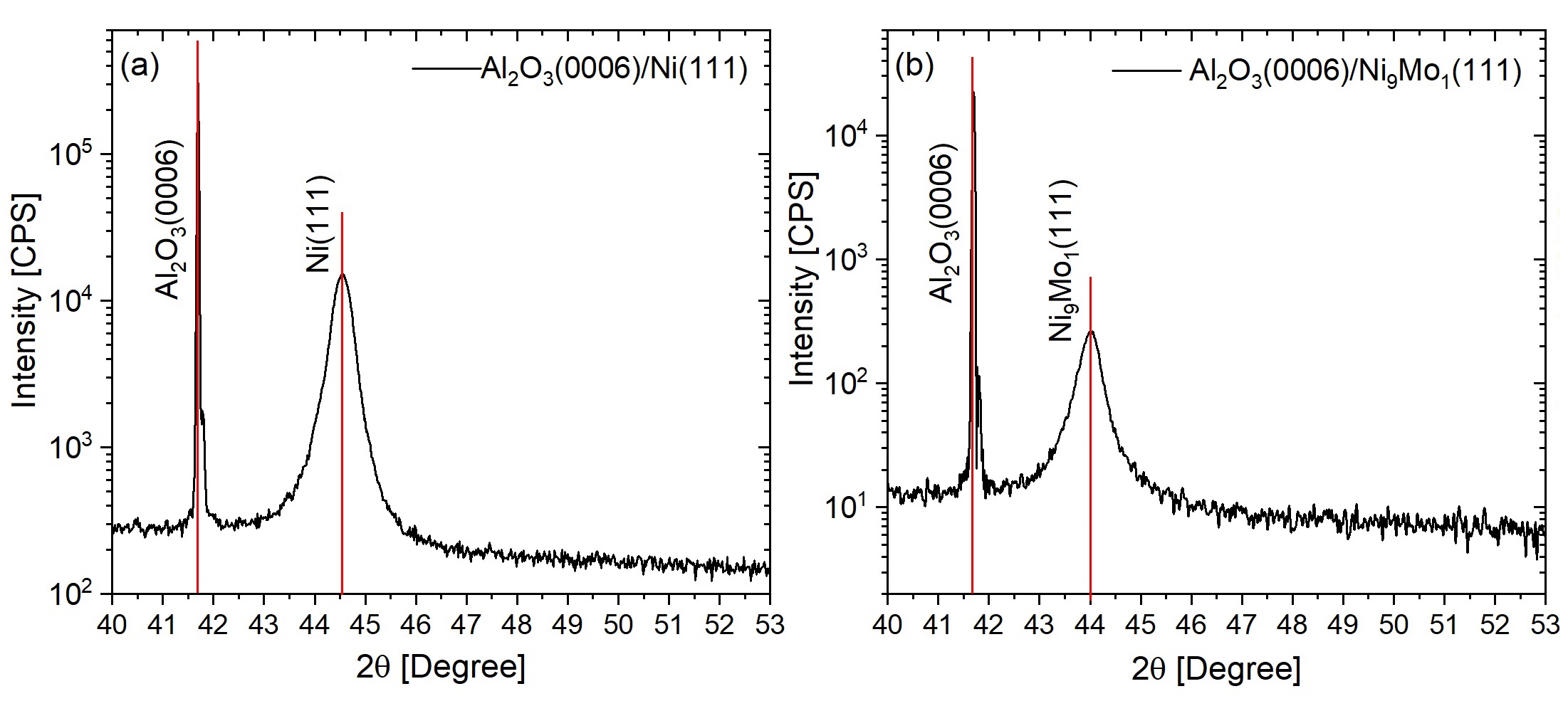
The TM films were deposited at RT on 1 mm thick Al2O3(0001) substrates using a CEVP magnetron sputtering chamber with a base pressure of 1.2 - 2 10-8 mTorr. The thick substrates were used to reduce the possibility of sample deformation which could affect the reflectivity measurements. The deposition of the TM films was performed using 99.9% pure Ni and Ni9Mo1 targets. A DC current of 0.1 A and a constant flow of pure Argon of 14 sccm were used to grow 80 nm of highly textured Ni(111) and Ni9Mo1(111) films at a rate of 0.02 nm s-1 in a plasma pressure of 2 mTorr (3 mTorr for Ni9Mo1). Figure 6 shows the X-ray diffraction (XRD) measurements of the deposited films acquired with a Bruker D8 Discover HRXRD with a Cu K monochromatic beam (40 kV, 40 mA). The scans show highly textured pure films oriented in the [111] direction for Ni and Ni9Mo1 films.
The samples were then transferred into a CVD system for the growth of graphene directly on Ni(111) and Ni9Mo1(111) films on 2 cm 2 cm substrates. Growth recipes similar to those reported by Patera et al. 29 were adapted to obtain epitaxial and rotated-domain graphene directly on the Ni film. For the Ni9Mo1 film, rotated-domain graphene was grown by first introducing pure H2 gas at a rate of 200 sccm to the CVD chamber with a base pressure of 2.7 10-6 mbar. Then, the CVD growth chamber was heated to 650 ∘C for 12 minutes and the sample was then exposed to C2H4 with a flow rate of 0.24 sccm and 40 minutes before it was cooled down to RT in vacuum. This approach reduces any oxidized TM back to a clean metallic surface before the growth of graphene.

The SEM images shown in Figure 7 illustrate the structure of epitaxial and rotated graphene on Ni with a surface coverage of %. Figure 7 (a) shows a homogenous monolayer of graphene on Ni. The darker grey regions in Figure 7 (b) are the differently oriented graphene domains, whereas the bright areas in Figure 7 (a) and (b) are the bare Ni film. The low-energy electron diffraction (LEED) in Figure 7 (c) shows the graphene’s hexagonal pattern epitaxially grown on the Ni(111) substrate. The () grown graphene structure is confirmed since no additional diffraction spots are observed in the LEED pattern.
IV.2 Raman Spectroscopy Measurements
RT Raman scans were taken at three different regions of each sample using a 532 nm excitation laser wavelength ( objective lens and m laser spot size). Before the measurements, the graphene was first transferred by a chemical etching process from the metallic films onto a Si substrate with a 300 nm thermally oxidized SiO2 layer. This approach overcome the fact that closely lattice-matched films lead to a loss in the resonance conditions for observing Raman spectra as a result of the strong chemical interaction between the graphene orbital and the d-states of Ni and Ni9Mo1 which also alters the graphene’s pz orbitals. Furthermore, the increase in the CC bond length to match the lattice of the FM leads to significant changes in the graphene’s phonon spectrum.22, 60 For the transfer process, the samples were cleaved into 5 mm 5 mm squares and HNO3, diluted to 5% for Ni9Mo1 and 10% for Ni, was used to etch the metallic films slowly while preserving the graphene layer.
IV.3 X-ray Magnetic Circular Dichroism (XMCD)
We carried out element selective XMCD measurements to detect and distinguish the magnetization in the graphene from that of the FM layer. The XMCD experiments were performed at 300 K at the SIM end station of the Swiss Light Source (SLS) at the Paul Scherrer Institut (PSI), Switzerland, using total electron yield (TEY) detection mode with 100% circularly polarized light. The rotated-domain graphene/Ni sample was set to an incident angle of 30∘ from the incoming X-ray beam. An electromagnet was fixed at 40∘ to the incoming X-ray beam and a magnetic field of 0.11 T was applied for 30 seconds in-plane to the surface of the samples to align the film magnetization along the beam direction. It was then reduced to 0.085 T during the X-ray absorption spectroscopy measurements. The intensity of the incident X-ray beam was measured with a clean, carbon free, gold mesh placed just before the sample position. This is particularly important for normalizing the signal at the C K-edge due to the presence of carbon on the surface of the X-ray optical components.
IV.4 Polarized Neutron Reflectivity
The PNR measurements were conducted at 10 K and 300 K, under an in-plane magnetic field of 0.5 T, using the Polref instrument at ISIS spallation neutron source (UK). The fitting of the data was done using the Refl1D 61 software package with preliminary fits done in GenX.62 Although Ni is ferromagnetic at RT, the 10 K measurements are expected to provide a better estimation of the induced magnetic moment due to the lower thermal excitations of the electron spin at low temperature. Both the 10 K and 300 K data sets were fitted simultaneously to provide further constraint to the fits. This is analogous to the isotropic contrast matching 63, 64 used in soft matter neutron reflectivity experiments. This is very important in this case due to the attempt to measure a thin layer of graphene within a limited total Q range for PNR. The PNR is sensitive to only part of the broad fringe from the graphene layer which acts as an envelope function on the higher frequency fringes from the thicker Ni layer underneath (the modelling methodology is discussed in detail in the SI).
Acknowledgements
We would like to thank the ISIS Neutron and Muon Source for the provision of beam time (RB1510330 and RB1610424). The data is available at the following DOIs https://doi.org/10.5286/ISIS.E.RB1510330 and https://doi.org/10.5286/ISIS.E.RB1610424. Part of this work was performed at the Surface and Interface Microscopy (SIM) beamline of the Swiss Light Source (SLS), Paul Scherrer Institut (PSI), Villigen, Switzerland.
Other data presented in this study are available from the corresponding author upon request.
References
- Karpan et al. [2007] V. M. Karpan, G. Giovannetti, P. A. Khomyakov, M. Talanana, A. A. Starikov, M. Zwierzycki, J. van den Brink, G. Brocks, and P. J. Kelly, Physical Review Letters 99 (2007).
- Weser et al. [2011] M. Weser, E. N. Voloshina, K. Horn, and Y. S. Dedkov, Physical Chemistry Chemical Physics 13, 7534 (2011).
- Zhang et al. [2015] J. Zhang, B. Zhao, Y. Yao, and Z. Yang, Scientific Reports 5, 1 (2015).
- Högl et al. [2020] P. Högl, T. Frank, K. Zollner, D. Kochan, M. Gmitra, and J. Fabian, Physical Review Letters 124, 136403 (2020).
- Meng et al. [2013] J. Meng, J. J. Chen, Y. Yan, D. P. Yu, and Z. M. Liao, Nanoscale 5, 8894 (2013).
- Song and Dai [2015] Y. Song and G. Dai, Applied Physics Letters 106 (2015).
- Xu et al. [2018] J. Xu, S. Singh, J. Katoch, G. Wu, T. Zhu, I. Žutić, and R. K. Kawakami, Nature Communications 9, 2 (2018).
- Hill et al. [2006] E. W. Hill, A. K. Geim, K. Novoselov, F. Schedin, and P. Blake, IEEE Transactions on Magnetics 42, 2694 (2006).
- Semenov et al. [2007] Y. G. Semenov, K. W. Kim, and J. M. Zavada, Applied Physics Letters 91, 153105 (2007).
- Sze and Ng [2006] S. M. Sze and K. K. Ng, Physics of Semiconductor Devices (Wiley, 2006).
- Pi et al. [2010] K. Pi, W. Han, K. M. McCreary, A. G. Swartz, Y. Li, and R. K. Kawakami, Physical Review Letters 104, 187201 (2010).
- Zhang et al. [2018] Y. Zhang, X. Sui, D. L. Ma, K. K. Bai, W. Duan, and L. He, Physical Review Applied 10, 1 (2018).
- Weser et al. [2010] M. Weser, Y. Rehder, K. Horn, M. Sicot, M. Fonin, A. B. Preobrajenski, E. N. Voloshina, E. Goering, and Y. S. Dedkov, Applied Physics Letters 96, 012504 (2010).
- Leutenantsmeyer et al. [2016] J. C. Leutenantsmeyer, A. A. Kaverzin, M. Wojtaszek, and B. J. van Wees, 2D Materials 4, 014001 (2016).
- Wang et al. [2015] Z. Wang, C. Tang, R. Sachs, Y. Barlas, and J. Shi, Physical Review Letters 114, 016603 (2015).
- Haugen et al. [2008] H. Haugen, D. Huertas-Hernando, and A. Brataas, Physical Review B 77, 115406 (2008).
- Tuan and Roche [2016] D. V. Tuan and S. Roche, Physical Review Letters 116, 106601 (2016).
- Dedkov and Voloshina [2015] Y. Dedkov and E. Voloshina, Journal of Physics: Condensed Matter 27, 303002 (2015).
- Fal’ko [2007] V. Fal’ko, Nature Physics 3, 151 (2007).
- Voloshina and Dedkov [2014] E. N. Voloshina and Y. S. Dedkov, Materials Research Express 1 1, 035603 (2014).
- Voloshina and Dedkov [2011] E. Voloshina and Y. Dedkov, in Physics and Applications of Graphene - Experiments, Vol. 499 (InTech, 2011) pp. 75–78.
- Dahal and Batzill [2014] A. Dahal and M. Batzill, Nanoscale 6, 2548 (2014).
- Dedkov and Fonin [2010] Y. S. Dedkov and M. Fonin, New Journal of Physics 12, 125004 (2010).
- Vita et al. [2014] H. Vita, S. Böttcher, P. Leicht, K. Horn, A. B. Shick, and F. Máca, Physical Review B - Condensed Matter and Materials Physics 90, 1 (2014).
- Marchenko et al. [2015] D. Marchenko, A. Varykhalov, J. Sánchez-Barriga, O. Rader, C. Carbone, and G. Bihlmayer, Physical Review B - Condensed Matter and Materials Physics 91, 1 (2015).
- Mertins et al. [2018] H. C. Mertins, C. Jansing, M. Krivenkov, A. Varykhalov, O. Rader, H. Wahab, H. Timmers, A. Gaupp, A. Sokolov, M. Tesch, and P. M. Oppeneer, Physical Review B 98, 1 (2018).
- Mendes et al. [2019] J. B. Mendes, O. Alves Santos, T. Chagas, R. Magalhães-Paniago, T. J. Mori, J. Holanda, L. M. Meireles, R. G. Lacerda, A. Azevedo, and S. M. Rezende, Physical Review B 99, 1 (2019).
- Karpan et al. [2008] V. M. Karpan, P. A. Khomyakov, A. A. Starikov, G. Giovannetti, M. Zwierzycki, M. Talanana, G. Brocks, J. van den Brink, and P. J. Kelly, Physical Review B 78, 195419 (2008).
- Patera et al. [2013] L. L. Patera, C. Africh, R. S. Weatherup, R. Blume, S. Bhardwaj, C. Castellarin-Cudia, A. Knop-Gericke, R. Schloegl, G. Comelli, S. Hofmann, and C. Cepek, ACS nano , 7901 (2013).
- Weatherup et al. [2014] R. S. Weatherup, H. Amara, R. Blume, B. Dlubak, B. C. Bayer, M. Diarra, M. Bahri, A. Cabrero-Vilatela, S. Caneva, P. R. Kidambi, M. B. Martin, C. Deranlot, P. Seneor, R. Schloegl, F. Ducastelle, C. Bichara, and S. Hofmann, Journal of the American Chemical Society 136, 13698 (2014).
- Kozlov et al. [2012] S. M. Kozlov, F. Viñes, and A. Görling, Journal of Physical Chemistry C 116, 7360 (2012).
- Wang et al. [2008] Y. Y. Wang, Z. H. Ni, Z. X. Shen, H. M. Wang, and Y. H. Wu, Applied Physics Letters 92 (2008), 10.1063/1.2838745, arXiv:0801.4595 .
- Chilres et al. [2013] I. Chilres, L. A. Jauregui, W. Park, H. Cao, and Y. P. Chen, in New Developments in Photon and Materials Research, edited by J. I. Jang (Nova Science Publishers, Incorporated, 2013) Chap. 19, pp. 553–595.
- Reina et al. [2008] A. Reina, H. Son, L. Jiao, B. Fan, M. S. Dresselhaus, Z. Liu, and J. Kong, The Journal of Physical Chemistry C 112, 17741 (2008).
- Malard et al. [2009] L. M. Malard, M. A. Pimenta, G. Dresselhaus, and M. S. Dresselhaus, Physics Reports 473, 51 (2009).
- Ferrari and Basko [2013] A. C. Ferrari and D. M. Basko, Nature Nanotechnology 8, 235 (2013).
- Cabrero-Vilatela et al. [2016] A. Cabrero-Vilatela, R. S. Weatherup, P. Braeuninger-Weimer, S. Caneva, and S. Hofmann, Nanoscale 8, 2149 (2016).
- Martin et al. [2015] M.-B. Martin, B. Dlubak, R. S. Weatherup, M. Piquemal-Banci, H. Yang, R. Blume, R. Schloegl, S. Collin, F. Petroff, S. Hofmann, J. Robertson, A. Anane, A. Fert, and P. Seneor, Applied Physics Letters 107, 012408 (2015).
- Dlubak et al. [2012] B. Dlubak, M.-B. Martin, R. S. Weatherup, H. Yang, C. Deranlot, R. Blume, R. Schloegl, A. Fert, A. Anane, S. Hofmann, P. Seneor, and J. Robertson, ACS nano 6, 10930 (2012).
- O’Brien and Tonner [1994] W. L. O’Brien and B. P. Tonner, Physical Review B 50, 12672 (1994).
- Eriksson et al. [1992] O. Eriksson, A. M. Boring, R. C. Albers, G. W. Fernando, and B. R. Cooper, Physical review. B, Condensed matter 45, 2868 (1992).
- O’Brien et al. [1994] W. L. O’Brien, B. P. Tonner, G. R. Harp, and S. S. P. Parkin, Journal of Applied Physics 76, 6462 (1994).
- Eriksson et al. [1990] O. Eriksson, B. Johansson, R. C. Albers, A. M. Boring, and M. S. S. Brooks, Physical Review B 42, 2707 (1990).
- Vaz et al. [2008] C. Vaz, J. Bland, and G. Lauhoff, Reports on Progress in Physics 71, 056501 (2008).
- Nakajima et al. [1999] R. Nakajima, J. Stöhr, and Y. U. Idzerda, Physical Review B 59, 6421 (1999).
- Chen et al. [1990] C. T. Chen, F. Sette, Y. Ma, and S. Modesti, Physical Review B 42, 7262 (1990).
- Söderlind et al. [1992] P. Söderlind, O. Eriksson, B. Johansson, R. C. Albers, and A. M. Boring, Physical Review B 45, 12911 (1992).
- Menezes et al. [2017] N. Menezes, V. S. Alves, E. C. Marino, L. Nascimento, L. O. Nascimento, and C. Morais Smith, Physical Review B 95, 1 (2017).
- Song et al. [2010] Y. J. Song, A. F. Otte, Y. Kuk, Y. Hu, D. B. Torrance, P. N. First, W. A. De Heer, H. Min, S. Adam, M. D. Stiles, A. H. MacDonald, and J. A. Stroscio, Nature 467, 185 (2010).
- Semenikhin et al. [2020] P. V. Semenikhin, A. N. Ionov, and M. N. Nikolaeva, Technical Physics Letters 46, 186 (2020).
- Skilling [2004] J. Skilling, AIP Conference Proceedings 735, 395 (2004), https://aip.scitation.org/doi/pdf/10.1063/1.1835238 .
- McCluskey et al. [2020] A. R. McCluskey, J. F. K. Cooper, T. Arnold, and T. Snow, Machine Learning: Science and Technology 1, 035002 (2020).
- Ma’Mari et al. [2015] F. A. Ma’Mari, T. Moorsom, G. Teobaldi, W. Deacon, T. Prokscha, H. Luetkens, S. Lee, G. E. Sterbinsky, D. A. Arena, D. A. MacLaren, M. Flokstra, M. Ali, M. C. Wheeler, G. Burnell, B. J. Hickey, and O. Cespedes, Nature 524, 69 (2015).
- Pai et al. [2010] W. W. Pai, H. T. Jeng, C. M. Cheng, C. H. Lin, X. Xiao, A. Zhao, X. Zhang, G. Xu, X. Q. Shi, M. A. Van Hove, C. S. Hsue, and K. D. Tsuei, Physical Review Letters 104, 1 (2010).
- Moorsom et al. [2014] T. Moorsom, M. Wheeler, T. Mohd Khan, F. Al Ma’Mari, C. Kinane, S. Langridge, D. Ciudad, A. Bedoya-Pinto, L. Hueso, G. Teobaldi, V. K. Lazarov, D. Gilks, G. Burnell, B. J. Hickey, and O. Cespedes, Physical Review B - Condensed Matter and Materials Physics 90, 1 (2014).
- Hansen et al. [1958] M. Hansen, R. P. Elliott, and F. A. Shunk, Constitution of Binary Alloys (McGraw-Hill, New York, 1958) p. 1305.
- Kienzle et al. [2011] P. Kienzle, J. Krycka, N. Patel, and I. Sahin, https://bumps.readthedocs.io/en/latest/ (2011), accessed: (28-09-2021).
- Mertins et al. [2004] H.-C. Mertins, S. Valencia, W. Gudat, P. M. Oppeneer, O. Zaharko, and H. Grimmer, Europhysics Letters (EPL) 66, 743 (2004).
- Céspedes et al. [2004] O. Céspedes, M. S. Ferreira, S. Sanvito, M. Kociak, and J. M. D. Coey, Journal of Physics: Condensed Matter 16, L155 (2004).
- Allard and Wirtz [2010] A. Allard and L. Wirtz, Nano Letters 10, 4335 (2010).
- Kienzle et al. [2017] P. Kienzle, B. Maranville, K. O’Donovan, J. Ankner, N. Berk, and C. Majkrzak, https://www.nist.gov/ncnr/reflectometry-software (2017), accessed: (28-09-2021).
- Björck and Andersson [2007] M. Björck and G. Andersson, Journal of Applied Crystallography 40, 1174 (2007).
- Majkrzak et al. [2003] C. F. Majkrzak, N. F. Berk, and U. A. Perez-Salas, Langmuir 19, 7796 (2003).
- Koutsioubas [2019] A. Koutsioubas, Journal of Applied Crystallography 52, 538 (2019).