Response of undoped cryogenic CsI to low-energy nuclear recoils
Abstract
The bright scintillation of pure CsI operated at liquid-nitrogen temperature makes of this material a promising dark matter and neutrino detector. We present the first measurement of its quenching factor for nuclear recoils. Our findings indicate it is indistinguishable from that for sodium-doped CsI at room temperature. Additional properties such as light yield, afterglow, scintillation decay properties for electron and nuclear recoils, and energy proportionality are studied over the 108-165 K temperature range, confirming the vast potential of this medium for rare-event searches.
I Introduction
An ongoing interest in characterizing the response of radiation detectors to low-energy nuclear recoils, induced by the elastic scattering of neutral particles, is traceable to the first direct search for Weakly Interacting Massive Particles (WIMPs) Ahlen et al. (1987), popular dark matter candidates. Neutrinos with energies below a few tens of MeV can scatter coherently from nuclei via the weak neutral current Freedman (1974), also producing few-keV nuclear recoils as the single outcome from this process. The recent observation of this so-called Coherent Elastic Neutrino-Nucleus Scattering (CENS) Akimov et al. (2017); Scholz (2017) has added thrust to a quest for new materials well-adapted to the detection of these subtle low-energy interactions.
Scintillating sodium-doped cesium iodide (CsI[Na]), operated at room-temperature, was chosen as the favored detector material for the first CENS measurement. A long list of virtues leading to its selection is described in Collar et al. (2015); Fields (2014). Among those is a large and essentially identical CENS cross-section for both Cs and I, a high light-yield, reduced afterglow, and a quenching factor (QF) of order 10 % in the few-keV nuclear recoil (NR) energy region of interest. This QF is the ratio between the light yield for NRs and that for electron recoils (ERs) of the same energy. A precise understanding of the energy dependence of the QF is of crucial importance in the interpretation of WIMP and CENS searches Collar et al. (2019).
Undoped CsI exhibits a large increase in light yield at liquid-nitrogen temperature, reaching a theoretical limit in light-conversion efficiency that exceeds 100 scintillation photons per keV of ER energy deposition Amsler et al. (2002); Moszynski et al. (2005, 2003); Nadeau (2015); Clark et al. (2018); Liu et al. (2016); Woody et al. (1990); Zhang et al. (2018); Mikhailik et al. (2015). This is close to three times the room-temperature yield of CsI[Na]. When monitored with silicon light sensors combined with state-of-the-art waveshifters able to maximize their quantum efficiency, the potential to detect NRs as low in energy as 1 keV appears to be within reach Baxter et al. (2020). This is a NR energy regime unprecedented for scintillators; one where the new physics beyond the Standard Model that is reachable via CENS concentrates Baxter et al. (2020). However, the assumption on which this promise is based is that the QF of this cryogenic material, unknown for NRs until now, is at least as favorable as for doped CsI at room-temperature.
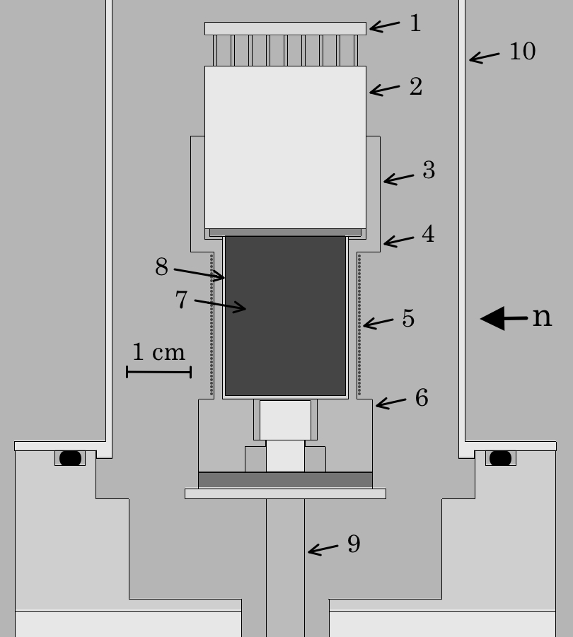
This work describes a first measurement of this QF in the temperature range 108-165 K, using the custom detector assembly in Fig. 1, exposed to monochromatic 2.25 MeV neutrons from a D-D generator. Neutrons scattering from the CsI crystal are detected by a Bicron 501A liquid scintillator cell with neutron/gamma discrimination capability. This cell is placed at a user-defined angle from the initial neutron trajectory, allowing to select the energy deposited by NRs in CsI. This experimental setup, data acquisition system (DAQ), and analysis method have been previously employed by us for NaI[Tl] and CsI[Na] room-temperature QF measurements. Details of these technical aspects are provided in Collar (2013a); Collar et al. (2015, 2019). In this new implementation, a PID algorithm was used to monitor the temperature at both ends of the CsI crystal and control the power injected into a heating element (manganin wire, Fig. 1) resulting in a temperature stability of 0.03 K, and a gradient across the crystal of 1.5 K, for all present runs.
In the second section of this paper we describe our QF measurements. The third section includes the determination of a number of additional quantities (light yield, afterglow, scintillation decay properties for ERs and NRs, and energy proportionality) of interest to assess the potential of this new material for rare event searches. Its extraordinary promise is emphasized in our conclusions.
II isolation of low energy nuclear recoils and QF measurement
The use of a small 7.24 cm3 CsI scintillator pro ensures that single-scatters dominate neutron interactions in the crystal. Multiple scatters make up for just 17-27% of the total, depending on the selected scattering angle, and are accounted for in simulations. A Hamamatsu R8520-506 cryogenic bialkali photomultiplier (PMT) was directly coupled to the sample using optical RTV. While operation of this PMT down to 87 K is possible, the lowest temperature of 108 K achieved in this study was limited by the cooling power of the horizontal-arm cryostat employed.
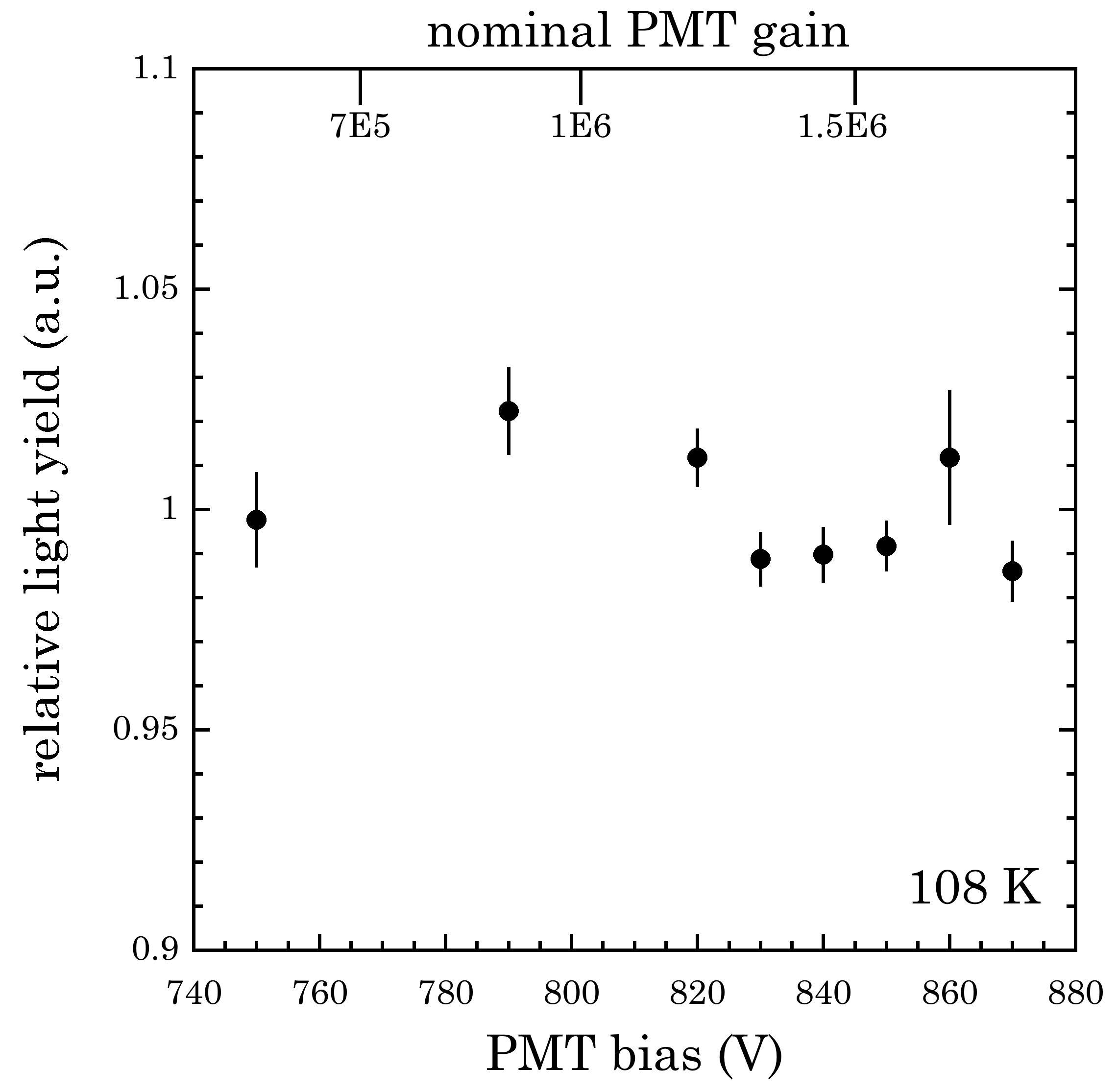
Among the lessons derived from our latest CsI[Na] experimentation is the impact on QF measurements of PMT saturation at high bias. As a preliminary precaution, we compared the PMT charge output for 241Am 59.5 keV gammas and for single photo-electrons (SPE) following the test procedure developed in Collar et al. (2019). The normalized ratio of these outputs provides a light yield in units of PE/keV. As in Collar et al. (2019), the energy reference used in the definition of the QF was given by this 241Am emission, assuming direct proportionality for lower ER energies. Tests over a range of PMT voltage biases were made at 108 K, a temperature corresponding to the maximum light yield observed in this work. As can be seen in Fig. 2 the chosen 820 V bias is well within the linear response of this PMT and away from saturation effects at the light levels involved in this study.
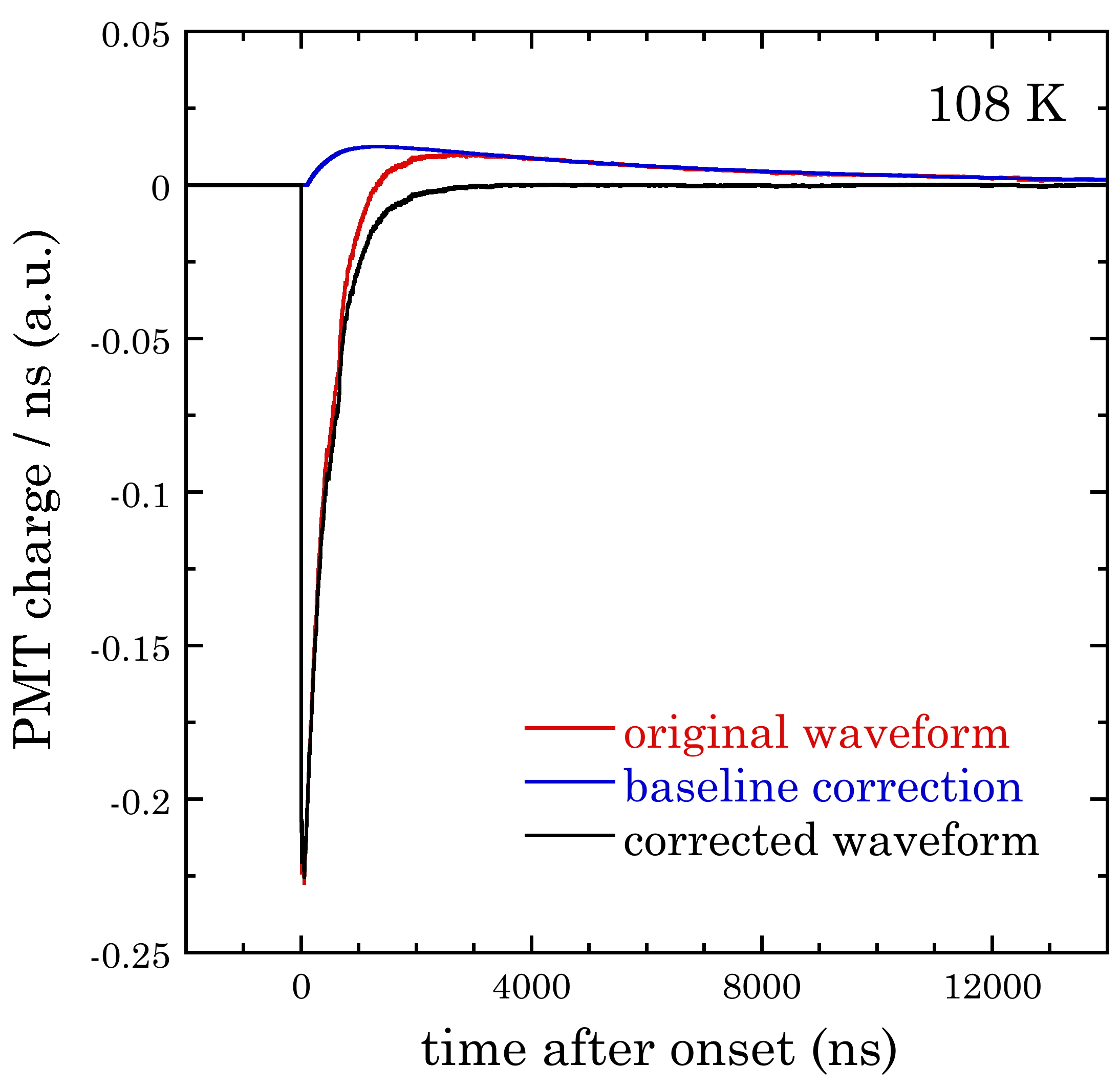
Incremental improvements to the MCNP-PoliMi simulations Pozzi et al. (2003) used in Collar et al. (2019) were made by accounting for subdominant inelastic neutron scattering through de-exitation gamma escape from CsI. Similarly to Collar et al. (2019), charge was integrated over the 3 s following the onset of scintillation signals. However, due to electrical safety concerns for metallic-envelope PMTs like the R8520-506, a positively-biased voltage divider was used. The resulting capacitive coupling to the DAQ produces a well-documented overshoot of the PMT signal Wright and Wright (2017). Uncorrected, this leads to an underestimation of the integrated charge carried by a scintillation signal. Remedial analysis techniques have been put forward in a number of affected experiments Acciarri et al. (2017); Abe et al. (2012); Zhang et al. (2019). The impact of this effect and its correction on our charge measurements can be assessed from Fig. 3. The corrective procedure uses an inverse high-pass algorithm (an offline pole-zero cancellation) to allow for accurate charge integration below the median waveform baseline. Special attention was paid to ensure that this average charge correction also applied to signals at lower energy. As expected, since the response of the output capacitor causing the overshoot depends on signal frequency, and not on amplitude, the magnitude of the integrated charge correction was found to be consistent at 18 for all energies.
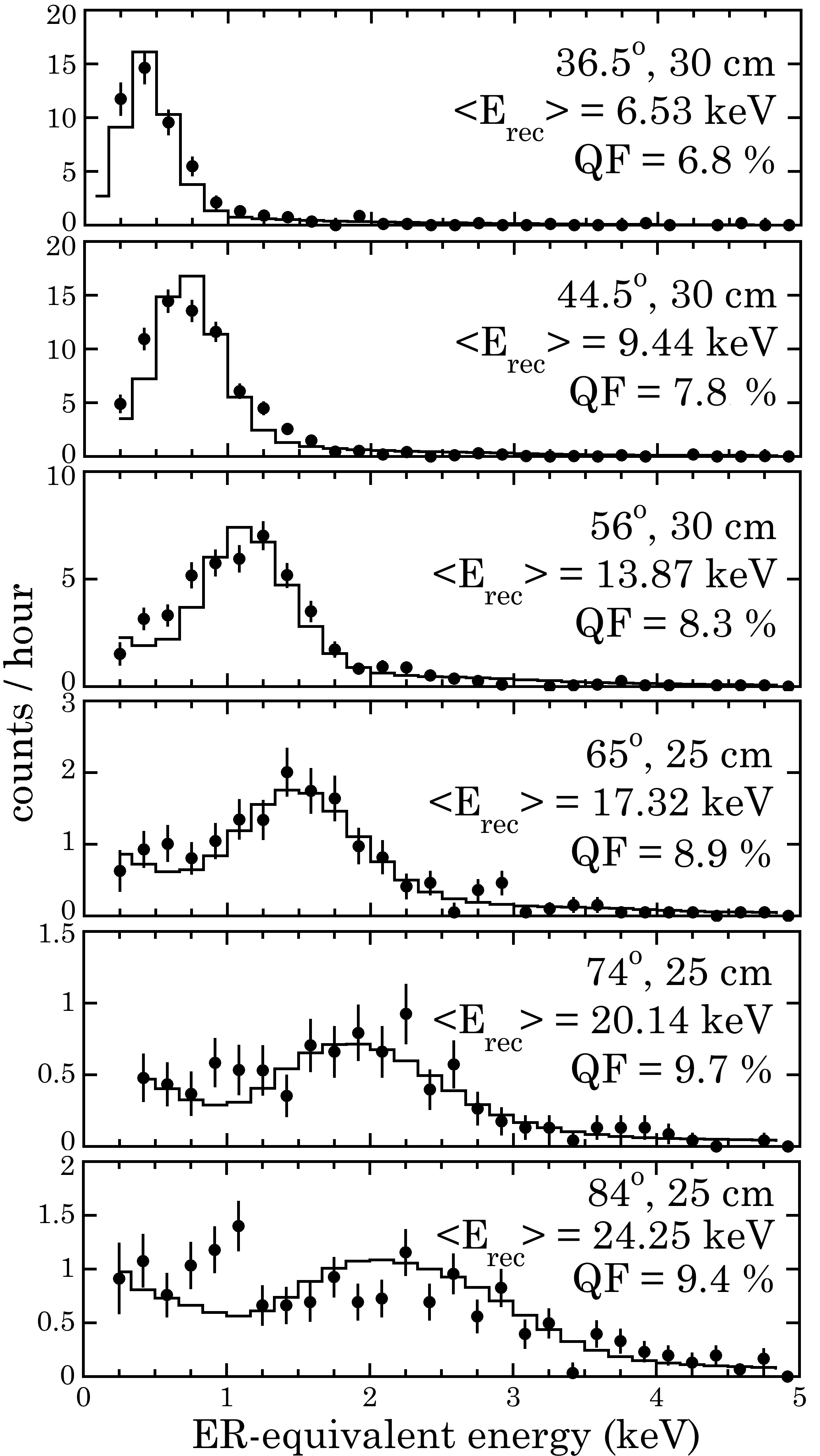
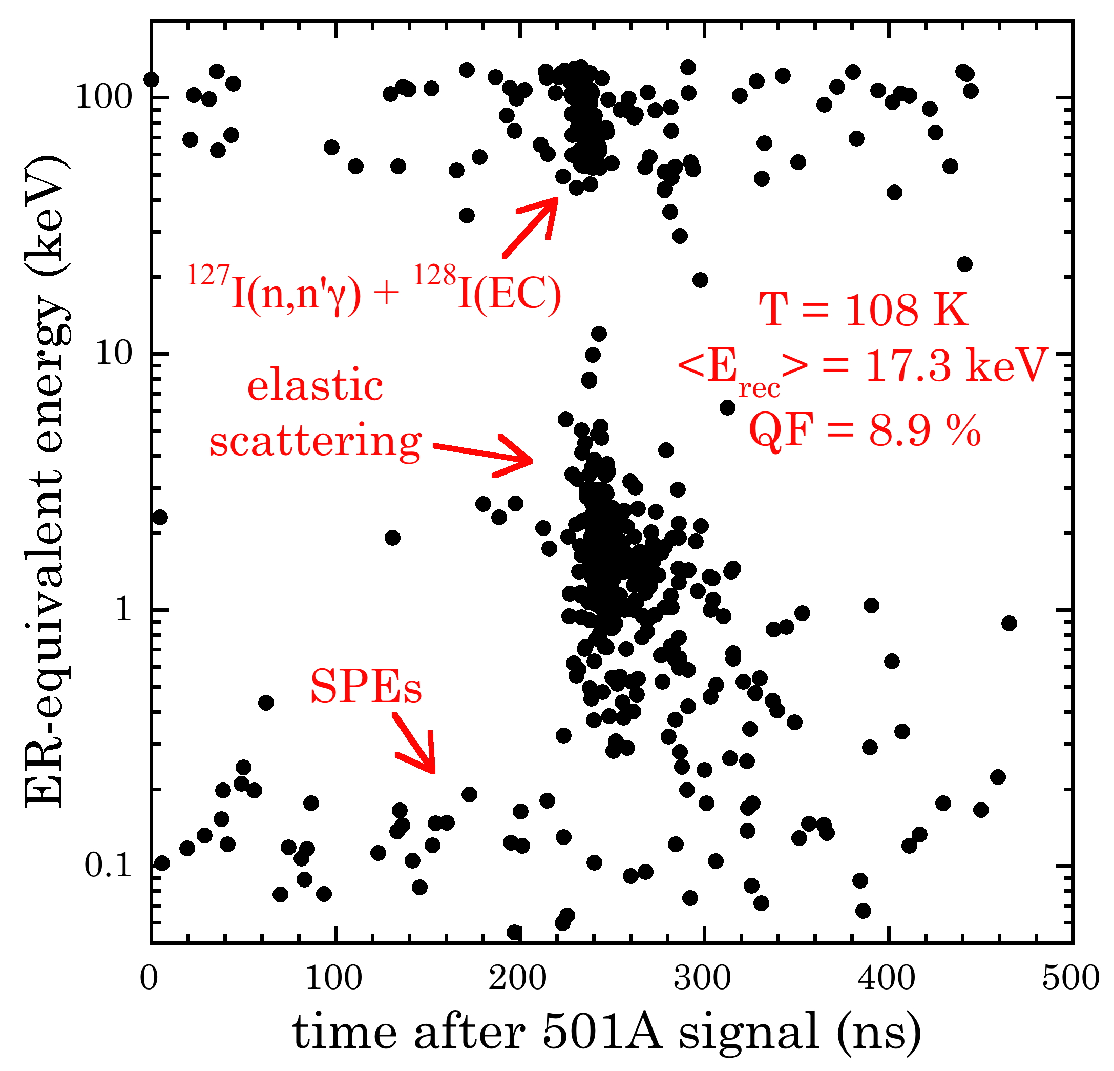
Also following Collar et al. (2019), we utilize an integrated rise-time (IRT) analysis Luo et al. (2014); Ronchi et al. (2009) to separate neutron- from gamma-induced events in the Bicron 501A backing detector. The resulting data quality is illustrated in Fig. 4 following rejection of initially-dominant gamma contaminations. The modest background of random coincidences between CsI and backing detector is removed by subtraction of the energy spectrum of events within the 105-225 ns time range of Fig. 4 from that for true coincidences concentrated within the 225-345 ns interval. The residual spectra of NR signals from elastic neutron scattering at 108 K are shown in Fig. 5, for each of the six scattering angles explored.
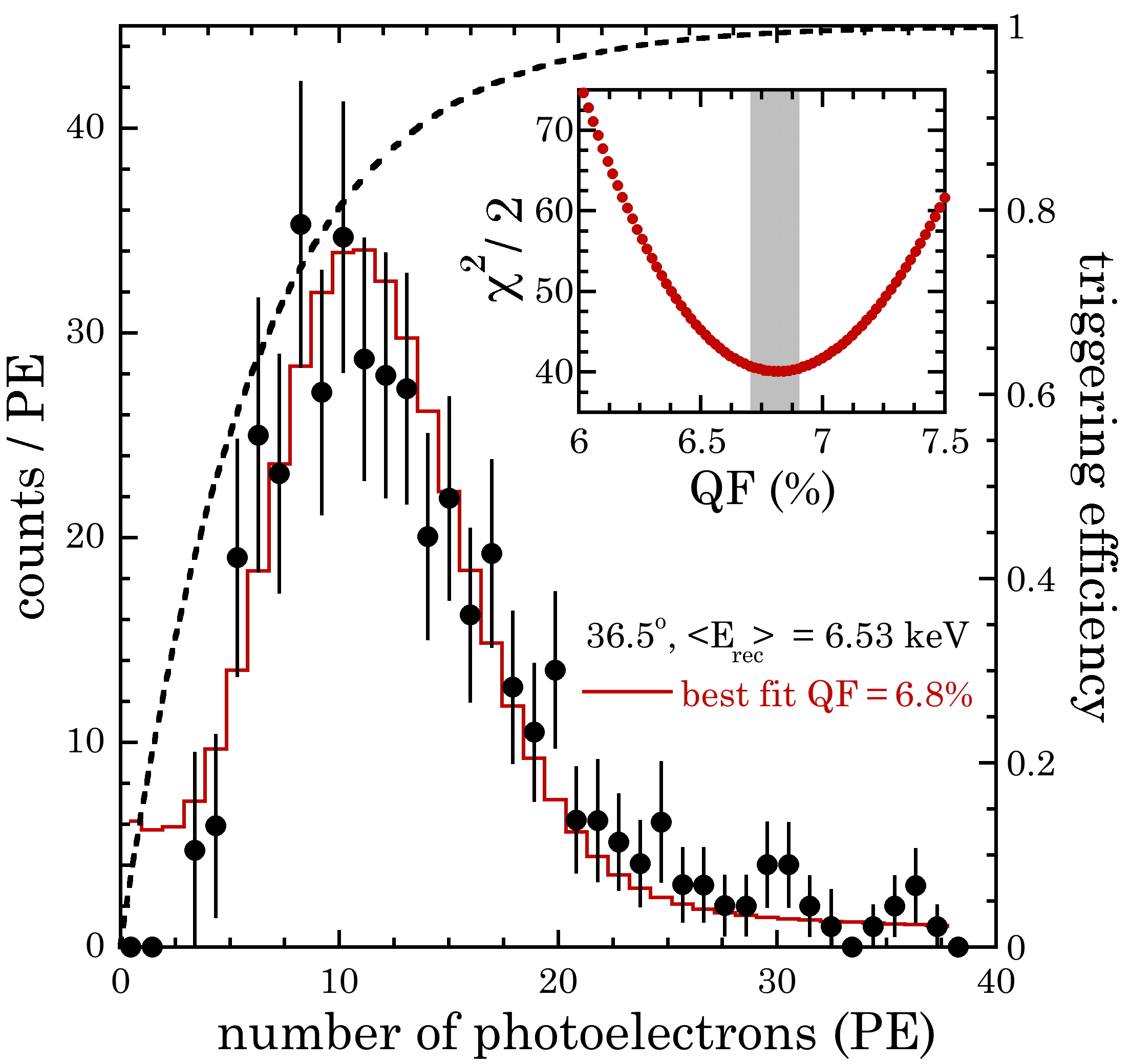
The extraction of a best-fit QF is accomplished identically to Collar et al. (2019): simulated energy depositions are translated to a corresponding number of photoelectrons (PE), accounting for Poisson smearing of PE statistics and the effect of the assumed QF. The obtained simulated distributions are then compared with the experimental residuals of Fig. 5, with a log-likelihood analysis selecting the most adequate QF. Fig. 6 illustrates this procedure for the lowest-energy NRs measured. The uncertainties in the QF values, manifested as vertical error bars in Fig. 7, combine the 1-sigma log-likelihood error and the small dispersion in the 241Am light yield. This energy reference was measured repeatedly during these runs. Horizontal error bars in Fig. 7 are akin to those in Collar et al. (2019), i.e., derived from the simulated spread in NR energies probed.
The totality of our QF measurements for pure CsI at 108 K are reported in Fig. 7. An excellent match to the modified Birks model developed for room-temperature CsI[Na] in Collar et al. (2019) is noticeable. At least from the point of view of the adiabatic factor included in that model this agreement is not surprising: the band gap on which this adiabatic factor depends is not expected to change significantly from room-temperature to 108 K Varshni (1967); Karo and Hardy (1968), an argument supported by observations in other cryogenic scintillators Spassky et al. (2015). However, this apparent constancy of the QF for NRs over the 108-295 K temperature range is in contrast with a reported factor of 7 increase with decreasing temperature in the QF for alpha particles, over the same range, for this material Nadeau (2015); Clark et al. (2018).
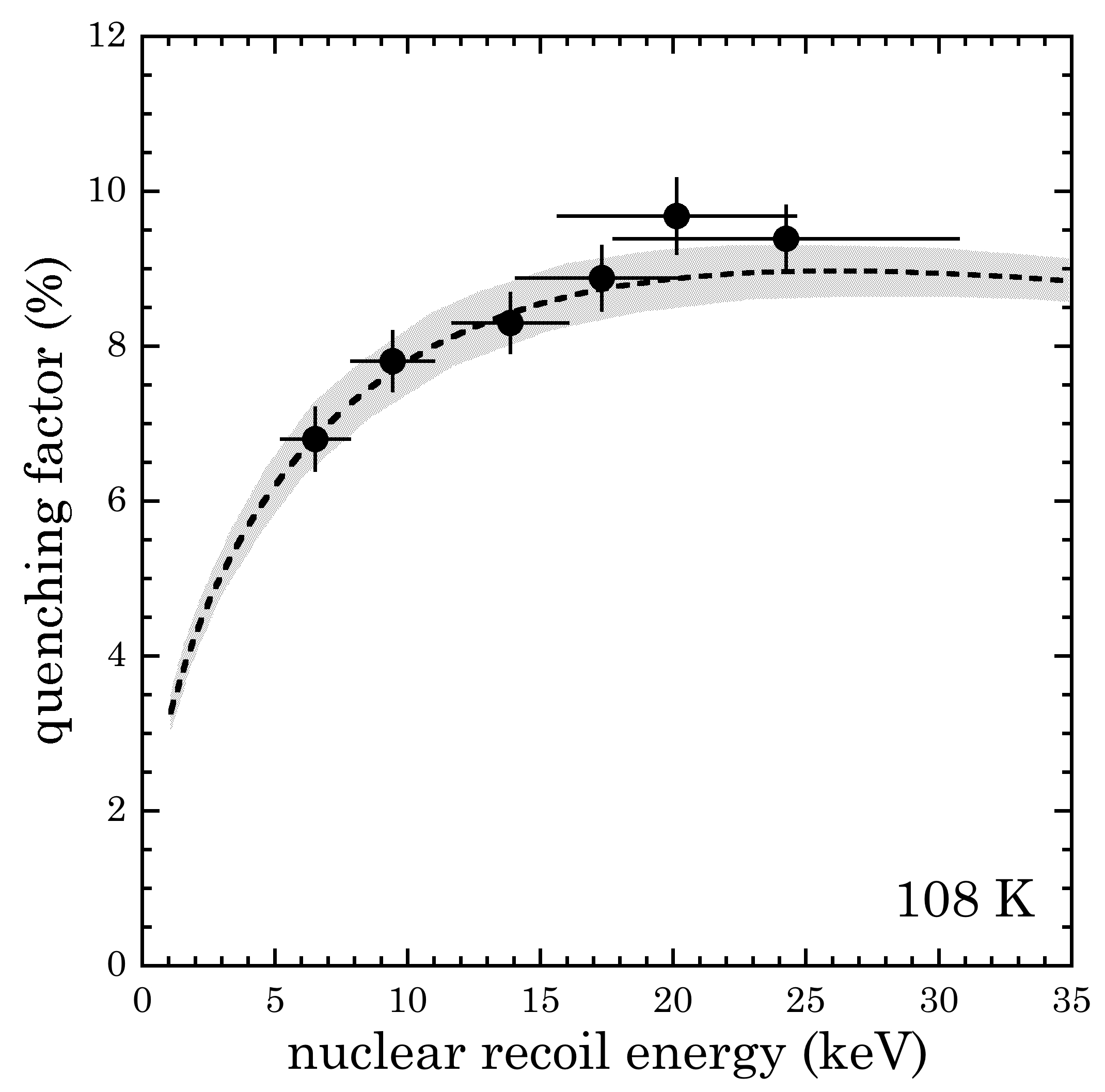
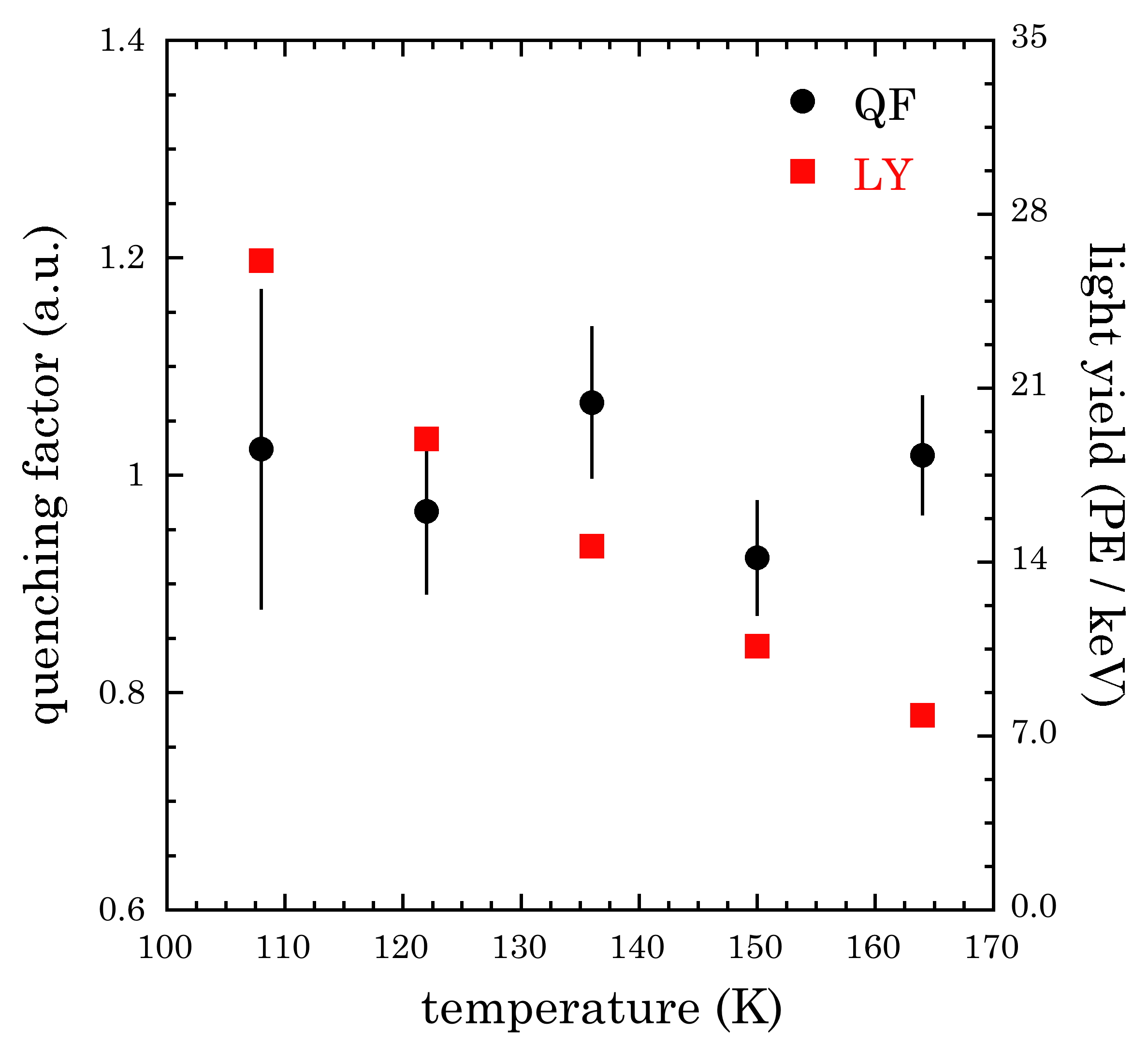
To confirm the observed independence of the QF on operating temperature, measurements at the 56∘ scattering angle (13.9 keV NR energy) were repeated for four additional temperatures, up to 165 K. The maximum temperature that could be explored was limited by the rapidly decreasing light yield. Fig. 8 shows the result of these measurements. As expected, no statistically-significant variation in the QF is visible, over a temperature range for which the overall light yield nevertheless more than tripled.
III ancillary measurements
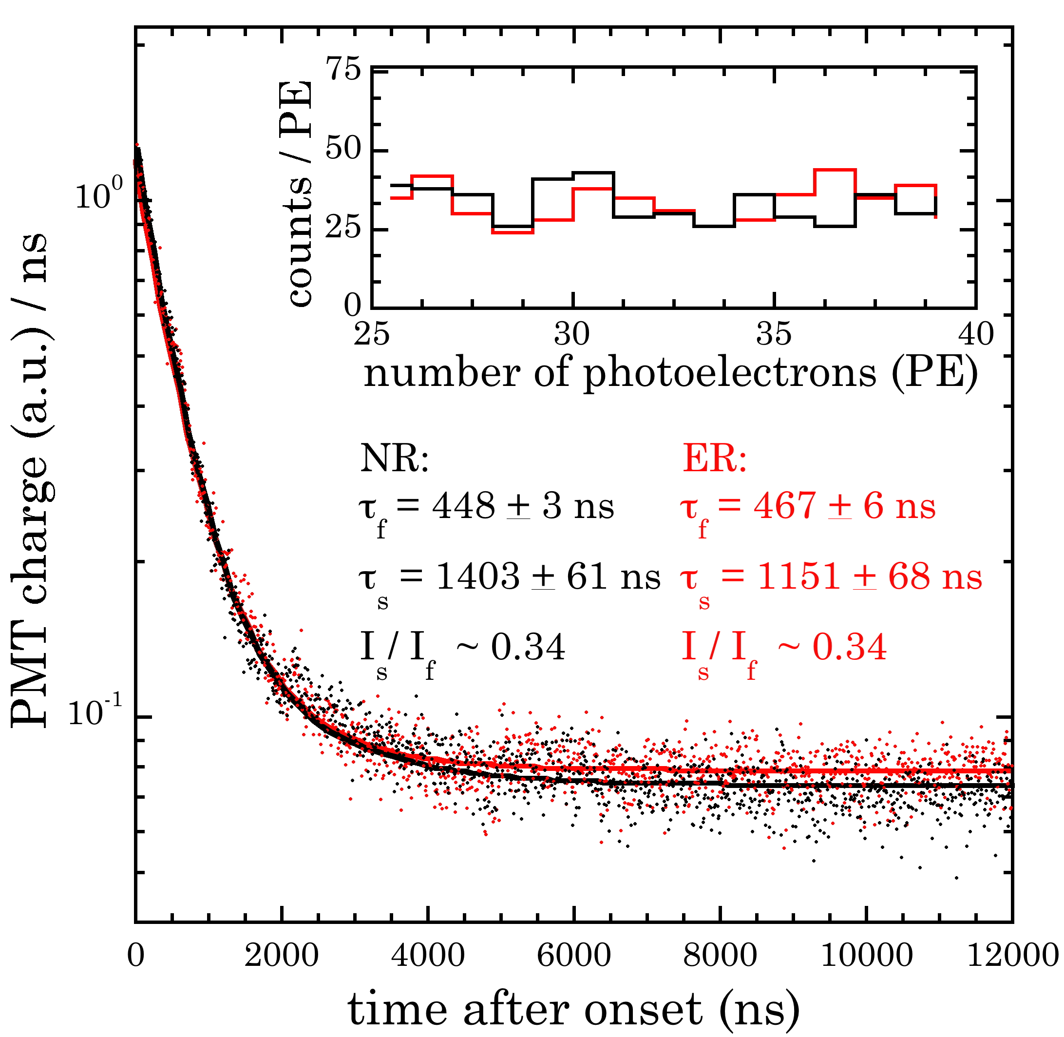
Small differences in the scintillation decay properties of ERs and NRs have been exploited in past experiments. Even when these are too subtle for event-by-event ER-NR discrimination they can still be applied to a large enough ensemble of events, statistically improving the sensitivity of a search for rare NR events Smith et al. (1996). To explore this possibility, a dedicated ER data set was collected containing Compton scatters from a collimated beam of 133Ba gammas impinging on the CsI crystal. Low-energy events were favored by triggering the DAQ on coincidences with a backing detector placed at a small angle with respect to the incoming beam Scholz (2017). Five hundred events were selected from this data set, and the same number from available NR data, with the criterion that both groups should have similar distributions in the number of PE registered per event (Fig. 9 inset, Collar et al. (2015)). This PE range selection corresponds to a NR energy of 15-25 keV. Waveforms within these two subsets were co-added, aligning all traces at the position of the first PE in each. The resulting artificial spike in PMT current at time-zero was removed from the analysis Collar et al. (2015). The average ER and NR traces thus obtained, shown in Fig. 9, were fitted allowing for fast and slow scintillation decay components Amsler et al. (2002); Woody et al. (1990). The PMT overshoot corrections for each data subset had identical decay components, to avoid introducing an artifact in the fits. This direct comparison between few-keV ER and NR events in CsI at 108 K shows only subtle differences, probably too difficult to exploit even for statistical ER-NR discrimination.
Separately, exposures to a variety of gamma-emitting radiosotopes were obtained in order to define the light yield proportionality of pure CsI at 108 K. A lowest-energy datapoint at 5.9 keV was acquired by placing an evaporated 55Fe source adjacent to the CsI crystal, in contact with its PTFE reflector. These results are displayed in Fig. 10, along with all other available similar data for this material Moszynski et al. (2005); Lu et al. (2015); Salakhutdinov and Efanov (2015). Attempts have been made to understand the considerable dispersion in these results as a function of operating temperature and of CsI sample origin Lu et al. (2015). Our measurements using Amcrys/Proteus stock pro show a characteristic absence of reduction in light yield below 30 keV ER energy, seen in other datasets. The deviation from the assumption made in the definition of the QF that direct proportionality exists below 59.5 keV ER energy, is modest for our data. As emphasized in Collar et al. (2019), this assumption is in any case immaterial as long as the NR energy scale it defines is applied consistently to both QF calibrations and in the interpretation of physics runs.
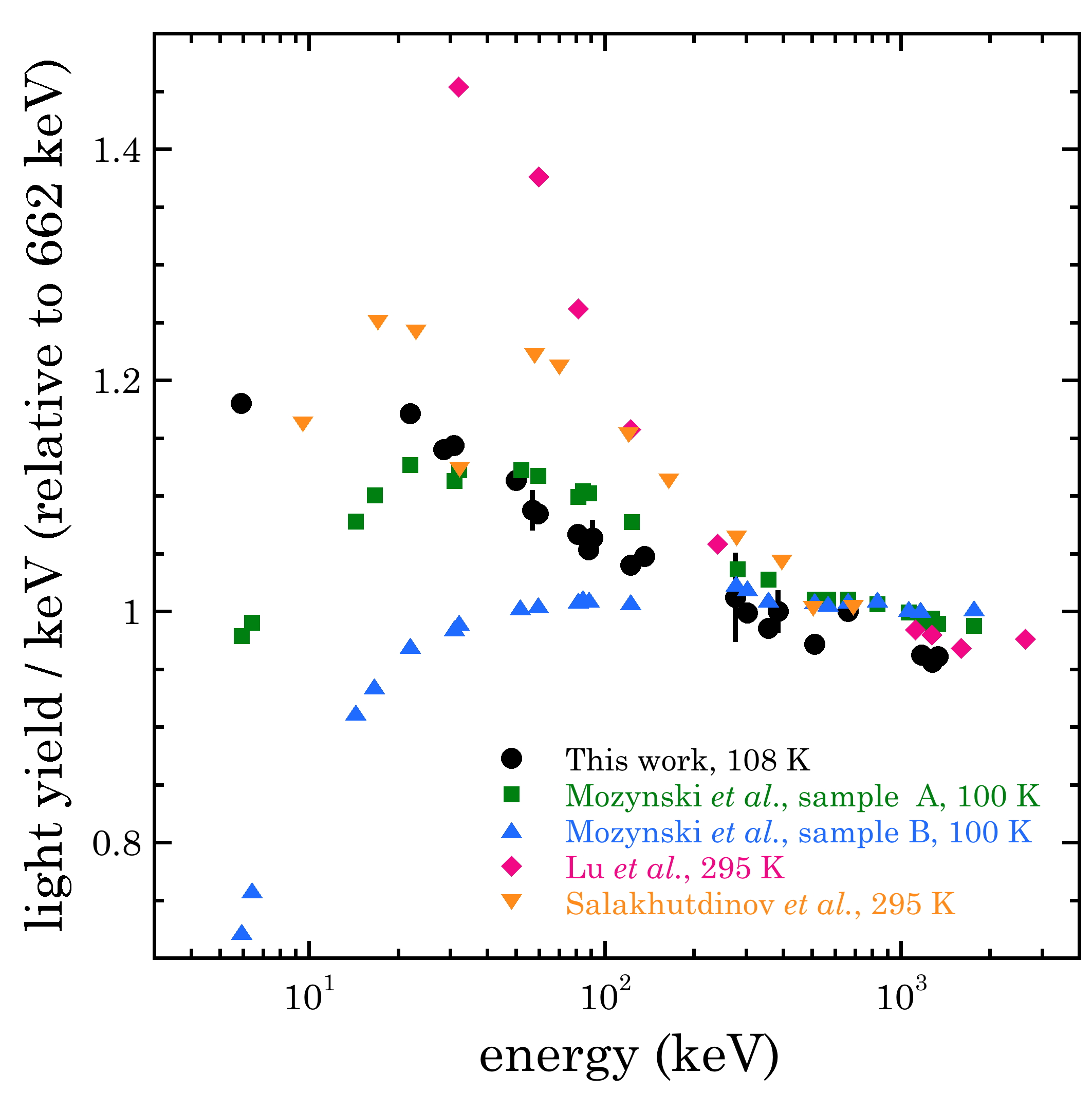
The light yield previously demonstrated for pure CsI at liquid nitrogen temperature (80 K) is in the range of 80-125 scintillation photons per keV at a reference ER energy of 662 keV Amsler et al. (2002); Moszynski et al. (2003); Clark et al. (2018); Liu et al. (2016), displaying a dependence on CsI stock Moszynski et al. (2005); Donghia (2016). The presently measured yield is 26.13 0.37 photoelectrons per keV at 108 K (Fig. 8). Accounting for the 25 quantum efficiency of R8520-506 PMTs at the 340 nm emission characteristic of cryogenic pure CsI Woody et al. (1990); Amsler et al. (2002), and a non-proportionality of 8.5 between 59.5 keV and 662 keV (Fig. 10), gives 96.3 1.4 scintillation photons per keV at 108 K, based on our data. Following the temperature trends observed in Amsler et al. (2002); Mikhailik et al. (2015); Woody et al. (1990); Zhang et al. (2018) and Fig. 8, an additional 10% increase is to be expected at the minimum 87 K operating temperature of present-day cryogenic bialkali PMTs. As emphasized in Zhang et al. (2018); Clark et al. (2018); Baxter et al. (2020); Liu et al. (2016), this uncommonly-high yield is ideal for low-energy NR detection.
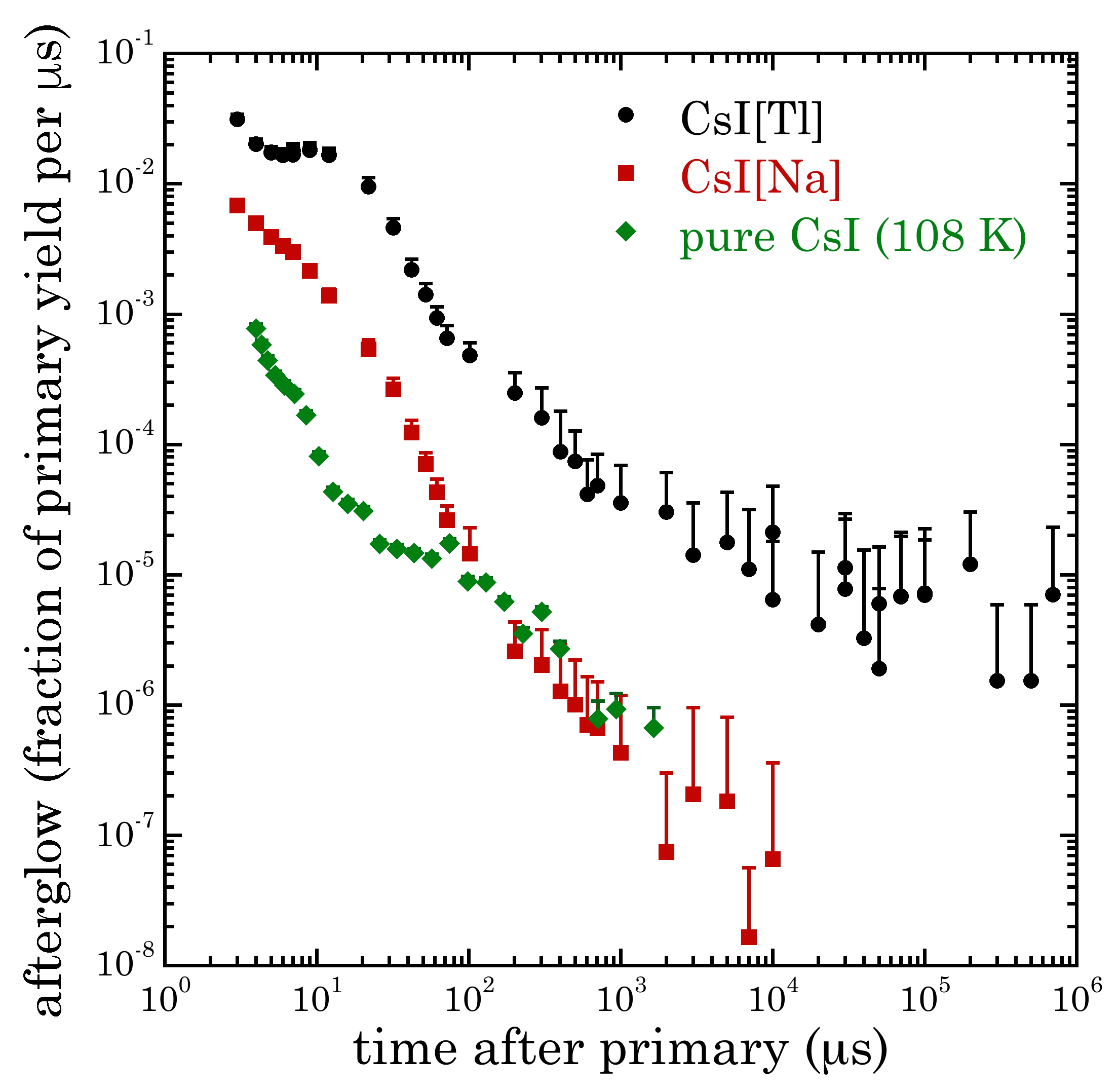
A final study was performed to quantify the afterglow (phosphorescence) of cryogenic pure CsI, thus far also an unknown quantity. These long-delayed few-PE emissions following a primary energy deposition can lead to a continuum of low-energy pulses that impede the identification of NRs, raising the effective threshold of the detector. The abatement of afterglow is of particular importance for CENS searches, performed without the benefit of a significant overburden, and therefore subject to frequent, energetic primaries from cosmic-ray traversal. CsI[Na] was preferred over CsI[Tl] during the first CENS measurement for this reason Collar et al. (2015): however, the removal of residual afterglow events still resulted in significant signal acceptance losses Akimov et al. (2017); Scholz (2017). Fig. 11 illustrates our results, following the same procedure as in Collar et al. (2015) (integration of afterglow over 1 s periods following a 1.5 MeV primary). The substantial further inhibition of this process for cryogenic CsI is immediately noticeable to an experimenter upon simple inspection of oscilloscope traces: this bodes well for its use in the detection of faint scintillation signals.
IV Conclusions
We have presented a first measurement of the quenching factor for low-energy nuclear recoils in undoped CsI at cryogenic temperature. Our results indicate that it is indistinguishable from that for room-temperature CsI[Na], further validating a physical response model developed for that scintillator in Collar et al. (2019), and the modified interpretation of the first CENS measurement described in that same publication. The combination of a sizeable and by now well-understood NR quenching factor, negligible afterglow, and light yield in excess of 100 scintillation photons per keV defines an exceptionally promising material for WIMP and CENS detection. Specifically, when combined with high quantum-efficiency light sensors, cryogenic CsI can provide a sensitivity to 1 keV nuclear recoils, a new frontier for scintillating materials Baxter et al. (2020). In the context of the high neutrino flux expected from the upcoming European Spallation Source, a small array of cryogenic CsI scintillators can provide an unprecedented sensitivity to physics beyond the Standard Model via CENS studies Baxter et al. (2020).
Additional work using a 88Y/Be photoneutron source and the QF measurement technique laid out in Scholz et al. (2016); Collar (2013b); Chavarria et al. (2016) is planned. This will probe nuclear recoils below 4.6 keV in CsI. A search for low-energy deviations from the model depicted in Fig. 7, stemming from Migdal-like processes Ibe et al. (2017); Kahn et al. (2020); Knapen et al. (2020), should be possible using this unique material.
V Acknowledgements
We are indebted to P. Parkhurst at Proteus Inc. for many useful consultations. This work was supported by NSF awards PHY-1806722, PHY-1812702, and by the Kavli Institute for Cosmological Physics at the University of Chicago through an endowment from the Kavli Foundation and its founder Fred Kavli.
References
- Ahlen et al. (1987) S. Ahlen, F. Avignone, R. Brodzinski, A. Drukier, G. Gelmini, and D. Spergel, Phys. Lett. B 195, 603 (1987).
- Freedman (1974) D. Z. Freedman, Phys. Rev. D 9, 1389 (1974).
- Akimov et al. (2017) D. Akimov et al., Science 357, 1123 (2017).
- Scholz (2017) B. Scholz, Ph.D. thesis, University of Chicago (2017), arXiv:1904.01155 .
- Collar et al. (2015) J. I. Collar, N. E. Fields, M. Hai, T. W. Hossbach, J. L. Orrell, C. T. Overman, G. Perumpilly, and B. Scholz, Nucl. Instr. Meth. A 773, 56 (2015).
- Fields (2014) N. Fields, Ph.D. thesis, University of Chicago (2014).
- Collar et al. (2019) J. I. Collar, A. R. L. Kavner, and C. M. Lewis, Phys. Rev. D 100, 033003 (2019).
- Amsler et al. (2002) C. Amsler, D. Grogler, W. Joffrain, D. Lindelof, M. Marchesotti, P. Niederberger, H. Pruys, C. Regenfus, P. Riedler, and A. Rotondi, Nucl. Instr. Meth. A 480, 494 (2002).
- Moszynski et al. (2005) M. Moszynski, M. Balcerzyk, W. Czarnacki, M. Kapusta, W. Klamra, P. Schotanus, A. Syntfeld, M. Szawcowski, and V. Kozlov, Nucl. Instr. Meth. A 537, 357 (2005).
- Moszynski et al. (2003) M. Moszynski, W. Czarnacki, W. Klamra, M. Szawlowski, P. Schotanus, and M. Kapusta, Nucl. Instr. Meth. A 504, 307 (2003).
- Nadeau (2015) P. Nadeau, Ph.D. thesis, Queen’s University (2015).
- Clark et al. (2018) M. Clark, P. Nadeau, S. Hills, C. Dujardin, and P. D. Stefano, Nucl. Instr. Meth. A 901, 6 (2018).
- Liu et al. (2016) J. Liu, M. Yamashita, and A. Soma, J. Instrum. 11, P10003 (2016).
- Woody et al. (1990) C. L. Woody, P. W. Levy, J. A. Kierstead, T. Skwarnicki, Z. Sobolewski, M. Goldberg, N. Horwitz, P. Souder, and D. F. Anderson, IEEE Trans. Nucl. Sci. 37, 492 (1990).
- Zhang et al. (2018) X. Zhang, X. Sun, J. Lu, and P. Lu, Radiat. Detect. Technol. Methods 2, 15 (2018).
- Mikhailik et al. (2015) V. B. Mikhailik, V. Kapustyanyk, V. Tsybulskyi, V. Rudyk, and H. Kraus, Phys. Status Solidi (b) 252, 804 (2015).
- Baxter et al. (2020) D. Baxter, J. I. Collar, P. Coloma, C. E. Dahl, I. Esteban, P. Ferrario, J. J. Gomez-Cadenas, M. Gonzalez-Garcia, A. R. L. Kavner, C. M. Lewis, F. Monrabal, J. Muñoz Vidal, P. Privitera, K. Ramanathan, and J. Renner, JHEP 2020, 1 (2020).
- Collar (2013a) J. I. Collar, Phys. Rev. C 88, 035806 (2013a).
- (19) Amcrys stock, Czochralski grown in dedicated furnaces for trace-level dopant concentration. Procured from Proteus Inc., Chagrin Falls, Ohio 44022, USA.
- Pozzi et al. (2003) S. A. Pozzi, E. Padovani, and M. Marseguerra, Nucl. Instr. Meth. A 513, 550 (2003).
- Wright and Wright (2017) A. Wright and T. Wright, The Photomultiplier Handbook (Oxford University Press, 2017).
- Acciarri et al. (2017) R. Acciarri et al., J. Instrum. 12, P08003 (2017).
- Abe et al. (2012) Y. Abe et al. (Double Chooz Collaboration), Phys. Rev. D 86, 052008 (2012).
- Zhang et al. (2019) H. Zhang, Z. Wang, Y. Zhang, Y. Huang, F. Luo, P. Zhang, C. Zhang, M. Xu, J. Liu, Y. Heng, C. Yang, X. Jiang, F. Li, M. Ye, and H. Chen, J. Instrum. 14, T08002 (2019).
- Luo et al. (2014) X. L. Luo et al., Nucl. Instr. Meth. A 767, 83 (2014).
- Ronchi et al. (2009) E. Ronchi, P.-A. Soderstrom, J. Nyberg, E. A. Sunden, S. Conroy, G. Ericsson, C. Hellesen, M. G. Johnson, and M. Weiszflog, Nucl. Instr. Meth. A 610, 534 (2009).
- Varshni (1967) Y. Varshni, Physica 34, 149 (1967).
- Karo and Hardy (1968) A. M. Karo and J. R. Hardy, J. Chem. Phys. 48, 3173 (1968).
- Spassky et al. (2015) D. Spassky, V. Nagirnyi, A. Savon, I. Kamenskikh, O. Barinova, S. Kirsanova, V. Grigorieva, N. Ivannikova, V. Shlegel, E. Aleksanyan, and et al., J. Lumin. 166, 195–202 (2015).
- Smith et al. (1996) P. Smith, G. Arnison, G. Homer, J. Lewin, G. Alner, N. Spooner, J. Quenby, T. Sumner, A. Bewick, J. Li, D. Shaul, T. Ali, W. Jones, N. Smith, G. Davies, C. Lally, M. van den Putte, J. Barton, and P. Blake, Phys. Lett. B 379, 299 (1996).
- Lu et al. (2015) X. Lu, Q. Li, G. A. Bizarri, K. Yang, M. R. Mayhugh, P. R. Menge, and R. T. Williams, Phys. Rev. B 92, 115207 (2015).
- Salakhutdinov and Efanov (2015) G. Salakhutdinov and D. Efanov, 58, 345 (2015).
- Donghia (2016) R. Donghia, Nuovo Cimento C 39, 276 (2016).
- Scholz et al. (2016) B. Scholz, A. Chavarria, J. Collar, P. Privitera, and A. Robinson, Phys. Rev. D 94 (2016).
- Collar (2013b) J. I. Collar, Phys. Rev. Lett. 110 21, 211101 (2013b).
- Chavarria et al. (2016) A. E. Chavarria et al., Phys. Rev. D 94, 082007 (2016).
- Ibe et al. (2017) M. Ibe, W. Nakano, Y. Shoji, and K. Suzuki, JHEP 2018, 1 (2017).
- Kahn et al. (2020) Y. Kahn, G. Krnjaic, and B. Mandava, (2020), arXiv:2011.09477 [hep-ph] .
- Knapen et al. (2020) S. Knapen, J. Kozaczuk, and T. Lin, (2020), arXiv:2011.09496 [hep-ph] .