Theory of Active Intracellular Transport by DNA-relaying
Abstract
The spatiotemporal organization of bacterial cells is crucial for the active segregation of replicating chromosomes. In several species, including Caulobacter crescentus, the ATPase ParA binds to DNA and forms a gradient along the long cell axis. The ParB partitioning complex on the newly replicated chromosome translocates up this ParA gradient, thereby contributing to chromosome segregation. A DNA-relay mechanism—deriving from the elasticity of the fluctuating chromosome—has been proposed as the driving force for this cargo translocation, but a mechanistic theoretical description remains elusive. Here, we propose a minimal model to describe force generation by the DNA-relay mechanism over a broad range of operational conditions. Conceptually, we identify four distinct force-generation regimes characterized by their dependence on chromosome fluctuations. These relay force regimes arise from an interplay of the imposed ParA gradient, chromosome fluctuations, and an emergent friction force due chromosome-cargo interactions.
The interior organization of bacterial cells is an essential prerequisite for several vital processes, ranging from chromosome and plasmid segregation to cell division Surovtsev and Jacobs-Wagner (2018). Dedicated active mechanisms ensure the rapid translocation and accurate localization of macromolecular objects, such as low-copy-number plasmids Toro and Shapiro (2010), protein clusters Schumacher et al. (2017); Roberts et al. (2012), and carboxysomes Savage et al. (2010). A prominent example is the translocation of the partition complex in bacteria such as Caulobacter crescentus. One copy of the partition complex — bound to the newly replicated chromosome — translocates rapidly from the old to the new cell pole, resulting in chromosome segregation Jensen and Shapiro (1999). The translocation of the chromosome-bound partition complex depends on a protein gradient: the partition complex follows an increasing amount of the ATPase ParA in the cell Ptacin et al. (2010); Schofield et al. (2010); Shebelut et al. (2010); Surovtsev et al. (2016a). However, the physical principles underlying this directed motion of the partition complex remain unclear.
The ATPase ParA belongs to the widely conserved ParABS partitioning system for chromosome and plasmid segregation Livny et al. (2007). The partitioning complex is a large centromere-like protein-DNA cluster consisting of interacting ParB proteins Livny et al. (2007); Broedersz et al. (2014); Mohl and Gober (1997); Murray et al. (2006); Breier and Grossman (2007). The ATPase ParA exists in an ADP- and ATP-bound form and its prefered location in the cell can change dependent on its nucleotide state Lutkenhaus (2012): As an ATP-bound dimer, ParA binds nonspecifically to DNA and, upon interaction with ParB, its ATPase activity is stimulated leading to detachment of ADP-bound ParA monomers into the cytosol. The interactions of ParA ATPases with the partition complex are necessary for its directed translocation Lutkenhaus (2012).
Various mechanisms have been proposed for force generation Ringgaard et al. (2009); Ptacin et al. (2010); Banigan et al. (2011); Shtylla and Keener (2012); Sugawara and Kaneko (2011), including a class of Brownian-ratchet models Lim et al. (2014); Hu et al. (2015, 2017a, 2017b); Walter et al. (2017). Specifically, a DNA-relay mechanism was suggested Lim et al. (2014); Wiggins et al. (2010), where DNA-bound ParA proteins relay the partition complex up a ParA concentration gradient by exploiting elastic fluctuations of the chromosome Lim et al. (2014); Surovtsev et al. (2016b). It has been argued using simulations, that this model can explain the experimentally observed translocation of the partition complex Lim et al. (2014); Surovtsev et al. (2016b). However, a theoretical description of the DNA-relay force that reveals the dependence of the force on key system parameters is still lacking.
Here, we present an analytic theory for force generation by the DNA-relay mechanism. We compute the relay force by evaluating the stochastic binding of DNA-bound ParA-like proteins to a cargo using a Master equation approach. Conceptually, the predicted relay force originates from the interplay of the ParA gradient, chromosome fluctuations, and an emergent friction force due to the interactions of chromosome-bound ParA proteins with the cargo. These contributions give rise to four distinct force generation regimes, depending on the strength of chromosomal fluctuations and the cytoplasmic friction on the cargo. We thus establish a theoretical framework to characterize the DNA-relay mechanism over a broad range of operational conditions, providing conceptual insight into active directed transport of ParB-like cargos for in vivo Lim et al. (2014); Ietswaart et al. (2014); Schumacher and Søgaard-Andersen (2017); Le Gall et al. (2016) and in vitro Vecchiarelli et al. (2014) settings.
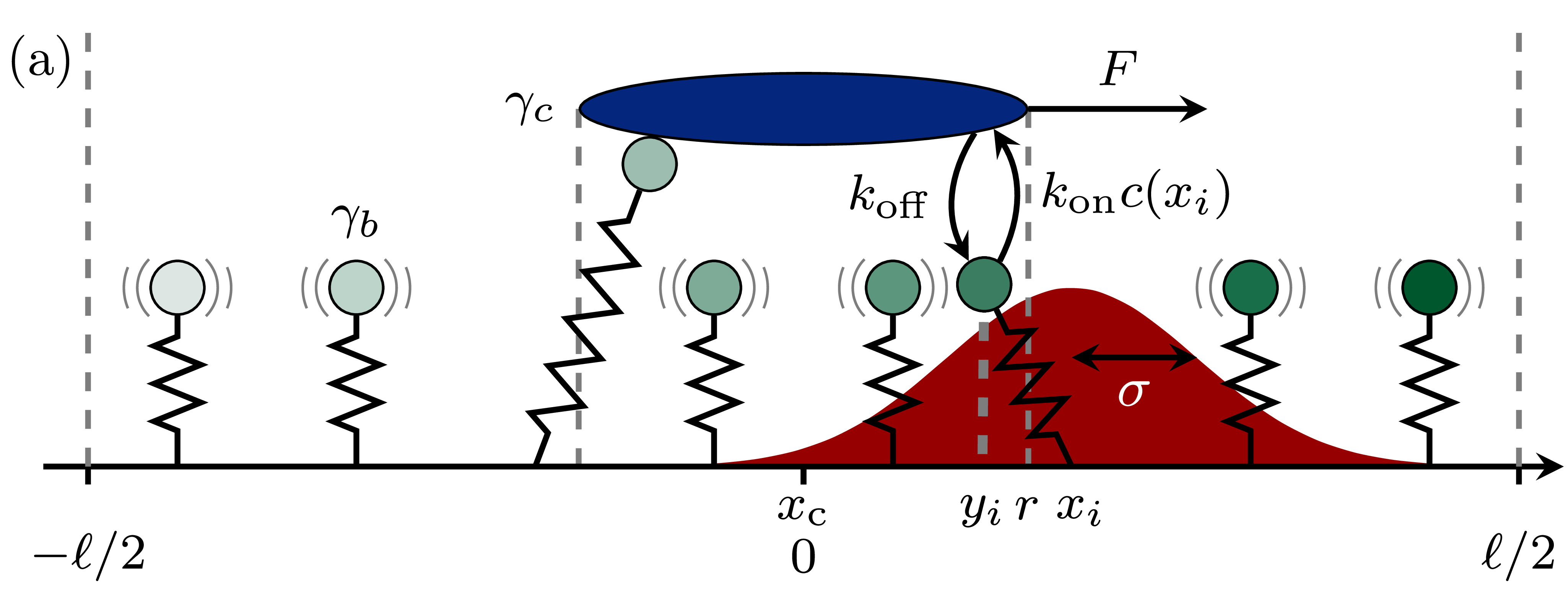
To elucidate force generation by the DNA-relay mechanism Lim et al. (2014); Surovtsev et al. (2016b), we study a minimal model obtained by reducing the full complexity of the partitioning system to key elements important for DNA-relaying (Fig. 1a). Our one-dimensional model consists of the cargo and ParA-bound chromosomal elements. To account for the chromosomal dynamics in a simplified manner, the chromosome is modelled as a set of fluctuating elastic springs. In ParABS-like partitioning systems, the ATPase ParA detaches from the chromosome at the cargo due to stimulation of ATP hydrolysis by ParB, and can only rebind to the chromosome upon ATP binding and dimerization. This dynamics results in a ParA gradient propagating with the cargo, as was shown for an in vitro reconstituted partitioning system Hwang et al. (2013); Vecchiarelli et al. (2014). Instead of modeling the ParA dynamics explicitly, we use this observation by imposing a co-moving ParA gradient on the cargo.
Specifically, the cargo is represented as a line segment of length with a reaction radius , and chromosomal regions are described in a coarse-grained way as a set of beads, equally spaced along a domain of length (Fig. 1a). Each bead is tethered to a fixed position by a spring with stiffness , thermally fluctuating with amplitude . The ParA concentration associated with a chromosomal bead at a distance from the cargo is set to . Cargo and chromosomal elements interact: beads within the reaction radius of the cargo bind with rate . Cargo-bound beads unbind with rate . Importantly, due to the elasticity of the DNA, cargo-bound chromosomal elements exert a force on the cargo. We describe the resulting cargo motion by an overdamped Langevin equation
| (1) |
where is the cargo position and the index runs over all cargo-bound chromosomal elements with rest position and bead position . The white noise term satisfies and , and is the friction coefficient of the cargo in the cytoplasm.
Our goal is to calculate the steady-state DNA-relay force on the cargo for a co-moving ParA gradient. To compute the steady-state DNA-relay force using a finite chromosomal domain of size , we employ periodic boundary conditions, such that there are always chromosomal elements the cargo could interact with Sup . For , the limited number of chromosomal elements becomes important, allowing us to study finite system size effects. In contrast, if , this model is effectively identical to one with an infinite system size.
To facilitate further theoretical analysis we recast variables and system parameters in a non-dimensional form using the system size as a characteristic length, , and the unbinding time as characteristic time scale, . Using this non-dimensionalized form, we identify four key parameters that dictate the system’s dynamics: The binding propensity characterizes the on/off kinetics between the cargo and ParA; the concentration gradient describes the asymmetry of the ParA gradient on the chromosome; sets the magnitude of chromosomal fluctuation relative to system size; and the cargo friction coefficient provides a measure for how susceptible the cargo is to DNA-relay forces Sup .
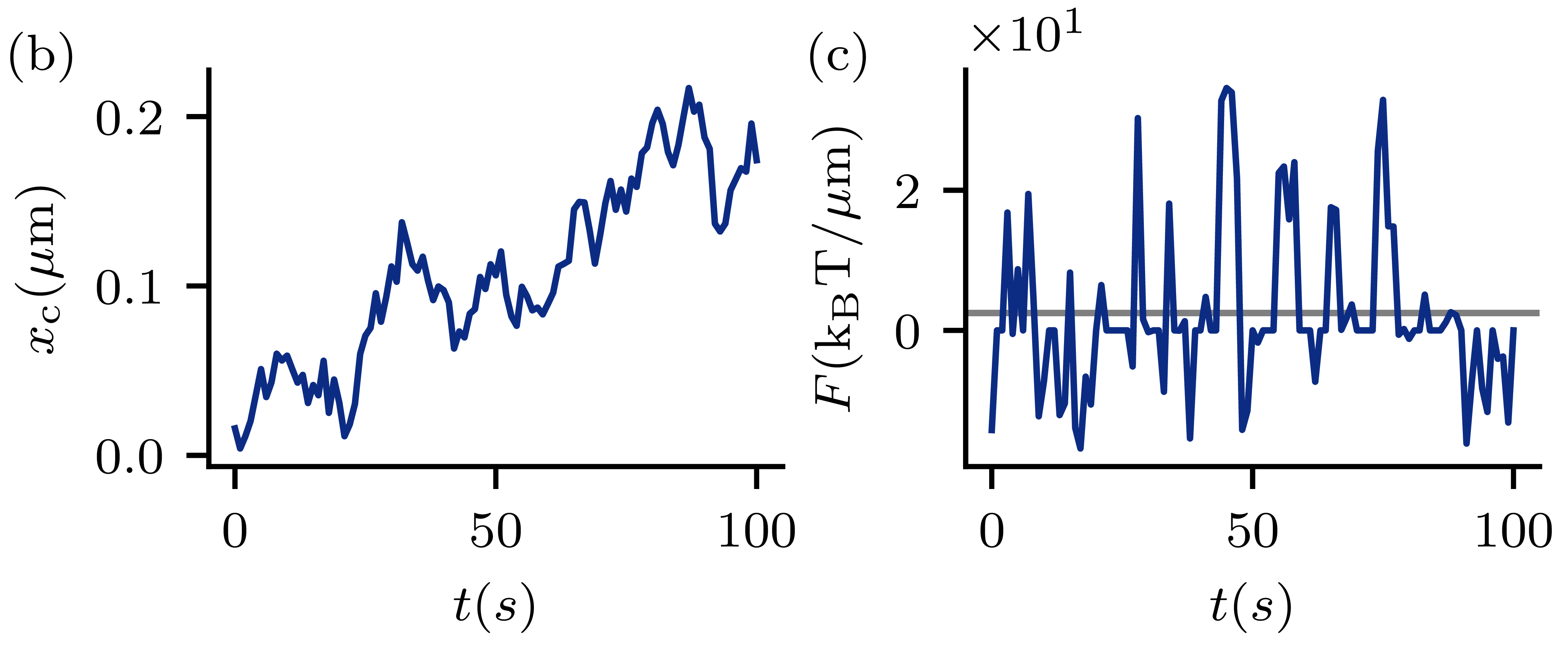
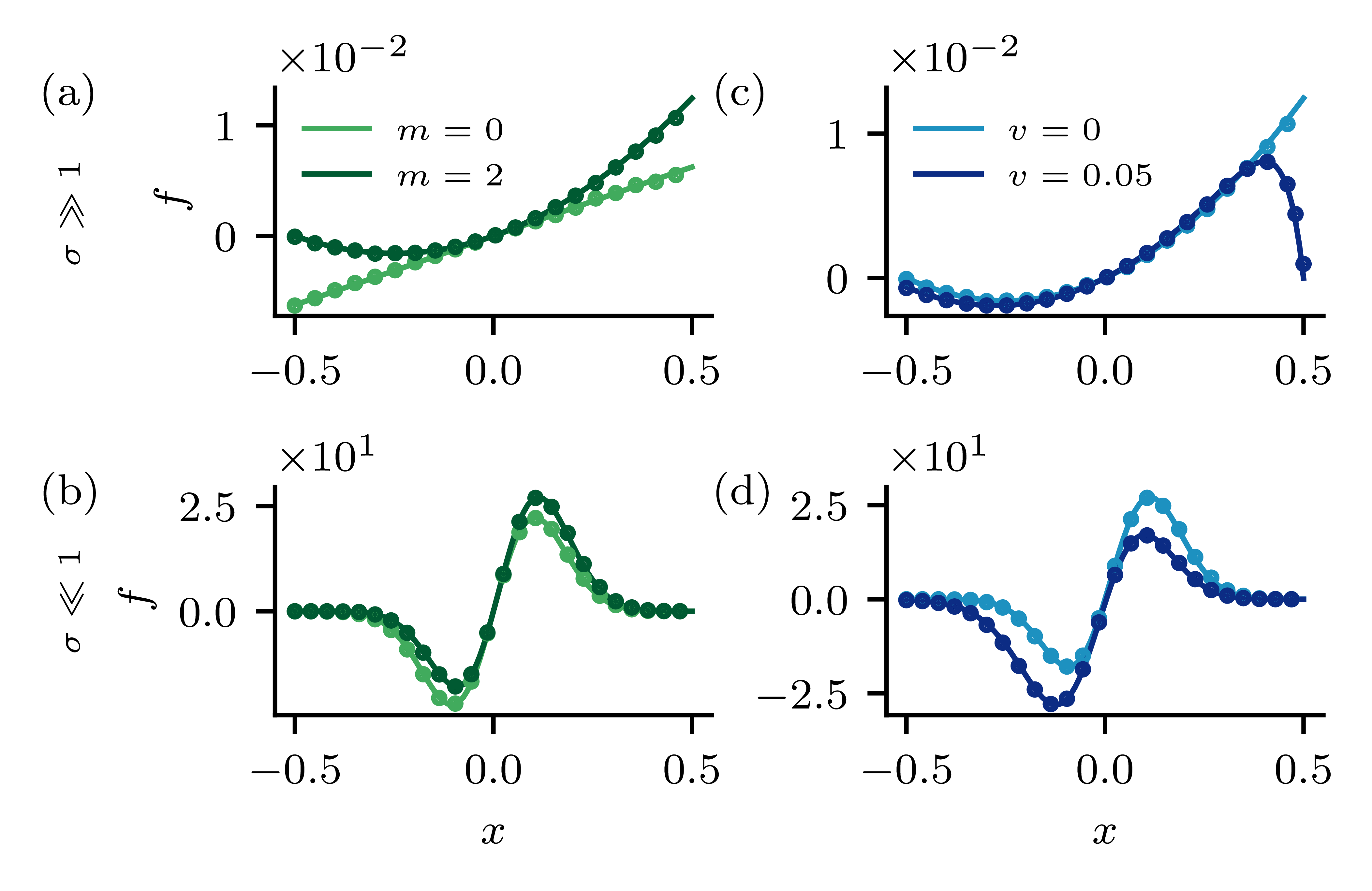
Using Brownian dynamics simulations (Fig. 1b,c) we find distinct force-generation regimes depending on the magnitude of chromosomal fluctuations and the cytoplasmic friction coefficient of the cargo, each characterized by a different dependence on (Fig. 2). While we observe maximal force under stalling conditions (), the system’s behavior changes drastically for a moving cargo (finite ). Interestingly, in this parameter range we find a maximum in the force at intermediate , suggesting an optimal operating regime for this transport mechanism.
To provide conceptual insight into the DNA-relay mechanism, we develop an analytical theory to calculate the relay force on the cargo. Specifically, we derive an approximation for the relay force
| (2) |
which reveals how microscopic system parameters control the DNA-relay mechanism. To obtain an explicit analytical expression, we consider the average relay force, and use a continuum approximation
| (3) |
We moved to the cargo frame of reference, introduced the density of cargo-bound chromosomal elements with a rest position and binding position at the cargo, and defined the force density
| (4) |
Thus, the relay force can be understood by studying the force density , for which we need to calculate .
The dynamics of the density is described by
| (5) |
For a static cargo (), the temporal change in is determined only by a gain and a loss term, corresponding to binding to and unbinding from the cargo. For a binding event, a chromosomal bead needs to move within the reaction radius of the cargo. We describe the position of an unbound bead as a Gaussian random variable with mean and variance . The probability that a bead with rest position is at position is thus given by the Gaussian probability density function (Fig. 1a). This is justified under weak chromosome-cargo interactions, i.e. whenever the decorrelation time is much smaller than the binding time . A binding event takes place stochastically with a rate , accounting for the finite density of chromosomal elements available for binding, where denotes the dimensionless ParA concentration. The total density of cargo-bound chromosomal beads with rest position can be obtained by integrating the density over all possible binding positions on the cargo:
| (6) |
Unbinding is described by a constant detachment rate, set by the last term in Eq. (5). Finally, when the temporal evolution of also includes an advection term to account for cargo motion.
We expect the weak-binding limit () to be the biologically relevant parameter regime in this model, because of the high ParA turnover rate caused by ParB-induced ATP hydrolysis of ParA and subsequent detachment of ParA from the cargo Vecchiarelli et al. (2010). Henceforth, we thus consider only this limit, for which saturation effects of the cargo by bound chromosomal elements are negligible. For completeness, we provide our results for the strong-binding limit Sup and find that the conceptual insights gained from the weak-binding limit largely apply.
Having established a theoretical framework to study force generation by DNA-relaying, we first consider the case of a static cargo (). Put simply, we compute the stalling force of the cargo. This static case allows us to study basic features of the force generation mechanism and provides insights that will also be relevant for the moving cargo scenario. We first calculate the steady-state solution of Eq. (5), and with this an expression for the steady-state force density Sup :
| (7) |
This expression for the force density constitutes one of our key findings and allows us to understand how the DNA-relay force is generated and how it depends on system parameters.
The force density encodes the contribution of a chromosomal element with rest position to force generation. Intuitively, this force density is determined by the interplay between how likely it is for a chromosomal element to bind to the cargo and how much force is exerted on the cargo in this configuration. In the limit , chromosomal beads exhibit strong fluctuations, and without a ParA gradient () every bead thus has approximately the same binding probability. Here, only the distance of a chromosomal element from the cargo matters for force generation and therefore the force density increases linearly with the distance of the bead from the cargo (Fig. 3a, light green). Because of the symmetry of , forces exerted on the cargo from chromosomal elements positioned behind and in front of the cargo cancel, such that no net force is generated. By contrast, if the ParA concentration on the beads increases towards the right , beads in front of the cargo are more likely to bind to the cargo than those behind. Hence, the force density profile becomes asymmetric, resulting in a net positive force (Fig. 3a, dark green). In the regime there is a non-uniform probability for chromosomal beads to bind to the cargo. While chromosomal elements far from the cargo are less likely to bind, they generate the largest force contribution. Consequently, the force density peaks at an intermediate position between the cargo edge and the system boundary (Fig. 3b). Again, in the presence of a ParA gradient becomes asymmetric, resulting in a net force on the cargo. In all cases, our analytical predictions for the force density are in accord with Brownian dynamics simulations.
Having analyzed the steady-state force density , we next evaluate the cargo stalling force in the weak-binding limit using Eq. (3):
| (8) |
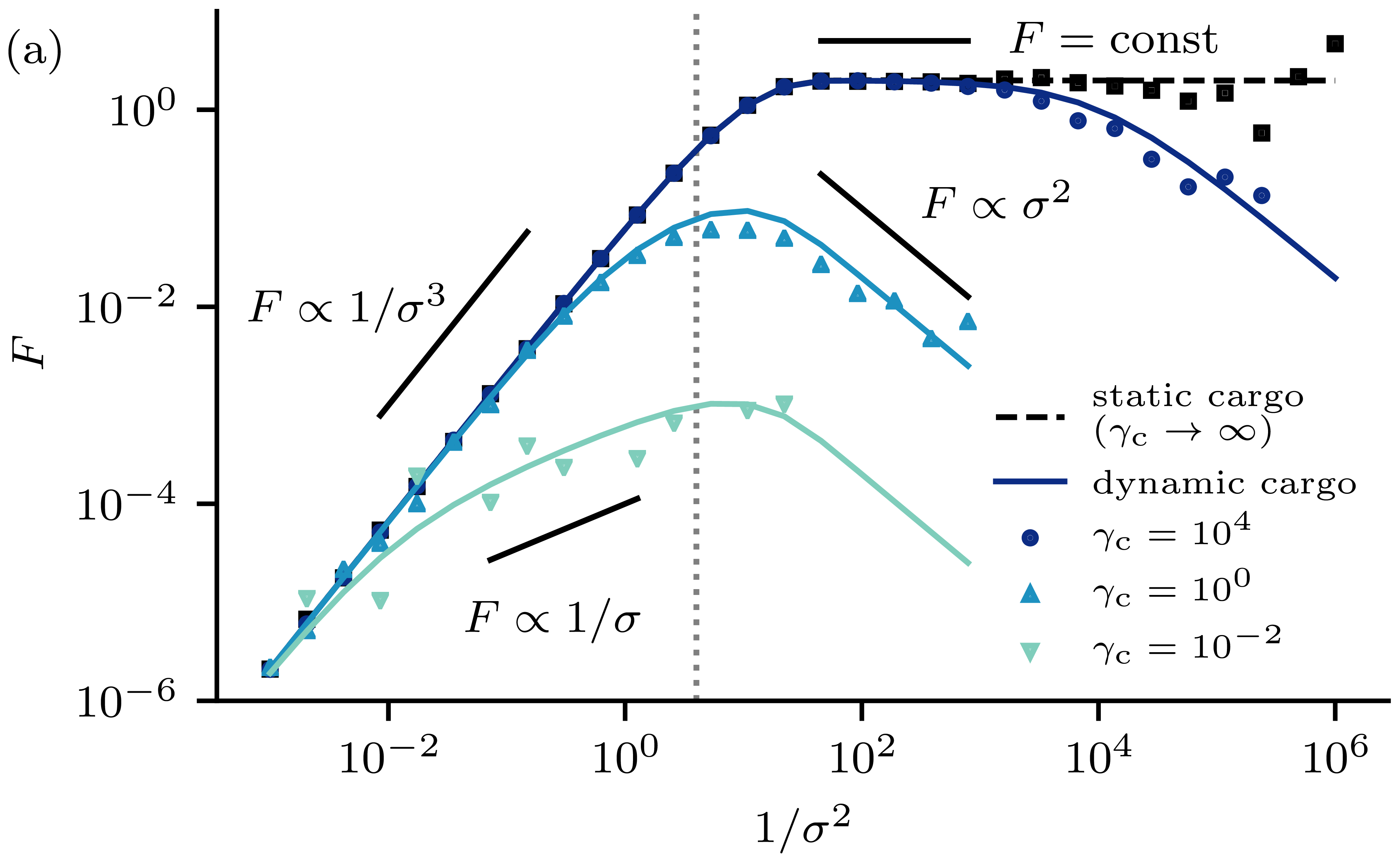
Upon performing this integral, we obtain the dependence of the cargo stalling force on (Fig. 2). Remarkably, for we find that is independent of . Upon increasing , more chromosomal elements are recruited to contribute to force generation. However, this increase in participation is precisely compensated by the softening of the springs resulting in a stiffness independent DNA-relay force . For , we obtain . Here, the finite size of the system affects force generation. Due to the limited number of beads, the softening of the springs can not be compensated anymore by an increased amount of beads interacting with the cargo. Therefore, the force on the cargo decreases.
To understand force generation for a dynamic cargo, we first consider the case of a cargo that moves with an imposed velocity . To this end, we study the steady-state force density, which determines the relay force . We calculate the steady-state solution of Eq. (5) for a fixed velocity and obtain the corresponding force density using Eq. (4). We observe that, for , weight of the binding profile is relocated from the leading (right) to the lagging (left) side of the cargo (Fig. 3c,d, dark blue). This can be understood intuitively: In the case of a dynamic cargo, the forward movement of the cargo and the finite time a chromosomal bead is attached to the cargo (on average ), result in an increased amount of chromosomal beads pulling the cargo backwards.
Interestingly, we find that a moving cargo experiences the force
| (9) |
which has two contributions: the static relay force and an additional force term linear in . This term can be interpreted as an emergent friction force with the friction coefficient , where denotes average number of cargo bound beads for a static cargo Sup .
Next, we use this result for imposed motion to obtain the DNA-relay force exerted on a cargo that moves autonomously due to diffusion and the interactions with ParA-bound beads. First, we self-consistently determine the velocity of a self-propelled cargo using force balance . From this analysis, we obtain an explicit expression for the generated force associated to this translocation velocity
| (10) |
Interestingly, the force on an autonomously moving cargo can be entirely calculated from quantities obtained for a static cargo.
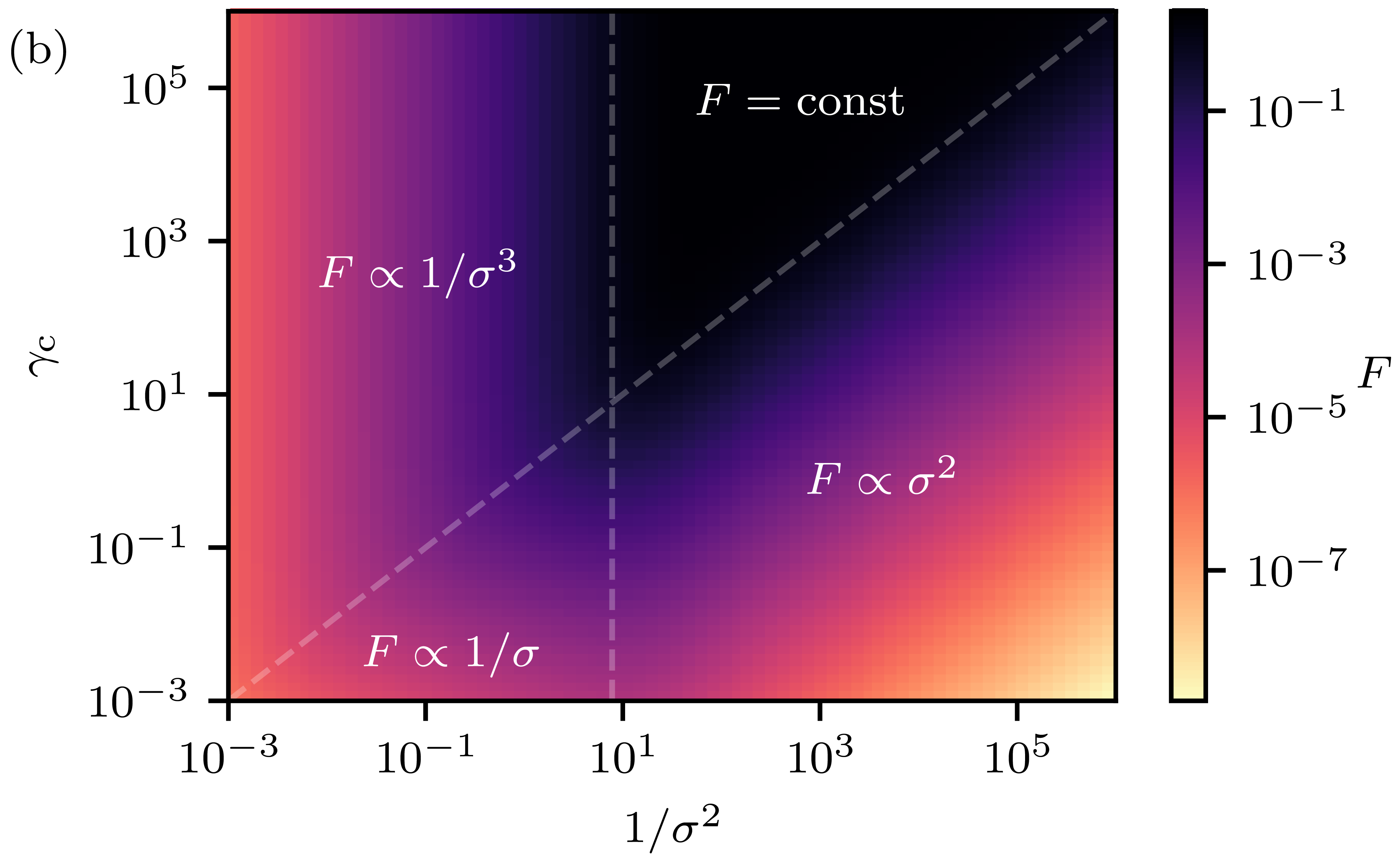
The interplay of self-propulsion and emergent friction force gives rise to four distinct force generation regimes, as depicted in the phase diagram in Fig. 2b. As in the static limit, we can distinguish force generation for small and large chromosomal fluctuations. Importantly however, the qualitative dependencies on the strength of the chromosome fluctuations can differ because of the emergent friction force. In the limit where the cytoplasmic friction dominates the emergent friction, , the dynamic relay force is well approximated by the static relay force (Fig. 2a, black line). Upon lowering the cytoplasmic friction slightly, the emergent friction only reduces force generation for small . Here, the -dependence of the emergent friction, , combines with the constant static cargo force to (Fig. 2a, dark blue line). Upon lowering further the emergent friction also influences the regime . For this parameter regime, the decrease in driving and friction force with increasing combine to (Fig. 2a, light blue line). In the limit , we find that the relay force vanishes, as for a static cargo. In all cases, we find that our analytical predictions agree well with the Brownian dynamics simulations.
Our work complements previous studies on numerically and phenomenologically modeling cargo motion in ParABS-like systems Lim et al. (2014); Jindal and Emberly (2019); Hu et al. (2015, 2017b, 2017a); Surovtsev et al. (2016b); Bergeler and Frey (2018) by providing an analytical microscopic theory for force generation by DNA-relaying. It is still debated whether the main contribution to force generation in ParABS systems derives from chromosome elasticity (DNA-relay force) Wiggins et al. (2010); Lim et al. (2014); Hu et al. (2015, 2017b, 2017a) or chemophoresis Jindal and Emberly (2015); Sugawara and Kaneko (2011); Walter et al. (2017). We contribute to this open question by developing a quantitative mechanistic theory. Our analytical predictions for the dependence of the DNA-relay force on microscopic parameters could be tested in in vitro experiments with a stiffness controlled DNA-carpet Vecchiarelli et al. (2014). In future work, our framework can serve as a starting point for further investigations of force generation in ParABS systems with complex ParA dynamical patterns Ringgaard et al. (2009) and non-equilibrium activity in the chromosome Gnesotto et al. (2018); Weber et al. (2012). Our theory might also be useful more generally for macroscopic cargo translocation driven by stochastic interactions Sabass and Schwarz (2010); Srinivasan and Walcott (2009).
Acknowledgements.
This research was funded by the Deutsche Forschungsgemeinschaft (DFG, German Research Foundation, Project 269423233 - TRR 174). C.H. thanks the Max Planck Institute for the Physics of Complex Systems, Dresden (Germany) for hospitality.References
- Surovtsev and Jacobs-Wagner (2018) I. V. Surovtsev and C. Jacobs-Wagner, Cell 172, 1271 (2018).
- Toro and Shapiro (2010) E. Toro and L. Shapiro, Cold Spring Harb Perspect Biol 2, a000349 (2010).
- Schumacher et al. (2017) D. Schumacher, S. Bergeler, A. Harms, J. Vonck, S. Huneke-Vogt, E. Frey, and L. Søgaard-Andersen, Dev Cell 41, 299 (2017).
- Roberts et al. (2012) M. A. J. Roberts, G. H. Wadhams, K. A. Hadfield, S. Tickner, and J. P. Armitage, Proc Natl Acad Sci U S A 109, 6698 (2012).
- Savage et al. (2010) D. F. Savage, B. Afonso, A. H. Chen, and P. A. Silver, Science 327, 1258 (2010).
- Jensen and Shapiro (1999) R. B. Jensen and L. Shapiro, Proceedings of the National Academy of Sciences 96, 10661 (1999).
- Ptacin et al. (2010) J. L. Ptacin, S. F. Lee, E. C. Garner, E. Toro, M. Eckart, L. R. Comolli, W. Moerner, and L. Shapiro, Nat Cell Biol 12, 791 (2010).
- Schofield et al. (2010) W. B. Schofield, H. C. Lim, and C. Jacobs-Wagner, EMBO J 29, 3068 (2010).
- Shebelut et al. (2010) C. W. Shebelut, J. M. Guberman, S. van Teeffelen, A. A. Yakhnina, and Z. Gitai, Proceedings of the National Academy of Sciences 107, 14194 (2010).
- Surovtsev et al. (2016a) I. V. Surovtsev, H. C. Lim, and C. Jacobs-Wagner, Biophys J 110, 2790 (2016a).
- Livny et al. (2007) J. Livny, Y. Yamaichi, and M. K. Waldor, J Bacteriol 189, 8693 (2007).
- Broedersz et al. (2014) C. P. Broedersz, X. Wang, Y. Meir, J. J. Loparo, D. Z. Rudner, and N. S. Wingreen, Proc Natl Acad Sci U S A 111, 8809 (2014).
- Mohl and Gober (1997) D. A. Mohl and J. W. Gober, Cell 88, 675 (1997).
- Murray et al. (2006) H. Murray, H. Ferreira, and J. Errington, Molecular Microbiology 61, 1352 (2006).
- Breier and Grossman (2007) A. M. Breier and A. D. Grossman, Molecular Microbiology 64, 703 (2007).
- Lutkenhaus (2012) J. Lutkenhaus, Trends Microbiol 20, 411 (2012).
- Ringgaard et al. (2009) S. Ringgaard, J. van Zon, M. Howard, and K. Gerdes, Proc Natl Acad Sci U S A 106, 19369 (2009).
- Banigan et al. (2011) E. J. Banigan, M. A. Gelbart, Z. Gitai, N. S. Wingreen, and A. J. Liu, PLoS Comput Biol 7, e1002145 (2011).
- Shtylla and Keener (2012) B. Shtylla and J. P. Keener, J Theor Biol 307, 82 (2012).
- Sugawara and Kaneko (2011) T. Sugawara and K. Kaneko, Biophysics 7, 77 (2011).
- Lim et al. (2014) H. C. Lim, I. V. Surovtsev, B. G. Beltran, F. Huang, J. Bewersdorf, and C. Jacobs-Wagner, Elife 3, e02758 (2014).
- Hu et al. (2015) L. Hu, A. G. Vecchiarelli, K. Mizuuchi, K. C. Neuman, and J. Liu, Proc Natl Acad Sci U S A 112, 201505147 (2015).
- Hu et al. (2017a) L. Hu, A. G. Vecchiarelli, K. Mizuuchi, K. C. Neuman, and J. Liu, Biophys J 112, 1489 (2017a).
- Hu et al. (2017b) L. Hu, A. G. Vecchiarelli, K. Mizuuchi, K. C. Neuman, and J. Liu, Plasmid 92, 12 (2017b).
- Walter et al. (2017) J.-C. Walter, J. Dorignac, V. Lorman, J. Rech, J.-Y. Bouet, M. Nollmann, J. Palmeri, A. Parmeggiani, and F. Geniet, Phys Rev Lett 119, 028101 (2017).
- Wiggins et al. (2010) P. A. Wiggins, K. C. Cheveralls, J. S. Martin, R. Lintner, and J. Kondev, Proc Natl Acad Sci U S A 107, 4991 (2010).
- Surovtsev et al. (2016b) I. V. Surovtsev, M. Campos, and C. Jacobs-Wagner, Proc Natl Acad Sci U S A 113, E7268 (2016b).
- Ietswaart et al. (2014) R. Ietswaart, F. Szardenings, K. Gerdes, and M. Howard, PLoS Comput Biol 10, e1004009 (2014).
- Schumacher and Søgaard-Andersen (2017) D. Schumacher and L. Søgaard-Andersen, Annu Rev Microbiol 71, 61 (2017).
- Le Gall et al. (2016) A. Le Gall, D. I. Cattoni, B. Guilhas, C. Mathieu-Demazière, L. Oudjedi, J.-B. Fiche, J. Rech, S. Abrahamsson, H. Murray, J.-Y. Bouet, and M. Nollmann, Nat Commun 7, 12107 (2016).
- Vecchiarelli et al. (2014) A. G. Vecchiarelli, K. C. Neuman, and K. Mizuuchi, Proc Natl Acad Sci U S A 111, 4880 (2014).
- Hwang et al. (2013) L. C. Hwang, A. G. Vecchiarelli, Y.-W. Han, M. Mizuuchi, Y. Harada, B. E. Funnell, and K. Mizuuchi, The EMBO Journal 32, 1238 (2013).
- (33) See Supplemental Material at [URL will be inserted by publisher] for a detailed derivation .
- Vecchiarelli et al. (2010) A. G. Vecchiarelli, Y.-W. Han, X. Tan, M. Mizuuchi, R. Ghirlando, C. Biertümpfel, B. E. Funnell, and K. Mizuuchi, Molecular Microbiology (2010).
- Jindal and Emberly (2019) L. Jindal and E. Emberly, PLOS ONE 14, e0218520 (2019).
- Bergeler and Frey (2018) S. Bergeler and E. Frey, PLOS Computational Biology 14, e1006358 (2018).
- Jindal and Emberly (2015) L. Jindal and E. Emberly, PLoS Comput Biol 11, e1004651 (2015).
- Gnesotto et al. (2018) F. S. Gnesotto, F. Mura, J. Gladrow, and C. P. Broedersz, Reports on Progress in Physics 81, 066601 (2018).
- Weber et al. (2012) S. C. Weber, A. J. Spakowitz, and J. A. Theriot, Proceedings of the National Academy of Sciences 109, 7338 (2012).
- Sabass and Schwarz (2010) B. Sabass and U. S. Schwarz, Journal of Physics: Condensed Matter 22, 194112 (2010).
- Srinivasan and Walcott (2009) M. Srinivasan and S. Walcott, Physical Review E 80, 046124 (2009).