Direct measurement of key exciton properties: energy, dynamics and spatial distribution of the wave function
Keywords: condensed matter physics; exciton physics; many-body physics; time-resolved photoemission spectroscopy; ultrafast dynamics; quasi-particle interactions; semiconductors
Abstract
Excitons, Coulomb-bound electron-hole pairs, are the fundamental excitations governing the optoelectronic properties of semiconductors. While optical signatures of excitons have been studied extensively, experimental access to the excitonic wave function itself has been elusive. Using multidimensional photoemission spectroscopy, we present a momentum-, energy- and time-resolved perspective on excitons in the layered semiconductor WSe2. By tuning the excitation wavelength, we determine the energy-momentum signature of bright exciton formation and its difference from conventional single-particle excited states. The multidimensional data allows to retrieve fundamental exciton properties like the binding energy and the exciton-lattice coupling and to reconstruct the real-space excitonic distribution function via Fourier transform. All quantities are in excellent agreement with microscopic calculations. Our approach provides a full characterization of the exciton properties and is applicable to bright and dark excitons in semiconducting materials, heterostructures and devices.
Excitons, bound electron-hole quasi-particles carrying energy and momentum but no net charge, are fundamental excitations of semiconductors and insulators arising from light-matter interaction[1]. An initial excitonic polarization induced by a light field (often referred to as coherent excitons and e.g. detected by optical absorption spectroscopy) rapidly loses coherence with the driving field and dephases into a population of incoherent excitonic states[2, 3]. The generated excitons propagate in solid-state materials through diffusion[4, 5] and eventually release their energy e.g. in the form of luminescence (photon), lattice excitation (phonon) or dissociation into single charged quasi-particles[6, 7, 8, 9]. Understanding exciton physics is of capital importance for advanced photonic and optoelectronic applications including photovoltaics. Layered transition metal dichalcogenide (TMDC) semiconductors exhibit rich exciton physics even at room temperature due to strong Coulomb interaction[10]. Excitons in TMDCs feature large oscillator strength[11] and their inter- and intra-band dynamics have been extensively investigated[12, 13, 14]. Moreover, strong spin-orbit coupling and broken inversion symmetry in each crystalline trilayer lead to a locking between spin, valley and layer degrees of freedom, which started a surge of valley physics studies [15, 16, 17].
A large portion of the research on excitonic phenomena in TMDCs adopts optical spectroscopic techniques[10, 12, 14, 15, 16, 18, 19, 20, 21], which only access bright excitonic transitions with near-zero momentum transfer. While techniques such as time-resolved THz spectroscopy also allow probing optically dark excitons via internal quantum transitions[13], finite-momentum excitons which lie outside the radiative light cone remain inaccessible to such methods. This limitation is overcome by time- and angle-resolved photoemission spectroscopy (trARPES), a spectroscopic tool accessing excited states, including excitons, in energy-momentum space and on ultrafast timescales[17, 22, 23, 24]. Here, we reveal the characteristics of the excitonic wave function in the photoemission signal of the prototypical layered TMDC semiconductor 2H-WSe2 and establish that all fundamental exciton properties are encoded in the trARPES signal’s energy, time, and momentum dimensions: the exciton binding energy, its self-energy as a measure of the exciton-lattice coupling, as well as the real-space distribution of the excitonic wave function.
Fig. 1(a) depicts the experimental scheme of trARPES employing femtosecond near-infrared (NIR) pump and extreme ultraviolet (XUV) probe pulses combined with two types of photoelectron analyzers: a hemispherical analyzer (HA) and a time-of-flight momentum microscope (MM). The whole setup allows us to measure the 3D time-dependent electronic structure in a given energy-momentum-plane with high counting statistics using the HA, and alternatively resolve both in-plane momentum directions yielding a 4D photoemission signal of the entire valence band with the MM[22, 25]. Fig.1 (b-d) and (e-g) show snapshots of the 3D and 4D data with 1.55 eV excitation, respectively, at three selected time delays: i) prior to optical excitation, showing the ground-state band structure of WSe2 from the Brillouin zone (BZ) center (only shown in the MM data) to the BZ boundary K points (b,e); ii) upon optical excitation resonant with the A exciton absorption (the first excitonic state), featuring excited-state signal at the and valleys (c,f); and iii) at fs after optical excitation, with excited-state signal mostly at the valleys (d,g). In the following, we identify the excitonic features in the excited-state photoemission signal and quantify the exciton properties retrieved from the energy, time and momentum dimensions of the 4D trARPES signal.

Photoemission signature of excitons.
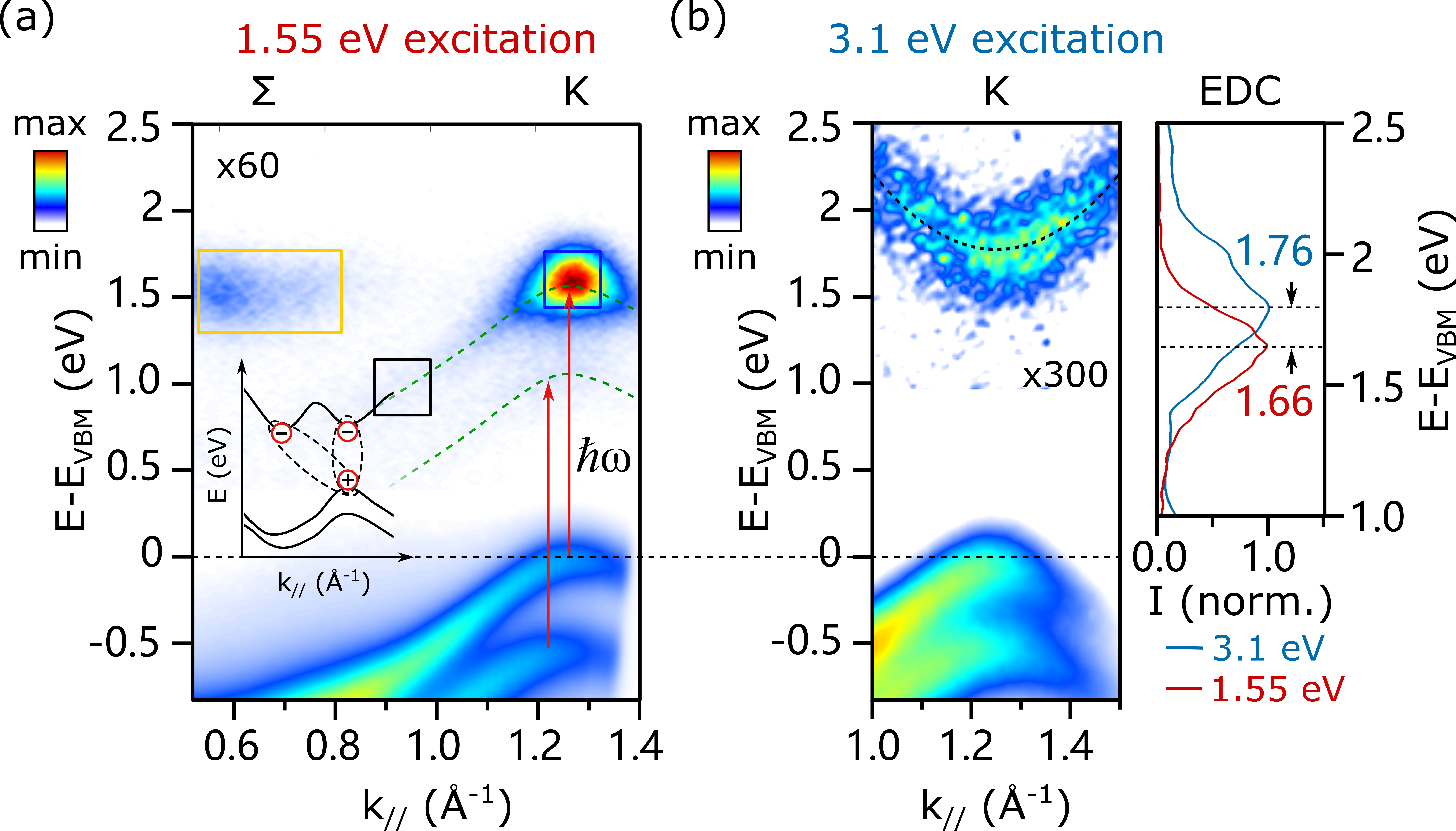
During the photoemission of an electron bound in an exciton, the electron-hole interaction diminishes, i.e. the exciton breaks up, as a single-particle photoelectron is detected while a single-particle hole is left behind in the material[23]. To identify the signature of the excitonic electron-hole interaction in photoemission spectroscopy, we compare the signal of excitons with that of single-particle excited states. For generating excitons, we excite with 1.55 eV photons ( bandwidth = 43 meV), in resonance with the low-energy side of the A-excitonic absorption of bulk WSe2[12]. Fig.2(a) shows the excited-state signal integrated in the first 25 fs after pump-probe overlap. The data reveals a vertical transition at the K point through an excited-state signal localized in energy and momentum. In contrast, the above-band-gap excitation with 3.1 eV photon energy generates a population higher in the conduction band, which rapidly redistributes to all bands and valleys of the lower conduction band (the equivalent scenario applies to the holes in the valence band). Fig.2(b) shows this excited-state signal in the K valley 100 fs after excitation, where carriers have redistributed in energy and momentum. This signal resembles the dispersion of a single-particle band with an effective mass of , in good agreement with electronic band structure calculations[26]. The energetic positions of the excitonic and single-particle states at the K point are determined as the center of mass of the energy distribution curves (EDCs), see Fig.2(b). The excited-state signal upon resonant excitation of the A exciton is centered below the center of the single-particle band. Such a signal below the single-particle band has been predicted as photoemission signature of excitons and the energy difference can be associated with the exciton binding energy [27, 3, 2, 9]. A calculation of the A-exciton binding energy in bilayer WSe2 based on the screened Keldysh-like potential (see SI for details) yields , in very good agreement with the experimental value. It is important to note that we retrieve the exciton binding energy directly from measuring the absolute energies of many-body and single-particle states with a single photoemission experiment, in contrast to combining different experimental methods[18] or by comparing photoemission signals with electronic structure calculations[24]. The observation that the excitonic binding energy is measurable as energy loss of the photoelectron confirms that the hole final states indeed are identical to single-particle holes, which further implies that the localized hole of the exciton transforms to Bloch-like single-particle states during the photoemission process.
To set the stage for discussing the exciton dynamics, we emphasize a signal appearing as replicas of the upper (VB1) and lower (VB2) spin-orbit split valence bands in Fig.2(a) shifted by the photon energy . This signal only appears during temporal pump-probe overlap and we attribute it to a photon-dressed electronic state due to coherent coupling to the optical driving field. Since the employed s-polarized pump light (polarization parallel to the sample surface) suppresses laser-assisted photoemission, the experimental configuration selectively probes the coherent excitonic polarization induced by the pump field [2, 3, 28].

Formation and decay dynamics of bright excitons.
The bright A excitons at the K point are not the lowest-energy excitons in WSe2 but can relax their energy further by scattering in momentum space. We extract the quasiparticle dynamics within three regions of interest (ROIs) from the trARPES data in Fig.2(a), representing the coherent excitonic replica of VB1, the excitonic state at the K valley, and the valley population. The respective time traces in Fig.3(a) reflect three types of quasi-particle dynamics: the dephasing of the coherent excitonic polarization (black), the buildup and relaxation of a bright exciton population at (blue) and the carrier accumulation of dark states at (yellow).
The observed carrier dynamics imply the following microscopic processes as sketched in Fig.3(b). First, the interaction of the initial valence band state with the near-resonant optical light field creates a coherent excitonic polarization (dashed line), which quickly dephases into an optically bright exciton population , offset by the pump detuning . The decoherence process occurs with the pure dephasing time . These bright excitons undergo rapid scattering into the optically dark -point state on the timescale . We model these processes and the photoemission signals from these states into the continuum final states and using a five-level extension to the optical Bloch equations [29, 30] (OBE, see SI). Based on a multivariate least-squares fitting procedure, we can describe the dynamics of coherent and incoherent exciton contributions, obtaining a coherent exciton dephasing time of fs and a population lifetime for the bright A-exciton population of fs. The extracted dephasing time corresponds well to microscopic calculations[31, 14].

To evaluate the mechanism governing the bright exciton scattering, we performed ab initio calculations of the single-particle self-energy of WSe2. At low excitation densities, the electron self-energy is dominated by electron-phonon interaction which is computed using density functional perturbation theory (DFPT), taking into account the electronic screening of the lattice motion (see SI). The imaginary part of the momentum-resolved self-energy is shown in Fig.3(c) encoded by the color scale. From the calculation, we obtain at the conduction band minimum and at the valence band maximum of the valleys. While a rigorous description of exciton-phonon coupling requires treatment on the basis of excitonic eigenstates, in the weak coupling limit, i.e., small self-energy renormalization due to the electron-hole interaction, the exciton-phonon self-energy is dominated by its incoherent contribution[32]. In this case, the exciton-phonon interaction can be approximated as sum of the single-particle-phonon interactions. Our calculated value + meV agrees with the experimental exciton self-energy determined according to . This agreement with theory shows that the exciton lifetime provides a quantitative measure of the strength of its interaction with the lattice and supports the assumption of a dominating incoherent self-energy contribution.
Momentum- and real-space distribution of A excitons.
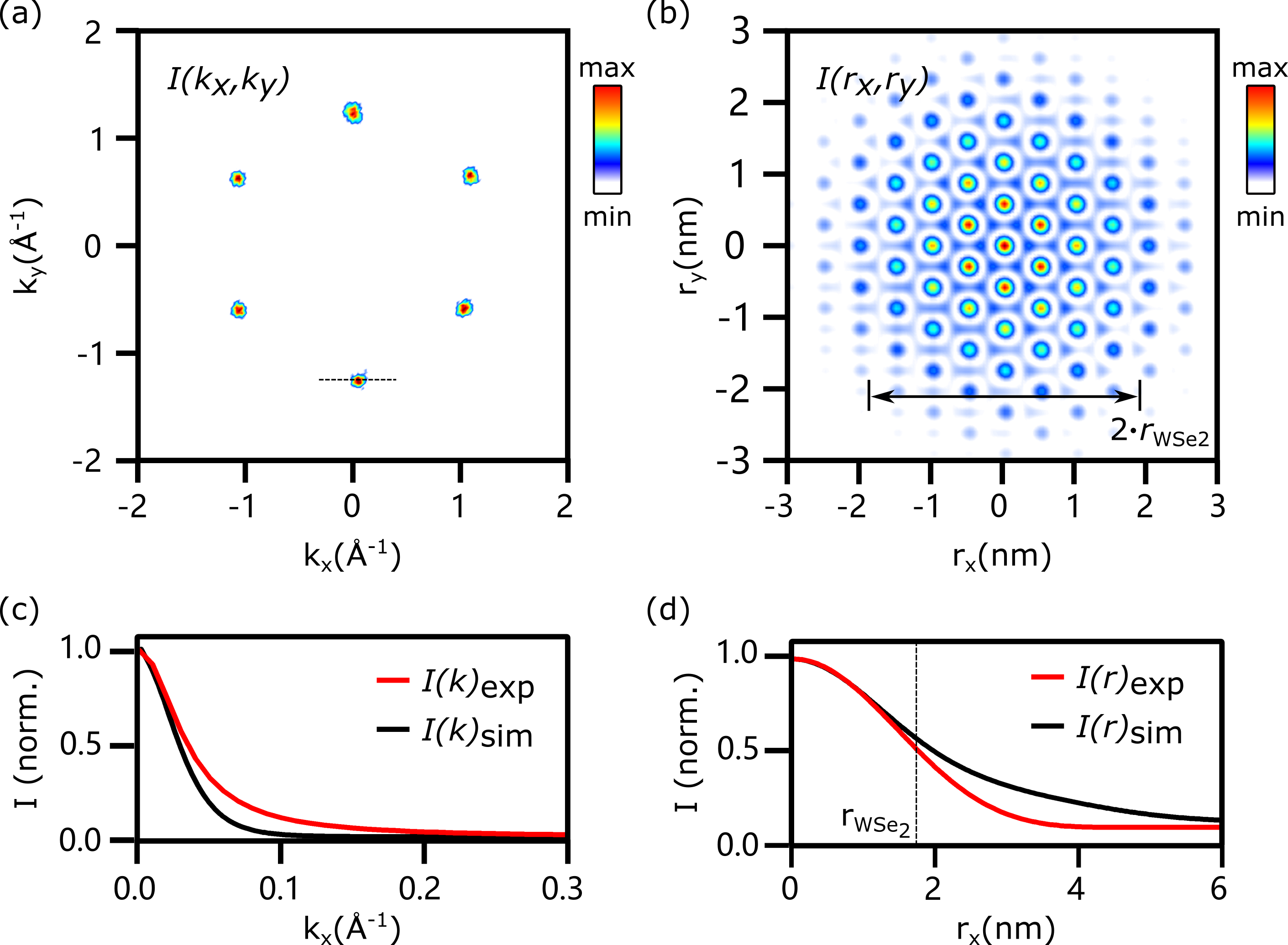
Our 4D trARPES data not only provides the energy-momentum dynamics of excitons but also contains direct amplitude information about exciton wave functions. In Fig.4(a), we display the early-time excited-state momentum distribution of the K valleys, by integrating in energy over the CB. Signals from other valleys are filtered out in order to focus on the A excitons (see SI). The total photoemission intensity is proportional to the squared transition dipole matrix element, , which connects the initial state wave function to the photoemission final state , via the polarization operator . Here, A is the vector potential of the light field and p is the momentum operator. Within the plane wave approximation (PWA) for the final state, the matrix element takes the form
| (1) |
where k is the wave vector of the photoionized electron. According to Eq.1, the matrix element is proportional to the amplitude of the Fourier transform (FT) of the initial state wave function. Therefore, the momentum distribution of the photoemission signal can be used to retrieve the real-space probability density of the electron contribution to the two-particle excitonic wave function, i.e., the modulus-squared wave function, , with a suitable assumption for the missing phase information.

A similar reconstruction of electronic wave functions from ARPES spectra has been previously demonstrated for occupied molecular orbitals in the ground state of crystalline organic films and chemisorbed molecular monolayers[33, 34]. Here, we extend this technique into the time domain and apply it to reconstruct the excitonic wave function in WSe2. Assuming a constant phase profile across the BZ as a lower-limit wave function extension (see SI), we retrieve the exciton probability density via 2D FT as shown in Fig.4(a-b). The reconstruction exhibits a broad isotropic real-space exciton distribution carrying high-frequency oscillations, corresponding to the hexagonal periodic lattice structure of WSe2. To resolve the isotropic exciton wave function envelope more clearly, the 1D real-space carrier distribution without the oscillatory pattern is shown in Fig.4(d), obtained by FT of only one of the six K valleys, yielding a value of nm for the excitonic Bohr radius.
To verify the method of reconstructing excitonic wave functions, we performed microscopic calculations of trARPES spectra. The momentum-resolved description of the exciton is based on a many-particle treatment of the Coulomb interaction between electron-hole pairs and the exciton-phonon scattering dynamics[3] (see SI). The momentum distributions of the bright K-excitons calculated within the PWA for the final state is shown in Fig.4(c). We find a very good agreement to the experimental momentum distribution curve (MDC) taken along the dashed line in Fig.4(a)), supporting our assumption that the trARPES spectrum contains the fingerprints of the excitonic wave function and justifying the use of the PWA. Furthermore, the calculated real-space exciton distribution in Fig. 4(d) shows good agreement to our experimental results, yielding a very similar excitonic Bohr radius of nm. This agreement demonstrates the consistency of the experimentally retrieved exciton binding energy and Bohr radius and additionally suggests the validity of the assumption of a constant phase, which provides the FT-limited (lower-bound) exciton distribution. While the excitonic Bloch state is invariant under global and valley-dependent phase renormalization, we find that valley-local phase variations in momentum space can lead to broadening of the exciton probability distribution. In the SI, we reconstruct the real-space exciton density distribution with non-constant intervalley and intravalley phase profiles, where we find a broadened exciton distribution in the case of an intravalley varying phase. Therefore, we note here that the real-space reconstruction of the exciton density with a constant phase is suitable for topologically trivial solid-state wave functions. However, the winding of the phase in topologically non-trivial materials leads to an additional expansion of the carrier density distribution, requiring explicit momentum-dependent phase information. In general, the phase of the excitonic wave function might additionally be reconstructed through iterative phase retrieval algorithms[35]. We envision that future developments will allow retrieving the phase as well as orbital information of excitonic wave functions by utilizing dichroic observables[36, 37, 38] in trARPES.
In this work, we provide a comprehensive experimental characterization of an excitonic state with trARPES. The interactions governing the formation of this prototypical many-body state are observable as energy renormalization in comparison to single-particle states, while its interaction strength with other quasi-particles is reflected in the excited state’s lifetime. These quantities are intimately connected to the real and imaginary parts of the many-body state’s self-energy and our approach establishes experimental access to these elusive quantities. Moreover, we retrieve real-space information of the excitons by Fourier transform of its momentum distribution, establishing the measurement of wave function properties of transient many-body states with 4D photoemission spectroscopy. Our approach is applicable to all exciton species occurring in a wide range of inorganic and organic semiconductors, van der Waals heterostructures and devices. Its extension to other many-body quasi-particles in solids appears straightforward.
Data availability We provide the full experimental dataset as well as the details of the data analysis on the data repository Zenodo. Also, we provide the source code of our data analytics on GitHub.
References
- [1] Frenkel Jacov. On the transformation of light into heat in solids. I Phys. Rev. 1931;37:17.
- [2] Perfetto E, Sangalli D, Marini A, Stefanucci G. First-principles approach to excitons in time-resolved and angle-resolved photoemission spectra Phys. Rev. B 2016;94:245303.
- [3] Christiansen Dominik, Selig Malte, Malic Ermin, Ernstorfer Ralph, Knorr Andreas. Theory of exciton dynamics in time-resolved ARPES: Intra-and intervalley scattering in two-dimensional semiconductors Phys. Rev. B 2019;100:205401.
- [4] Wang Ke, De Greve Kristiaan, Jauregui Luis A, et al. Electrical control of charged carriers and excitons in atomically thin materials Nat. Nanotechnol. 2018;13:128–132.
- [5] Kaviraj Bhaskar, Sahoo Dhirendra. Physics of excitons and their transport in two dimensional transition metal dichalcogenide semiconductors RSC Adv. 2019;9:25439–25461.
- [6] Palummo Maurizia, Bernardi Marco, Grossman Jeffrey C. Exciton radiative lifetimes in two-dimensional transition metal dichalcogenides Nano Lett. 2015;15:2794–2800.
- [7] Yuan Long, Wang Ti, Zhu Tong, Zhou Mingwei, Huang Libai. Exciton dynamics, transport, and annihilation in atomically thin two-dimensional semiconductors J. Phys. Chem. Lett. 2017;8:3371–3379.
- [8] Christiansen Dominik, Selig Malte, Berghäuser Gunnar, et al. Phonon sidebands in monolayer transition metal dichalcogenides Phys. Rev. Lett. 2017;119:187402.
- [9] Steinhoff Alexander, Florian Matthias, Rösner Malte, Schönhoff Gunnar, Wehling TO, Jahnke Frank. Exciton fission in monolayer transition metal dichalcogenide semiconductors Nat. Commun. 2017;8:1166.
- [10] Wang Gang, Chernikov Alexey, Glazov Mikhail M, et al. Colloquium: Excitons in atomically thin transition metal dichalcogenides Rev. Mod. Phys. 2018;90:021001.
- [11] Wang Gang, Marie Xavier, Gerber I, et al. Giant enhancement of the optical second-harmonic emission of WSe2 monolayers by laser excitation at exciton resonances Phys. Rev. Lett. 2015;114:097403.
- [12] Li Yilei, Chernikov Alexey, Zhang Xian, et al. Measurement of the optical dielectric function of monolayer transition-metal dichalcogenides: MoS2, Mo Se2, WS2, and WSe2 Phys. Rev. B 2014;90:205422.
- [13] Pöllmann Christoph, Steinleitner Philipp, Leierseder Ursula, et al. Resonant internal quantum transitions and femtosecond radiative decay of excitons in monolayer WSe2 Nat. Mater. 2015;14:889–893.
- [14] Selig Malte, Berghäuser Gunnar, Raja Archana, et al. Excitonic linewidth and coherence lifetime in monolayer transition metal dichalcogenides Nat. Commun. 2016;7:13279 .
- [15] Zeng Hualing, Dai Junfeng, Yao Wang, Xiao Di, Cui Xiaodong. Valley polarization in MoS2 monolayers by optical pumping Nat. Nanotechnol. 2012;7:490–493.
- [16] Mak Kin Fai, He Keliang, Shan Jie, Heinz Tony F. Control of valley polarization in monolayer MoS2 by optical helicity Nat. Nanotechnol. 2012;7:494–498.
- [17] Bertoni Roman, Nicholson Christopher W, Waldecker Lutz, et al. Generation and evolution of spin-, valley-, and layer-polarized excited carriers in inversion-symmetric WSe2 Phys. Rev. Lett. 2016;117:277201.
- [18] Park Soohyung, Mutz Niklas, Schultz Thorsten, et al. Direct determination of monolayer MoS2 and WSe2 exciton binding energies on insulating and metallic substrates 2D Mater. 2018;5:025003.
- [19] Yao Kaiyuan, Yan Aiming, Kahn Salman, et al. Optically discriminating carrier-induced quasiparticle band gap and exciton energy renormalization in monolayer MoS2 Phys. Rev. Lett. 2017;119:087401.
- [20] Klein J, Kerelsky A, Lorke M, et al. Impact of substrate induced band tail states on the electronic and optical properties of MoS2 Appl. Phys. Lett. 2019;115:261603.
- [21] Chernikov Alexey, Berkelbach Timothy C, Hill Heather M, et al. Exciton binding energy and nonhydrogenic Rydberg series in monolayer WS2 Phys. Rev. Lett. 2014;113:076802.
- [22] Puppin Michele, Deng Yunpei, Nicholson CW, et al. Time-and angle-resolved photoemission spectroscopy of solids in the extreme ultraviolet at 500 kHz repetition rate Rev. Sci. Instrum. 2019;90:023104.
- [23] Weinelt Martin, Kutschera Michael, Fauster Thomas, Rohlfing Michael. Dynamics of exciton formation at the Si (100) c (4 2) surface Phys. Rev. Lett. 2004;92:126801.
- [24] Madéo Julien, Man Michael KL, Sahoo Chakradhar, et al. Directly visualizing the momentum forbidden dark excitons and their dynamics in atomically thin semiconductors Science 2020;370:1199-1204.
- [25] Maklar Julian, Dong Shuo, Beaulieu Samuel, et al. A quantitative comparison of time-of-flight momentum microscopes and hemispherical analyzers for time-resolved ARPES experiments Rev. Sci. Instrum. 2020;90:023105.
- [26] Wickramaratne Darshana, Zahid Ferdows, Lake Roger K. Electronic and thermoelectric properties of few-layer transition metal dichalcogenides J. Chem. Phys. 2014;140:124710.
- [27] Rustagi Avinash, Kemper Alexander F. Photoemission signature of excitons Phys. Rev. B 2018;97:235310.
- [28] Perfetto E, Stefanucci G. Ultrafast creation and melting of nonequilibrium excitonic condensates in bulk WSe2 arXiv:2011.11967 2020.
- [29] Knoesel E, Hotzel A, Wolf M. Ultrafast dynamics of hot electrons and holes in copper: Excitation, energy relaxation, and transport effects Phys. Rev. B 1998;57:12812.
- [30] Ueba Hiromu, Gumhalter Branko. Theory of two-photon photoemission spectroscopy of surfaces Prog. Surf. Sci. 2007;82:193–223.
- [31] Raja Archana, Selig Malte, Berghauser Gunnar, et al. Enhancement of Exciton–Phonon Scattering from Monolayer to Bilayer WS2 Nano Lett. 2018;18:6135–6143.
- [32] Marini Andrea. Ab initio finite-temperature excitons Phys. Rev. Lett. 2008;101:106405.
- [33] Offenbacher Hannes, Lüftner Daniel, Ules Thomas, et al. Orbital tomography: Molecular band maps, momentum maps and the imaging of real space orbitals of adsorbed molecules Electron. Spectrosc. Relat. Phenom. 2015;204:92–101.
- [34] Puschnig Peter, Berkebile Stephen, Fleming Alexander J, et al. Reconstruction of molecular orbital densities from photoemission data Science. 2009;326:702–706.
- [35] Jansen Matthijs, Keunecke Marius, Düvel Marten, et al. Efficient orbital imaging based on ultrafast momentum microscopy and sparsity-driven phase retrieval New J. Phys. 2020;22:063012.
- [36] Wießner M, Hauschild D, Sauer C, Feyer V, Schöll A, Reinert F. Complete determination of molecular orbitals by measurement of phase symmetry and electron density Nat. Commun. 2014;5:41–56.
- [37] Beaulieu Samuel, Schusser Jakub, Dong Shuo, et al. Revealing Hidden Orbital Pseudospin Texture with Time-Reversal Dichroism in Photoelectron Angular Distributions Phys. Rev. Lett. 2020;125:216404.
- [38] Schüler Michael, Pincelli Tommaso, Dong Shuo, et al. Bloch Wavefunction Reconstruction using Multidimensional Photoemission Spectroscopy arXiv 2021;2103.17168.
Acknowledgments: This work was funded by the Max Planck Society,the European Research Council (ERC) under the European Union’s Horizon 2020 research and innovation and the H2020-EU.1.2.1. FET Open programs (Grant Nos. ERC-2015-CoG-682843, ERC-2015-AdG-694097, and OPTOlogic 899794), the Max Planck Society’s Research Network BiGmax on Big-Data-Driven Materials-Science, and the German Research Foundation (DFG) within the Emmy Noether program (Grant No. RE 3977/1), through SFB 951 ”Hybrid Inorganic/Organic Systems for Opto-Electronics (HIOS)” (Project No. 182087777, projects B12 and B17), the SFB/TRR 227 ”Ultrafast Spin Dynamics” (projects A09 and B07), the Research Unit FOR 1700 ”Atomic Wires” (project E5), and the Priority Program SPP 2244 (project 443366970). D.C. thanks the graduate school Advanced Materials (SFB 951) for support. S.B. acknowledges financial support from the NSERC-Banting Postdoctoral Fellowships Program. T.P. acknowledges financial support from the Alexander von Humboldt Foundation. Author contributions: S.D., M.P., S.B., T.P., C.W.N., M.D., Y.D., Y.W.W., M.W., L.R., and R.E. designed, prepared, and performed the experiment. T.P., M.P, R.P.X., and R.E. performed the OBE fitting. D.C., M.S., E.M., and A.K. performed the calculation of trARPES spectrum. H.H. and A.R. calculated the single-particle self-energy. R.P.X. developed the 4D data processing code. All authors contributed to the manuscript. Competing Interests: The authors declare that they have no conflict of interest.