Beam particle tracking with a low-mass mini time projection chamber in the PEN experiment111Dedicated to the memory of Andrey Korenchenko
Abstract
The international PEN collaboration aims to obtain the branching ratio for the pion electronic decay , aka , to a relative precision of or better. The PEN apparatus comprises a number of detection systems, all contributing vital information to the PEN event reconstruction. This paper discusses the design, performance, and Monte Carlo simulation of the mini time projection chamber (mTPC) used for pion, muon, and positron beam particle tracking. We also review the use of the extracted trajectory coordinates in the analysis, in particular in constructing observables critical for discriminating background processes, and in maximizing the fiducial volume of the target in which decay event vertices can be accepted for branching ratio extraction without introducing bias.
keywords:
Time Projection Chamber , Tracking , Lepton Universality , Electroweak , Standard ModelNuclear Instruments and Methods in Physics Research A \runauthC.J. Glaser et al. \jidprocs \jnltitlelogoNucl. Instr. and Methods A
, , , ,
1 Introduction and motivation
Since the 1930s, the fields of experimental particle and nuclear physics have relied on different means to record and analyze trajectories of charged particles under study. Over the years, diverse designs for charged particle tracking detectors have emerged, and were put to use. All of them operate on the principle that a charged particle moving through matter (historically in a liquid or gas, more recently also including solids, primarily semiconductor crystals) ionizes the atoms near its trajectory. The ionization pattern is recorded with the aim to reconstruct the charged particle’s trajectory for further event analysis. Current advancement on the high energy and precision frontiers requires ever more sophisticated tracking detectors and techniques. The simple cloud chambers of the 1930s, crucial for the discovery of the muon and positron, are no longer adequate for present day experiments in nuclear and particle physics.
In the late 1970s, David Nygren [1] developed the time projection chamber (TPC), a sophisticated gas-filled tracking chamber capable of reconstructing a charged particle’s trajectory in all three dimensions, by combining the features of a multiwire proportional chamber and a drift chamber. In its many design variations, the TPC has become a reliable and precise charged particle tracking device, sufficiently fast for many applications, often used in magnetic spectrometers.
TPCs have been used in numerous experimental projects over the past more than forty years. Typically, one coordinate is determined from the hit anode wire, another from the signal attenuation along the resistive wires, and the third from the drift time of electrons in the gas. In early applications, TPC sense wire output signals have been fed to charged coupled devices (CCDs) for storage prior to being read by ADCs, or directly to flash ADCs. In pursuit of minimizing dead time and increasing the acquired information, more recent experiments have chosen to digitize TPC electrode waveforms. Time projection chambers vary in size, from small table-top devices to large detectors of many meters across, used in high energy experiments.
The international PEN collaboration, led by the University of Virginia group, has designed and built a small, low-mass, mini time projection chamber (mTPC), for use within a much larger, complex detector system. The PEN mTPC is the primary subject of this paper.
The Standard Model (SM) provides an exceptionally precise calculation of the pion electronic decay branching ratio, :
| (1) |
This level of theoretical precision, unmatched among decays of other mesons, provides unique opportunities for tests of SM predictions. The decay, , connects the pion pseudoscalar state to the vacuum. The strong helicity suppression of the decay makes this process uniquely sensitive to a class of pseudoscalar (), or -loop coupled, non- contributions, arising from new, “beyond Standard Model” (BSM) physics, undetectable in analogous, helicity-unsuppressed leptonic decays, such as the , or . At the precision of , probes the , , and BSM mass scales up to 1000, 20, and 60 TeV, respectively, sets competitive limits on the violation of electron-muon universality, on certain SUSY partners, neutrino sector anomalies and massive sterile neutrinos (for a survey of rare pion decays see, e.g., Ref. [5]). We note that lepton universality, axiomatic in the SM, may be in question following recent LHCb results [6]. Sadly, the current value [7, 8] lags in precision behind the theoretical SM evaluations by an order of magnitude. The objective of the PEN experiment is to obtain the pion electronic decay branching ratio, with a relative uncertainty of or better.
This paper discusses the design and performance of the mini time projection chamber, as well as its use in the PEN analysis. Section 2 reviews the PEN detector system. Section 3 discusses the design and construction of the mTPC. Section 4 explains and documents the mTPC performance in extracting the trajectory coordinates from raw electrode signals. Section 5 discusses the generation of realistic mTPC synthetic data using Monte Carlo (MC) simulation methods. Section 6 demonstrates the use of the mTPC information in the PEN analysis with a focus on ensuring reliable calibration of the detection efficiency.
2 The PEN experiment and detector system
The PEN data were acquired in the E1 beam area of the Paul Scherrer Institute (PSI) Ring Cyclotron, Switzerland, during three runs, from 2008 through 2010, for a total of approximately 25 weeks of in-beam data acquisition, using the apparatus described below. The data analysis is in an advanced stage for all relevant decay channels in parallel. The main analysis is blinded. The unblinding of the result is planned after the completion of a full set of papers describing the analysis (of which this is one), and of the final analysis pass with fine parameter/cut adjustments, minimizing the total uncertainty, plus ensuring that all the analyzed decay observables are mutually fully consistent in a comprehensive analysis with independent checks.
Since , the goal precision of PEN, relative to the observed number of pion decays, is of . Even though many uncertainties, such as those associated with the number of stopped pions, and the solid angle acceptances for the two channels, cancel when building the ratio , the high level of precision still results in stringent demands on the accurate treatment of various subtle effects, such as pion and muon decays in flight and radiative corrections. One of the key supporting analysis tasks is beam particle tracking, accomplished trough the use of the PEN mTPC, optimized for small size, low mass, high double-pulse resolution, as well as easy installation and alignment.
The detector system used for the PEN experiment, schematically shown in Fig. 1, is an upgraded version of the one previously used in the PIBETA experiment [9].

The PIBETA project studied rare pion and muon decays, focusing on the pion beta decay, , in measurement runs from 1999 to 2004 [10, 11, 12]. Both PIBETA and PEN have studied decays at rest, with the beam pions stopping in an active target. During PEN data taking, the incoming pion beam momentum varied in the range between 71.5 MeV/ and 83.5 MeV/, depending on the run period. The main PEN detector components used in Runs 2 and 3 are listed below.
-
1.
A forward beam counter (BC) and active degrader (AD), both made of polyvinyltoluene (PVT), were used for beam particle discrimination. The 3.67 m separation between BC and AD was used to calculate the velocity of the particles and thus their mass, using the known beam momentum.
-
2.
A mini time projection chamber was used for the determination of the pion trajectory in all three dimensions.
-
3.
A cylindrical active target (AT) made of fast PVT, with both radius and length equal to 15 mm, was used to measure the energy depositions of the incoming pion and its charged decay products.
-
4.
Two concentric cylindrical multi-wire proportional chambers (MWPC1, 2) tracked charged decay particles emerging from the target. The MWPC1, 2 anode wires were located at and 120 mm, respectively. Track polar angle, , is used to define the acceptance of the spectrometer for the signal and normalization decay channels, thus controlling a significant source of systematic between the two processes.
-
5.
Tightly surrounding MWPC2 was a hodoscope detector (PH) consisting of 20 identical PVT staves, each 4 mm thick, used for particle identification and timing. Thanks to strong separation between minimum ionizing positrons from weak decays, and highly ionizing protons from interactions of pions in the central detector region, the PH detector provided for efficient suppression of the copious hadronic background.
-
6.
A spherical, 240-module pure CsI electromagnetic calorimeter surrounded the target and tracking detectors, covering sr. The crystal shapes were hexagonal and pentagonal truncated pyramids.
The recorded detector signals are primarily used to identify, characterize, and discriminate between the signal decay, and the normalization process, . In addition to their primary uses outlined above, individual PEN detectors are used to check calibrations in the other detectors.
Main recorded event types were triggered with a signal in the plastic hodoscope and a minimum energy in the CsI calorimeter. Events were only recorded within a window extending between 50 ns before, and 220 ns after the time of the pion coming to rest in the active target. Events with low energy recorded in the CsI, overwhelmingly due to positrons from muon decays, were prescaled by factors ranging from 8 to 256 depending on the positron-induced electromagnetic shower energy, and on the run period.
The incoming beam contained pions, positrons and muons, most of which can be separated by using the particle energy loss in the upstream beam detector BC, and time of flight (TOF) between BC and AD. The reconstructed pion trajectory is used to determine the pion stop location, which is in turn used to construct methods of cleanly separating the main pion decay mode from the rare decay mode of interest, .
3 The PEN mTPC design and construction
Beam particle tracking prior to the stopping of pions in the target was performed in Run 1 by a four-piece wedge degrader detector made of PVT. To improve on the wedge degrader, for Runs 2 and 3 the PEN collaboration designed and built low-mass mini time projection chambers, discussed below.
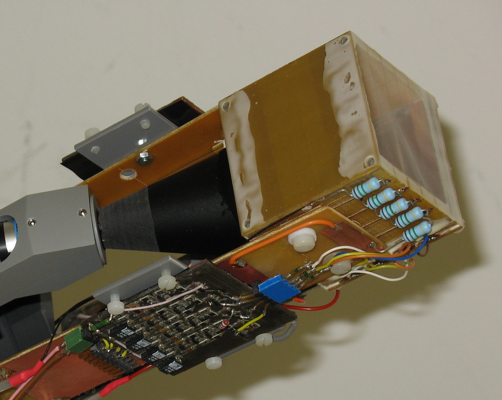
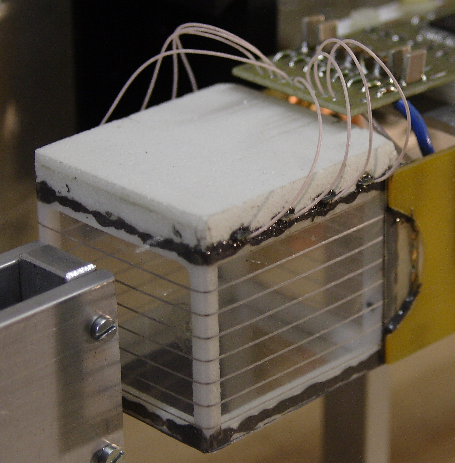
The mTPC used in PEN Run 2, shown in Fig. 2, was a low-mass cubic box with dimensions mm3. The geometry was studied using the CERN GARFIELD [13] drift chamber simulation package with the goal to optimize the electric field homogeneity in the drift region, and to equalize gas amplification for the four anode wires. Further modifications and improvements to the field cage were implemented in the second generation mTPC for Run 3, shown in Fig. 3. We first focus on the Run 2 device, and then highlight the differences in the Run 3 version.


The Run 2 chamber’s active volume was subdivided by a grid into drift and proportional regions. The drift region dimensions were mm3. The homogeneous electric field in the drift region was shaped by the cathode electrode, the separating grid, and eight field shaping electrodes affixed to the inner surfaces of the side walls. The drift plate was at kV with respect to ground (anode) potential, and the drift velocity 1.8 cm/s. The thickness of the Mylar windows perpendicular to the beam was 20 m, and the potential electrodes were made of copper wire, 0.1 mm in diameter. The grid was made of 0.1 mm bronze wires with 2 mm spacing. Above the separating grid was the 7 mm wide amplification region with the four anode wires arranged perpendicular to the beam direction, with a spacing of 10 mm. The 40 mm long anodes were made of Nichrome (NiCr) wire, 12 m in diameter, with the resistance of 60 /cm. The mTPC was filled with a gas mixture of 90% argon and 10% methane at atmospheric pressure (slow bubblers). No connectors were placed directly on the mTPC. All electric connections for signal, power, and HV cables were made by soldering. The voltage dividers for the field shaping electrodes were placed outside the detector. The signals from the two sides of all four anode wires were fed through to a CAEN V1720 unit where they were digitized into waveforms at the sampling rate of 250 MS/s.
A modified version of the detector was used in Run 3 [14]. Notable differences include significantly reduced mass, 12.0 mm wire spacing instead of 10.0 mm, and elimination of the grid wires. Its lower mass allowed the placement of the detector further downstream, closer to the active target. Photographs of the two mTPCs are shown in Fig. 4.


The basic mechanisms of signal development in a TPC are well understood. In a first stage the electrons from the track of some ionising particle drift in a more or less constant drift field towards the anode plane. The observed drift time allows one to calculate the position coordinate in the direction of the drift field (vertical, or , for PEN mTPCs). In most cases the anode consists of thin sense wires where the avalanches, developing in the field, result in the necessary signal gain. After reaching a wire the secondary electrons create electric signals which propagate towards both wire ends. The signal exponential fall time constant reflects the velocity at which the cloud of gas ions moves away from the anode. The electron signals are split between the two anode ends with a ratio inversely proportional to the wire resistances between the ends and the charge arrival location on the wire. Thus, the observed signal amplitude ratio may allow reconstruction of that location along the anode coordinate (horizontal, , for PEN mTPCs).


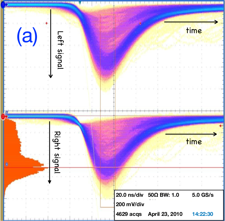
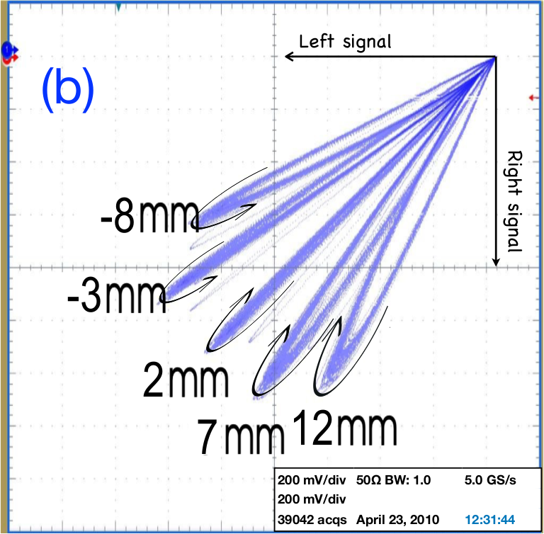
Signal shapes are distorted by reflections at the amplifier inputs and as a result the tails of the two signals gradually lose their position dependence, as illustrated in Fig. 5. The figure shows oscilloscope pictures of the left and right signals of an anode wire of the Run 2 mTPC recorded as the detector was irradiated with a collimated 55Fe X-ray source at different locations along the anode. The energy distribution peaks around 5.9 keV, about double the energy loss of our beam pions, a Landau distribution with mean of 3.2 keV and peak probability of 2.2 keV per wire. Part (a) shows the individual left and right signals for central source location. The D0M Ampl-8.3 amplifier [15] has leading/trailing edge of 7 ns. The histogram on the left shows the energy distribution obtained from the signal integral in the indicated gate around the peak. Part (b) shows the correlation between left and right signal, varying the source position along the wire in steps of 5 mm. Note that the signals are separated by ten or more standard deviations, corresponding to a position resolution well below a mm, i.e., (1%) of the wire length. This value includes the unknown contribution of the 55Fe source collimator. We also note that charge division does not only affect the amplitudes, but also the signal shapes. Signals follow a loop starting and ending at the origin at the top right. The direction of time evolution through the loops is indicated by the curved arrows at the signal peaks, clockwise on the right side and counter-clock on the left. Central hits result in identical signals which land on the diagonal (not shown in the figure). For off-center hits the amplitude ratio deviates most from unity during the rise time. At later times the position dependence gradually fades away, so the loops return to the diagonal. For this reason the tracking algorithm optimized for off-line analysis only uses the first 30 ns of the recorded wave forms.
Signal development in TPCs with meters long sense wires, used in high-energy experiments, is much more affected by reflections and cross-talk. In those situations numerical calculations have been quite successful in describing the experimental observations [16]. In this analysis we have followed a more phenomenological approach based on accurate calibrations of the measured data, and adjusting the PEN experiment Monte Carlo simulation to those.


The geometrical arrangements deployed in PEN Runs 2 and 3, respectively, are shown in Fig. 6. In Run 2, the mTPC was separated from the target by about 37 mm, with a beam collimator in between. In Run 3, the mTPC-target separation was about 10 mm, with no collimator between them, leading to an improved resolution of the decay vertex.
4 Particle tracking and event reconstruction with the PEN mTPC
As indicated in the preceding section, the relevant event-level mTPC information is stored in the form of waveform arrays (aka records, traces), one for each end of the four anode wires. Waveform traces for a typical event are shown in Fig. 8.


This section presents the strategies used to search for signals in the anode waveforms, their combination into tracks, the determination of the associated trajectory coordinates, and relation to beam particle identification.
After subtraction of appropriately averaged baseline levels from the raw waveforms, the geometric means of the left and right waveform bins are constructed bin-wise for each anode wire, and the result is differentiated as shown in Fig. 8. The peaks in the resulting distributions are used as a measure of the anode arrival times. The signal amplitudes at both ends of a wire reflect the energy deposition and their ratio is a measure of the coordinate of a hit (Fig. 9a).


The anode wire number defines the coordinate along the pion trajectory. The wire spacing was measured with an accuracy of 0.1 mm. The anode arrival times are used to calculate the drift time and thus the coordinate. A valid hit in the degrader detector, which was separated by 6 mm (Run 2) and 8 mm (Run 3) of air from the mTPC’s front face, provided the start signal for the drift time. The correlation between the calibrated mTPC signal drift times and the track coordinates for anode wire 4 in Run 2 is shown in Fig. 9b.
Once anode hits have been found, straight tracks are reconstructed, based on the following principles. Each hit can be used for one track only. Tracks are searched in order of the number of hits per track, beginning with fully efficient tracks. Collinearity tests are used to remove hit combinations that do not belong to a single beam particle. In the case of fully efficient tracks the straight trajectory defined by and must agree with the one defined by and . Collinearity is tested by comparing the locations at mean and the slopes in and , so for :
| (2) | ||||
| (3) |
where indices denote anode wires. Because of multiple small-angle scattering and intrinsic detector resolution, the above collinearity tests are not exactly satisfied in practice. Nevertheless, the two expressions center at or close to zero for valid tracks. Analogous expressions are constructed for the deduced values of the track. Figure 10 shows distributions of the two collinearity tests for both and .
The linear independence of the two collinearity observables is illustrated in Fig. 11.


Correlations do appear for large deviations from 0, which are relatively rare, as would be expected in case of a kink in the trajectory. For small deviations there is no such correlation, consistent with just the broadening due to intrinsic detector resolution. Collinearity tests may fail when there are two particles in the beam, when one of the wires doesn’t fire, if the tracked particle scatters, or in rare cases through waveform distortions due to -electron presence. Thus, a series of tests on the detected wire hit coordinates are applied in order to identify and select valid tracks, applying the following procedure:
-
1.
The full set of observed space points is scanned for a combination that satisfies the condition where is widened from 10 to 50 mm2 in steps of 10 mm2 until a solution is found. The corresponding cut circles are drawn in red in Fig. 11(a). Once a combination of values is found to satisfy one of the above cuts, it is saved and removed from the set. Search for tracks continues among the remaining wire hits until all are sorted into track candidates, or have failed the col test.
-
2.
The values corresponding to the saved track candidates in step 1 are analyzed next using analogous criteria: , again with from 10 to 50 mm2 in steps of 10 mm2. The circle corresponding to the first of these tests, , is shown in Fig. 11(b). At this stage, a track candidate consists of a set of four wire hit coordinate pairs that independently satisfy a and a cut.
-
3.
Sets of track candidate and hit coordinates found in the above two steps are further subjected to the final condition for a valid track: . This last test is indicated by the red line in Fig. 11(c).
If no track with signals from all four wires is found, the beam trajectory may still be reconstructed. In such situations, new collinearity observables are evaluated based on the signals of three wires only. For instance, in the absence of , a new collinearity test can be constructed:
| (4) |
Similar tests can be made in case a different wire didn’t report. Finally, if still no tracks are found, combinations of just two hits are searched, with the condition that the trajectory crosses AD and AT. However, for events with fewer than four anode wires reporting analyzable signals, the ability to identify trajectory kinks is reduced. Once tracks have been found, the corresponding trajectories are evaluated by linear interpolation. Figure 12 shows a representative set of reconstructed pion trajectories in horizontal and vertical projections.


In addition to reconstructing the pion beam trajectories, discussed above, the mTPC (along with the AD) provides independent confirmation of correct identification of the three beam particle types. The dissimilar velocities of the , , and sharing the same momentum, result in different energy losses in the chamber gas, thus producing distinct amplitude spectra, as illustrated in Fig. 13.


Hence, beam particle identification (PID), accomplished mainly through the BC–AD TOF observable, is independently validated by the qualitative agreement between the measured and Monte Carlo predicted energy deposited in mTPC gas by the three particle types. We note that a detailed simulation of the anode charge collection, e.g., accounting for delta electrons, was not implemented as the effects cancel (apart from an inconsequential rescaling) in the anode charge asymmetries (Fig. 9), and ultimately do not affect the evaluation of . The relevant instrumental effects are incorporated through smearing, discussed in Sec. 5.
As seen above, mTPC beam tracking provides the projected stopping coordinates for each beam pion that stops and decays in the AT. The -stop coordinate is determined as follows. The energy of a beam pion as it reaches the active degrader, , is calculated from the BC–AD time of flight. To obtain the pion’s energy as it reaches the target, and thus its range in the target, is reduced by the observed energy deposited by the pion in the degrader. In this way the pion stop position is predicted in three dimensions. Accurate prediction of the location of the stopped pion in the target is critical to controlling the PEN analysis systematics, primarily because it allows accurate attribution of fractions of observed target energy to the pion and positron, respectively, thus increasing the reliability of detection of the intermediate muon in the decay chain . Reliable sorting of decay types, or , is the main systematic challenge in the measurement. This point is further revisited in Sec. 6.
PEN analysis allows for independent calibration of mTPC tracking using the two MWPCs. Specifically, the distribution of pion stopping coordinates predicted by mTPC, BC and AD data, has been cross-calibrated using decay positron trajectories observed with the MWPCs. Horizontal (constant ) and vertical (constant ) tracks through the MWPCs provide an independent calibration of the mTPC/TOF-predicted stopping coordinates, as shown in Fig. 14.


Detection efficiency for a given mTPC anode wire has been determined by observing the fraction of valid tracks for which that wire registers a signal when the remaining three anode wires have fired. The four mTPC wire detection efficiencies are tabulated in Tab. 1 for PEN Runs 2 and 3. We note that the A1 wire failed completely through the second half of Run 3, and the A2 wire failed near the end of Run 3. For this reason the mTPC wire efficiencies for Run 3 are calculated and listed only for the first half of that run. We also note that the two wires that eventually failed were less efficient from the start; the actual cause of the problem has not been identified.
| Anode wire | Run 2 | Run 3 |
|---|---|---|
| 1 | 99.590 .004% | 95.84 .04% |
| 2 | 99.288 .006% | 96.52 .02% |
| 3 | 99.095 .007% | 98.76 .01% |
| 4 | 99.794 .003% | 99.43 .01% |
5 Monte Carlo simulation of the mTPC response
Monte Carlo simulations producing highly realistic synthetic events are needed in order to reach the precision goal of the PEN data analysis. Detailed detector geometries and responses are implemented using the Geant4 toolkit[17], supplemented by the PEN C++ code. The simulation uses waveform kernels obtained by taking an average of numerous observed detector responses, properly normalized and shifted, as illustrated in Fig. 15, to faithfully simulate details of the detector response.


Synthetic waveforms are constructed using the and trajectory coordinates, along with the energy deposited in the regions instrumented by the individual wires. In the process, Monte Carlo values of the and coordinates are appropriately smeared to account for the resolution of the detector. The waveforms are constructed by using a linear function in for the time bins, and two separate linear functions in for the anode left and right signal amplitudes, with opposite signs of their respective slopes. Simulated synthetic waveforms, which include appropriate smearing due to the measured detector response, are used to obtain the simulated pion stopping distribution, which, in turn, must agree with the measured data. Unlike the measured, simulated waveforms are accompanied by information on the actual (“known” or “true”) , , and coordinate values of the simulated pion as it passes by each mTPC wire. Of course, the reconstructed values are not quite the same as those of the “known” observables due to the statistical nature of detector smearing. Results of this procedure are illustrated in Figs. 16 and 17.


Fig. 16 demonstrates the absence of correlation between the smeared (reconstructed) and known values of an anode wire coordinates. It also affirms the superior resolution in , derived from charge drift time in gas, compared to , derived from charge splitting of the anode signals. There is a caveat associated with the smearing of the stopping distribution, as follows. The simulation was constructed so as to match the shape and rms of the stopping position in both the and coordinates (as well as ). This can be achieved in two ways. The first is to generate the pion in the simulation with wider and momentum component distributions. The second is to smear the simulated detector response of the mTPC. Both methods will have the same effect of broadening the stopping distribution. However, the two methods will produce different spectra in the downstream detectors, specifically in vertex quality which also uses the information from the MWPC. The best way to produce highly realistic stopping distributions is to construct the simulated waveforms not with the known positions of and , but rather using the known values plus a random variable representing the smearing due to the detector resolution. The known, reconstructed, and measured pion stopping and distributions are compared for a set of Run 2 data in Fig. 17. Comparing the smeared and reconstructed to the known Geant stopping , values provides a full measure of the mTPC tracking resolution at the pion stop location in the AT. The standard deviation values, displayed in the residuals plots in Fig. 17, were obtained by Gaussian fits. Even though the track position resolution at the measurement (wire) location is much better, as seen in a closer look at the track collinearity tests below, the pion stopping position affects the branching ratio analysis more directly.
After smearing, the simulated mTPC space points fully reflect the observed track coordinate resolutions. This is confirmed in Fig. 18 by the good agreement between the typical measured and simulated distributions of the four collinearity tests.


This figure therefore best illustrates the intrinsic tracking resolution of the mTPC, averaged per anode wire. The definitions of and , given in (2) and (3), respectively, yield
| (5) |
where is the average wire tracking resolution in , per wire. Analogous expressions hold for . By averaging values derived from col1x and col2x, and doing the same for , we arrive at the best estimates for the average tracking resolution per wire in and , respectively. Values for the experimental standard deviations of the four collinearities were determined by fitting the measured distributions with Gaussian functions for the two PEN run periods separately. Thus obtained standard deviations, and the derived per-wire track position resolutions, are summarized in Tab. 2,
| Run 2 | 1.15 | 0.74 | 0.53 | 2.70 | |
|---|---|---|---|---|---|
| 0.63 | 0.48 | 0.32 | 1.06 | ||
| Run 3 | 1.27 | 0.82 | 0.59 | 2.42 | |
| 0.81 | 0.71 | 0.44 | 0.95 |
along with the observed resolutions for the predicted pion stopping coordinates in the AT, deduced from the and residuals as shown above (Fig. 17). These results are consistent with the earlier observation of superior tracking resolution in (based on drift time) than in (based on charge division). In all, the obtained resolution values are very good, slightly above 0.5 mm in , and below 0.5 mm in . We note that the Run 3 mTPC is somewhat worse than its predecessor in intrinsic tracking resolution at the wire locations. In spite of that, it is better than the Run 2 mTPC in terms and , resolution of the pion stopping coordinates in the AT, thanks to the more compact AD–AT geometry (Figs. 6 and 12), made possible by its significantly lower mass. Again, accurate knowledge of the pion stop coordinates, and therefore of the decay vertex, affects the branching ratio systematics more strongly, as we discuss next.
6 mTPC in the PEN analysis
The ability to reproduce the values of several beam-related observables is key to the reliable reconstruction and interpretation of the PEN measured events in the following analysis steps.
-
1.
Predicted stopping position of the pion in the target (mTPC plus BC–AD TOF), yields , the predicted energy deposited by the pion in the AT . Combined with the trajectory (MWPCs), enables a geometrical estimate of , the decay positron’s pathlength in the AT.
-
2.
Combining with the known stopping power d/d of ’s in PVT, yields , the predicted energy deposition by the decay positron in the target.
-
3.
Evaluating the target rest energy, , enables the discrimination of the two main decay channels of interest to PEN: and , without relying on information from the CsI calorimeter, as discussed below. Distinguishing these two processes in the measured data with high accuracy is the greatest challenge of the experiment.


The accuracy of the reconstruction of the positron trajectory, and thus its energy deposition inside AT, is illustrated in Fig. 19. Of crucial importance is the fact that the method doesn’t make use of the AT waveform which otherwise might introduce a dependence on pion decay time, a dangerous source of systematic error in . In the cases where the decay occurs relatively quickly, the resulting superposition (“pileup”) of the stopping (pion) and decay particle (muon, positron) overlapping signals in the target waveform makes it difficult to determine the energy deposited in the target by the positron as opposed to the pion or muon. The effect is illustrated with synthetic waveforms in Fig. 21. By relying on subtraction of predicted energy in the target, our method avoids pulse fitting, so there is no minimum pulse separation or minimum pulse amplitude requirement for a valid result.


For events with well separated stopping and decay particle signals in the target waveforms, we observe a strong linear correlation between the positron energy deposition from the AT signal, and its value from the reconstructed e+ path, as shown in Fig. 21.
An astute reader will notice two minor anomalies in two figures above. First, the shortest reconstructed pathlengths in Fig. 19 depart from the diagonal, trending slightly above the corresponding known values. These events are decays occurring close to the edge of the AT. The required minimum AT positron energy cut filters such reconstructed events asymmetrically, favoring longer pathlengths in the AT, thus producing the observed effect. The second anomaly concerns a slight high- tail in Fig. 21, another target edge effect. These rare events correspond to pion stops predicted to lie slightly outside the AT, or at its very edge, followed by a decay positron which turns back and traverses the full extent of the AT on its way to the calorimeter. Meanwhile, the actual pion stop occurs inside the AT, and the decay positron traverses a shorter pathlength through the AT. As seen in Fig. 12, in recognition of the (2 mm) mTPC track resolution in the target, the PEN analyzer accepts tracks that skirt or narrowly miss the AT.
The extraction of the positron energy loss in AT independent of the detailed AT waveform information is useful in separating the two main channels of the pion decay, and through the use of higher order observables. We recall , the target rest energy, obtained by subtracting the predicted energies deposited in the AT by the pion and the positron from for each event. For a event, this should result in . However, for the decay chain, MeV, the muon kinetic energy, as seen in Fig. 22.


The invariant mass variable shown in the figure is evaluated as , where is the sum of detected energy for the event in the apparatus, while is the vector sum of momenta associated with all showers ( is the best available measure of the energy of the unobserved neutrino in decay). Invariant mass defined in this way seamlessly accommodates radiative decays alongside the dominant variety with undetected/unseparated soft photon.
Several comments are appropriate here. First, without the combined mTPC and MWPC tracking, PEN analysis would not be able to evaluate the target rest energy for every event that passes other requirements valid for the signal and normalization decay channels (there are a relative few events that fail to produce valid tracks). If the analysis had to rely on the target waveforms alone, a waveform fit based separation would not be reliable for a large subset of closely spaced decay pulses, consequently greatly diminishing the number of usable events. A way around that is to use the , the total target energy instead of waveform fitting. As Fig. 22 amply demonstrates, that does not work nearly as well as using .
Second, while produces remarkably better decay separation compared to that achieved by , it is not the most discriminating variable between the and channels. That distinction belongs to , a more complicated variable that builds on ; its detailed discussion is beyond the scope of the present paper. The variable is introduced in [5], and will be further discussed in a forthcoming publication [18].
The effects on the final selection of events used for branching ratio analysis are seen in the final panel, Fig. 22(e).


Clearly, compared to , the mTPC-based cut suppresses the background in the “tail” by an additional factor of 5, also lowering the invariant mass threshold separating the “peak” and “tail” regions. Finally, unlike the cut, the cut does not significantly reduce the peak yield (the same applies to a cut). Combining all of these factors would restrict a non-mTPC analysis to the range above the in .
A way to avoid target waveform fitting altogether is to perform a branching ratio analysis based on event decay time which relies on the characteristic time signatures of the decay and the chain to separate the two processes. This approach is limited by corrections due to particle decays in flight. Muon decay in flight, , is particularly insidious, as it occurs at level, and has the same decay time signature as the main signal, similarly restricting the achievable precision to above . Again, in PEN the correction becomes tractable with the aid of mTPC-based cuts.
In addition to constructing highly discriminating observables, the mTPC is used to ensure that the pion stops well within the target for all events included in the analysis (top plots in Fig. 23). If a pion comes to a stop too close to the radial boundary of the target, mm, the muon produced in its decay may escape from the target volume, obscuring the rest energy determination. The exit of the decay muon from the target effectively removes its decay from detection, and leads to a deficit of counts of the decay chain positrons, used for normalization in the branching ratio determination. This deficit of recorded muon decay events systematically produces a higher branching ratio, as demonstrated in the bottom plots of Fig. 23, which show that the raw ratio , the main input in the branching ratio calculation, is stable for stopping radii within the first 9.5 mm (Run 2), or 12.5 mm (Run 3), a result that is not unexpected given the lower mass, more compact central detector setup in Run 3. Pion stopping position information in the target, deduced primarily from the mTPC beam tracking, is critical for the accurate evaluation of the simulated acceptance corrections for events outside the mm region for Run 3 (or mm for Run 2), necessary for their safe inclusion in the analysis. Including the large- events lowers the statistical uncertainty of the branching fraction evaluation, to the overall precision goal of . This is yet another essential contribution of the mTPC to the PEN analysis.
7 Conclusions
The mTPC is an efficient detector, used for beam particle tracking in the PEN experiment. Beam particle trajectories based on mTPC data are essential for constructing the pion stopping location in the active target. Precise knowledge of the stopping distribution plays a central role in the acceptance systematics for the primary () and normalization () decay processes at the experiment’s intended precision.
In addition, the mTPC beam trajectory information enables the construction of a number of observables critical for discriminating the primary and normalization processes. Reliable discrimination of the two, based on information other than the CsI calorimeter energy, is the central challenge of the experiment in terms of achieving its design precision.
The importance of the mTPC in PEN further extends to the basic evaluation of the branching ratio, , before corrections are applied. Event reconstruction and selection algorithms in PEN favor decays over the decay chain at the high tail of the beam pions’ radial stopping distribution, due to imbalances in the deposited energy in the target, or because of distortions of the decay time distributions. Restricting the accepted events only to low values notably reduces the overall event count. Thanks to the ultra-realistic simulation of the mTPC detector response, the higher events can be included in the branching ratio analysis without introducing undue bias. This significantly increases the accepted event statistics for the decay, keeping it consistent with the PEN overall precision goal.
Acknowledegments
The PEN collaborators gratefully acknowledge support from the US National Science Foundation, the Paul Scherrer Institute, and the Russian Foundation for Basic Research. The authors express their sincere gratitude to S.M. Korenchenko for his continued interest in this work, and thank S.N. Shkarovsky for the chamber field calculations. Finally, the authors dedicate this work to the memory of our friend and colleague Andrey Korenchenko, who developed the second version of this unique TPC, and took part in the testing and optimization of both chambers.
References
- [1] D. Nygren, PEP-198 (1975), and “Proposal for a PEP Facility based on the TPC”, PEP4-December 30 (1976).
- [2] W.J. Marciano and A. Sirlin, Phys. Rev. Lett. 71 (1993) 3629-3632.
- [3] M. Finkemeier, Phys. Lett. B 387 (1996) 391-394.
- [4] V. Cirigliano and I. Rosell, Phys. Rev. Lett. 99 (2007) 231801.
- [5] D. Počanić, E. Frlež and A. van der Schaaf, J. Phys. G: Nucl. Part. Phys. 41 (2014) 114002.
- [6] LHCb Collaboration: R. Aaiji et al., arXiv:2103.11769 (2021), and references therein.
- [7] A. Aguilar-Arevalo et al, Phys. Rev. Lett. 115 (2015) 071801.
- [8] P.A. Zyla et al. (Particle Data Group), Prog. Theor. Exp. Phys. 2020 (2020) 083C01.
- [9] E. Frlež et al., Nucl. Instrum. Methods Phys. Res. A 526 (2004) 300-347.
- [10] D. Počanić, et al., Phys. Rev. Lett. 93 (2004) 181803.
- [11] E. Frlež, et al., Phys. Rev. Lett. 93 (2004) 181804.
- [12] M. Bychkov, et al., Phys. Rev. Lett. 103 (2009) 051802.
- [13] R. Veenhof, Garfield, a drift-chamber simulation program, CERN program library entry W5050 (unpublished).
- [14] V.A. Baranov, et al. Phys. Part. Nuclei Lett. 9 (2012) 168.
- [15] G.D.Alexeev et al., Nucl. Instrum. Methods Phys. Res. A 462 (2001) 494-505.
- [16] P. Bock et al., JINST 7 (2012) 09003.
- [17] S. Agostinelli et al., Nucl. Instrum. Methods Phys. Res. A 506 (2003) 250.
- [18] C.J. Glaser, D. Počanić, M. Vitz, V.A. Baranov, A. van der Schaaf, in preparation.