samuelmensah@aims.ac.za 22institutetext: Department of Mathematical Sciences, Stellenbosch University, South Africa
Towards the Localisation of Lesions in Diabetic Retinopathy
Abstract
Convolutional Neural Networks (CNNs) have successfully been used to classify diabetic retinopathy (DR) fundus images in recent times. However, deeper representations in CNNs may capture higher-level semantics at the expense of spatial resolution. To make predictions usable for ophthalmologists, we use a post-attention technique called Gradient-weighted Class Activation Mapping (Grad-CAM) on the penultimate layer of deep learning models to produce coarse localisation maps on DR fundus images. This is to help identify discriminative regions in the images, consequently providing evidence for ophthalmologists to make a diagnosis and potentially save lives by early diagnosis. Specifically, this study uses pre-trained weights from four state-of-the-art deep learning models to produce and compare localisation maps of DR fundus images. The models used include VGG16, ResNet50, InceptionV3, and InceptionResNetV2. We find that InceptionV3 achieves the best performance with a test classification accuracy of 96.07%, and localise lesions better and faster than the other models.
keywords:
Deep Learning, Grad-CAM, Diabetic Retinopathy1 Introduction
There has been tremendous success in the field of deep learning, especially in the computer vision domain. This success may be attributed to the invention of Convolutional Neural Networks (CNNs). It has since been extended to specialised fields such as medicine [9]. With the help of transfer learning [16], pre-trained weights of state-of-the-art models can be used for the analysis of medical images. In this study, for example, we use deep learning backed with transferred weights of pre-trained models to classify Diabetic Retinopathy (DR) fundus images [6, 9]. Even though these models are able to achieve good performance, it can be difficult to understand the reasoning behind their discrimination processes. This is due in part to the fact that deep learning models have a nonlinear multilayer structure [5].
Recently, DR classification has received a lot of attention as it has been useful in the ophthalmology domain [6, 9]. However, ophthalmologists may not be able to evaluate the true performance of the model [5, 2]. We attempt to mitigate this issue by providing additional evidence of model performance [5] with the help of a post-attention technique called Gradient-weighted Class Activation Mapping (Grad-CAM) [12]. This technique can be seen as a Computer-Aided Diagnosis (CAD) tool integrated to increase the speed of diabetic retinopathy diagnosis.
The main contribution of this paper is to use Grad-CAM to generate coarse localisation maps from higher-level semantics of a CNN model, consequently aiding and speeding up the diagnosis process of diabetic retinopathy.
2 Data and Methodology
In this study, we used a publicly available labelled DR fundus image dataset from the Asia Pacific Tele-Ophthalmology Society (APTOS)111https://asiateleophth.org/ to train, validate and test the model. The dataset has 3662 DR fundus images categorised into a five-class scale of increasing severity, namely normal, mild, moderate, severe and proliferative [6, 1]. Figure 1 shows the distribution of classes in the dataset.
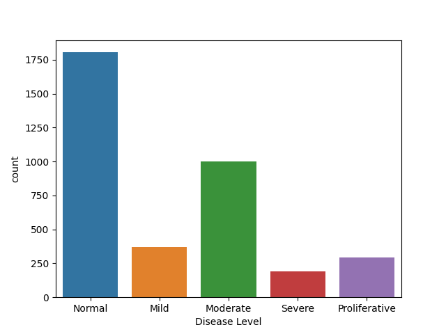
The plot shows that the data is imbalanced as the normal class has the highest number of instances. We resolve the class imbalance problem by incorporating weights of the classes into the loss function. In this case, the majority class was given a small weight while the minority classes were given larger weights. Intuitively, during training the model places more emphasis on the minority classes, giving higher penalties to misclassifications made by those minority classes, and less emphasis on the majority class [3, 8].
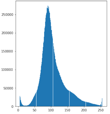
For image pre-processing, we used both vertical and horizontal flips for training data augmentation as well as standard normalisation of the data. We also resized all of the images to either or depending on the pre-trained model we want to use. An important pre-processing technique used in this study is Contrast Limited Adaptive Histogram Equalization (CLAHE) [18] because DR fundus images often suffer from low contrast issues [11]. Figure 2 shows a comparison of the intensity distribution of a DR fundus image before and after applying CLAHE.
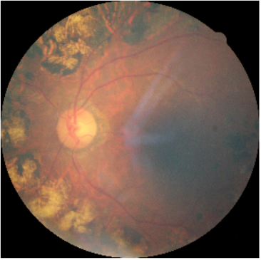 (a) Before CLAHE
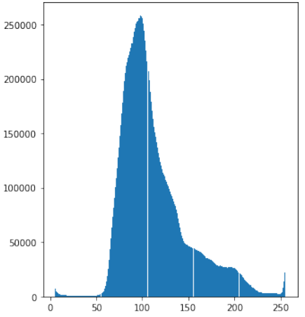 (b) Before CLAHE intensity distribution
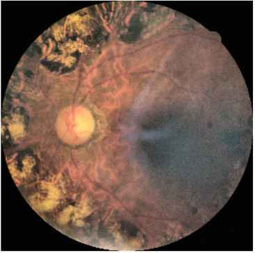 (c) After CLAHE
 (d) After CLAHE intensity distribution
It can be seen that the DR fundus image before CLAHE has more Gaussian (white) noise [11] making it potentially difficult to analyse. The Gaussian noise is reduced after adjusting the DR fundus image with CLAHE and its corresponding distribution is more normalised. This step is crucial because it can affect the sensitivity and specificity of the model [11].
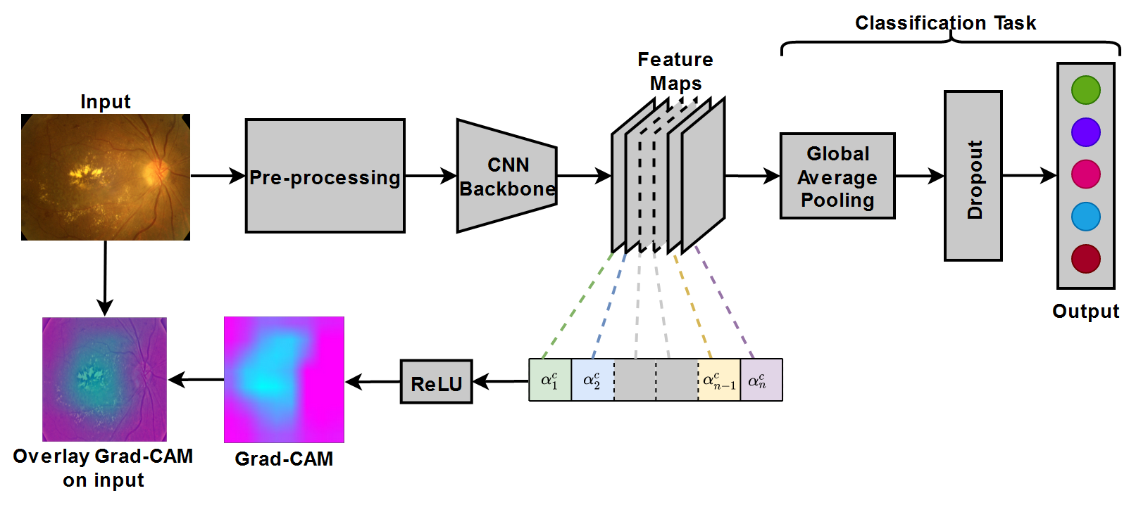
The pre-processed images are passed to the model which in this case has a CNN backbone. We consider and compare four CNN backbones in this study, namely VGG16 [13], ResNet50 [7], InceptionV3 [14] and InceptionResNetV2 [15]. The idea is to pass images through a pre-trained model and extract its feature maps at the last layer for classification. It should be noted that as images pass through a deep convolutional model, there exist trade-offs between losing spatial resolution and learning higher-level semantics, especially in the last convolutional layers. Neurons in those layers look for semantic class-specific information in the image, useful for discriminative purposes. Grad-CAM thus helps to interpret and explain individual components of a model [17, 12].
Grad-CAM is a technique used to produce visual explanations from decisions made by a CNN. Specifically, it uses gradients of a class concept in the penultimate layer to produce a coarse localisation map which helps to identify discriminative regions in an image. For every class , there are feature maps. To generate localisation maps, we first compute neuron importance weights by global-average-pooling gradients of the target with respect to the feature maps . It is important to note that is a spatial feature map hence it has width and height dimensions indexed by and [12]. Neuron importance weight is given by
| (1) |
Finally, we pass a linear combination of neuron importance weights and feature maps through a ReLU function to generate the coarse localisation map. The generated map can then be overlaid on top of the input image to identify the region(s) of interest. Grad-CAM is given by
| (2) |
Aside from using the feature maps to generate localisation maps, they are also used for discriminative purposes. In this study we replace the final layer with three successive layers, starting with a Global Average Pooling (GAP) layer, followed by a dropout (with 50% probability) layer and finally a dense layer. In summary, we feed a CNN model with a preprocessed DR fundus image. We then generate localisation maps using feature maps extracted from the CNN model. In addition, the feature maps are used to classify the DR fundus images in an image-level manner. Figure 3 shows a diagramme of this approach.
3 Results
In this section we evaluate the classification performance of the models and their ability to generate localization maps for DR fundus images. Models are trained using Keras with TensorFlow backend on an NVIDIA Tesla V100 GPU (available on the Centre for High Performance Computing [4]) for 15 epochs. We use the sigmoid activation function for the last layer and cross entropy as the loss function for training.
In this context, we use accuracy and Area Under Curve (AUC) as performance metrics over the test set. For every image we first predict a class and then generate the localisation map over the image. Table 1 shows the performance of the classification task.
| Model | Accuracy (%) | AUC | ||||
|---|---|---|---|---|---|---|
| Normal | Mild | Moderate | Severe | Proliferative | ||
| VGG16 | 95.31 | 0.97 | 0.64 | 0.77 | 0.62 | 0.78 |
| ResNet50 | 94.56 | 0.97 | 0.72 | 0.82 | 0.71 | 0.75 |
| InceptionV3 | 96.07 | 0.97 | 0.67 | 0.84 | 0.62 | 0.67 |
| InceptionResNetV2 | 94.39 | 0.96 | 0.77 | 0.81 | 0.68 | 0.69 |
We observe that InceptionV3 performed better than the other models as it had the highest accuracy of 96.05%. This is followed by VGG16, InceptionResNetV2 and ResNet50 in that order, however their performances are close to one other.
Finally, we randomly select some input images (see Figure 4) for demonstrative purposes. We pass the selected images through the InceptionV3 model (since it had the highest accuracy) to generate localisation maps and overlay the generated maps on the input images, as shown in Figure 4. We observe that the model generates a blurry image in Figure 4. The blurry effect can be attributed to the typical loss of spatial resolution as images pass through deep convolutional models. The blurry image in this case is the result of Grad-CAM. It may not be as useful until we map it on the input images. We see in Figure 4 that the Grad-CAM has highlighted regions of interest on the input images after mapping.
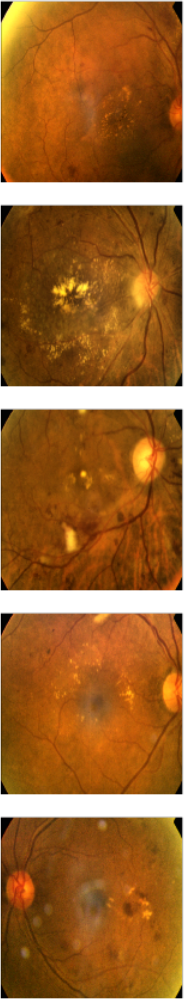 (a) Input
 (b) Grad-CAM
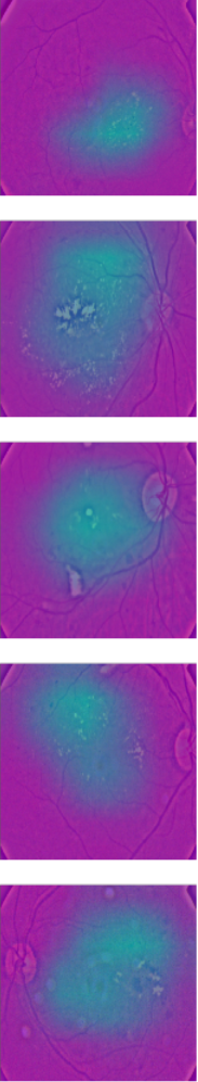 (c) Overlaid Grad-CAM
4 Conclusions
In this work we presented a technique which identifies regions of interest in DR fundus images and produces visual explanations for models. This could aid ophthalmologist in understanding the reasoning behind a model’s discriminative process and speed up diagnosis. We observe high performance for classification as well as potentially useful localisation maps. We observed that the performance of different models are very close to each other. In future, we intend to use in-training attention mechanism such as stand-alone self-attention [10] to classify and generate localisation maps. This is because in-training attention mechanisms overcome the limitation of losing spatial resolution.
Acknowledgement
We would like to thank German Academic Exchange Service (DAAD) for kindly offering financial support for this research. Also, we thank the Centre for High Performance Computing (CHPC) [4] for providing us with computing resource for this research.
References
- [1] American Academy of Ophthalmology: International Clinical Diabetic Retinopathy Disease Severity Scale Detailed Table, (2002)
- [2] Beede, E., Baylor, E., Hersch, F., Iurchenko, A., Wilcox, L., Ruamviboonsuk, P., Vardoulakis, L. M.: A human-centered evaluation of a deep learning system deployed in clinics for the detection of diabetic retinopathy. In Proceedings of the 2020 CHI Conference on Human Factors in Computing Systems (pp. 1-12), (2020).
- [3] Buda, M., Maki, A., Mazurowski, M. A.: A systematic study of the class imbalance problem in convolutional neural networks. Neural Networks, 106, 249-259, (2018).
- [4] CHPC, Centre for High Performance Computing, Department of Science and Technology, South Africa,https://www.chpc.ac.za/.
- [5] Gondal, W. M., Köhler, J. M., Grzeszick, R., Fink, G. A., Hirsch, M.: Weakly-supervised localization of diabetic retinopathy lesions in retinal fundus images. In 2017 IEEE International Conference on Image Processing (ICIP) (pp. 2069-2073), (2017).
- [6] Gulshan, V., Peng, L., Coram, M., Stumpe, M. C., Wu, D., Narayanaswamy, A., Kim, R.: Development and validation of a deep learning algorithm for detection of diabetic retinopathy in retinal fundus photographs. Journal of the American Medical Association (JAMA), 316(22), 2402-2410, (2016).
- [7] He, K., Zhang, X., Ren, S., Sun, J.: Deep residual learning for image recognition. In Proceedings of the IEEE Conference on Computer Vision and Pattern Recognition (pp. 770-778), (2016).
- [8] Johnson, J. M., Khoshgoftaar, T. M.: Survey on deep learning with class imbalance. Journal of Big Data, 6(1), 27, (2019).
- [9] Raghu, M., Zhang, C., Kleinberg, J., Bengio, S.: Transfusion: understanding transfer learning for medical imaging. In Advances in Neural Information Processing Systems (pp. 3347-3357), (2019).
- [10] Ramachandran, P., Parmar, N., Vaswani, A., Bello, I., Levskaya, A. Shlens, J.: Stand-alone self-attention in vision models. arXiv preprint arXiv:1906.05909 (2019).
- [11] Sahu, S., Singh, A. K., Ghrera, S. P., Elhoseny, M.: An approach for de-noising and contrast enhancement of retinal fundus image using CLAHE. Optics & Laser Technology, 110, 87-98, (2019).
- [12] Selvaraju, R.R., Cogswell, M., Das, A., Vedantam, R., Parikh, D., Batra, D.: Grad-cam: visual explanations from deep networks via gradient-based localization. In Proceedings of the IEEE International Conference on Computer Vision, pp. 618-626, (2017).
- [13] Simonyan, K., Zisserman, A.: Very deep convolutional networks for large-scale image recognition. arXiv preprint arXiv:1409.1556, (2014).
- [14] Szegedy, C., Liu, W., Jia, Y., Sermanet, P., Reed, S., Anguelov, D., Rabinovich, A.: Going deeper with convolutions. In Proceedings of the IEEE Conference on Computer Vision and Pattern Recognition (pp. 1-9), (2015).
- [15] Szegedy, C., Ioffe, S., Vanhoucke, V., Alemi, A.: Inception-v4, inception-resnet and the impact of residual connections on learning. arXiv preprint arXiv:1602.07261, (2016).
- [16] Tan, C., Sun, F., Kong, T., Zhang, W., Yang, C., Liu, C.: A survey on deep transfer learning. In International Conference on Artificial Neural Networks (pp. 270-279). Springer, Cham, (2018).
- [17] Zhou, B., Khosla, A., Lapedriza, A., Oliva, A., Torralba, A.: Learning deep features for discriminative localization. In Proceedings of the IEEE Conference on Computer Vision and Pattern Recognition, pp. 2921-2929, (2016).
- [18] Zuiderveld, K.: Contrast limited adaptive histogram equalization. Graphics Gems, 474-485, (1994).