Machine learning applications using diffusion tensor imaging of human brain: A PubMed literature review
Abstract
We performed a PubMed search to find 148 papers published between January 2010 and December 2019 related to human brain, Diffusion Tensor Imaging (DTI), and Machine Learning (ML). The studies focused on healthy cohorts (n = 15), mental health disorders (n = 25), tumor (n = 19), trauma (n = 5), dementia (n = 24), developmental disorders (n = 5), movement disorders (n = 9), other neurological disorders (n = 27), miscellaneous non-neurological disorders, or without stating the disease of focus (n = 7), and multiple combinations of the aforementioned categories (n = 12). Classification of patients using information from DTI stands out to be the most commonly (n = 114) performed ML application. A significant number (n = 93) of studies used support vector machines (SVM) as the preferred choice of ML model for classification. A significant portion (31/44) of publications in the recent years (2018-2019) continued to use SVM, support vector regression, and random forest which are a part of traditional ML. Though many types of applications across various health conditions (including healthy) were conducted, majority of the studies were based on small cohorts (less than 100) and did not conduct independent/external validation on test sets.
Keywords Diffusion Tensor Imaging Feature Extraction Machine Learning Deep Learning Support Vector Machine Classification
1 Introduction
Machine learning (ML) applications in medicine are being increasingly developed with the advancements made in deep learning. Numerous applications are developed using medical imaging [1, 2], electronic health records [3], and workflow data [4, 5], in addition to internet of things for healthcare applications [6]. Despite multiple criticisms surrounding the application of Artificial Intelligence (AI) in medicine [7], academic peer-reviewed contributions of ML applications in medicine are demonstrating significant future potential and raising hopes of the medical community [8].
In this review article, we turn our focus to ML applications of diffusion tensor imaging (DTI) of the human brain. Since its introduction in 1994, DTI analysis had been a part of different neuroimaging studies related to several brain diseases like Alzheimer’s disease [9, 10], traumatic brain injury [11], and depression [12, 13]. Due to the sensitivity of DTI towards microstructural tissues, it has gained popularity for the evaluation of White Matter (WM) architecture in both healthy and diseased adult and pediatric populations [14, 15, 16, 17]. DTI has also been used as the part of the multimodal magnetic resonance imaging (MRI) analysis or neuroimaging analysis. Various review studies related to neuroimaging and ML have are present in the literature [17, 18, 19, 20, 21, 22]. However, the usage of DTI in the light of its diverse ML applications for diseased and healthy populations has not yet been studied. Despite the limitations of DTI [23], it continues to be used in data-driven neuroimaging studies for developing ML applications.
In this paper, we review the neuroimaging studies indexed over the past decade in PubMed that involved the development of ML applications using human brain DTIs. We highlight each study’s application areas, ML tasks performed, reference standards, accomplishments, and any translational barriers.
2 Machine Learning (ML)
ML techniques are used to process input training data to learn patterns from the input data which represents experience to gain expertise through a computer program. The input data can be of various types e.g., imaging, text, continuous or discrete variables or a combination of these types. The training is performed using mathematical operations on the input data, such that trained model can be used to perform a specific task in the form of prediction. The mathematical operations for training are a part of the model or algorithm which is a well-defined process that enables learning the parameters/weights using the input data. Various applications of ML exist in the field of imaging analysis, text analysis, bioinformatics, and finance to name a few. Some applications of ML using medical imaging as input are detection of intracranial haemorrhage in CT images [24], classification of migraine patients into different categories using MRI [25], cell detection in microscopic imaging [26]. Examples of successful and popular ML algorithms include support vector machines (SVM), random forest (RF), artificial neural networks (ANN), Gaussian Mixture Model (GMM), Elastic Net Regression. Readers interested in ML terminology terms are referred to study by Liu et al. [27] for further details on ML applications within medicine.
With the advent of Deep Learning (DL) techniques, ML applications using medical images have gathered momentum in the recent years. Neuroimaging has been a great topic of interest for the ML community over several years. Existing reviews of ML related neuroimaging include a general review of progress and challenges in neuroimaging related ML [18], use of structural neuroimaging as a clinical predictor [19], neuroimaging based classification and related feature extraction [17], and systematic reviews of ML and neuroimaging studies for diseases like Alzheimer’s [20, 21]. Other review articles of ML applications involving specific diseases are related to glioma [22], pituitary tumors [28], Parkinson’s disease [29], neurodegenerative diseases [30], and schizophrenia [31]. A study focusing on ML-based tractography processed using diffusion imaging was conducted by Poulin et al. [32].
Owing to a long presence of DTI for neuroimaging research, we considered applications of ML that use DTI of human brain with or without other forms of imaging and non-imaging data. We reviewed PubMed literature from the last 10 years and focused on the spectrum of types of ML tasks that leveraged usage of DTI for various health conditions.
3 Diffusion Tensor Imaging: Basics and Usage
DTI is a form of diffusion weighted imaging (DWI) [33, 34]. DWI is a variant of magnetic resonance imaging (MRI) that produces images by using the diffusion rate of water within brain tissue and provides elaborate details about tissue microstructure. DWI can be acquired through existing MRI scanners thereby allowing for non-invasive acquisition; and does not require the use of contrast agents or radiotracers. In plain DWIs, diffusion is modeled by a scalar parameter or diffusion coefficient. However, as diffusion is three-dimensional phenomena, it can be modeled through diffusion tensor techniques (like DTI) which accounts for difference in diffusion of water and tissue molecules by providing information about the structural orientation and quantitative anisotropy of the molecules. Details of data acquisition and processing are elaborated by Le Bihan et al. [35] and Soares et al. [34]. The pipeline from the acquisition to the usage of DTI is complex and the typical steps are described below:
-
A.
Acquisition: For DTI modeling, DWI sequences are captured through axial (typical) slices (allowing zero gaps between the slices) of the entire brain at a low gradient pulse (usually b = 0 s/sq. mm) and at least in six non-collinear diffusion encoding directions (usually b = 800-1000 s/sq. mm). As in any MRI acquisition, the acquired signal is affected by several other acquisition parameters including, but limited to, field strength, field-of-view (FOV), slice thickness, echo time, and repetition time.
-
B.
Pre-processing and quality checks: Two major components involve: (i) visual verification of DWIs to note artifacts using general purpose image viewers, assuring the correctness of the acquisition parameters after importing the images, and conversion of raw image to specific image formats, and (ii) correction of eddy current and motion, performed as DWI sequences are characterized by low signal-to-noise (SNR) and motion artifacts. Additional and optional preprocessing can involve the removal of the skull, and other non-brain areas such as, muscle, skin, cerebrospinal fluid (CSF). etc. from the brain, registration, and normalization of the sequences.
-
C.
Processing: The heart of DTI processing lies in the estimation of diffusion tensor, characterized by the eigenvalues and eigenvectors, at each voxel. This is often followed by the estimation of various diffusion indices, metrics, or parameters such as fractional anisotropy (FA), mean diffusivity (MD), radial diffusivity (RD), axial diffusivity (AD). DTI also enables the three-dimensional estimation (using the principal eigenvector) of the trajectory and location of white matter tracts through a mechanism called tractography. Tractography can be used to create brain-wide mapping of neuronal connections among the anatomical regions (obtained automatically/semi-automatically/manually using structural MRI) of the brain to obtain a connectome.
-
D.
Quantitative Analysis: These analyses are often guided by the processing performed in step C. Summary measures of selected DTI indices can be extracted using anatomical regions or tracts for detecting groupwise voxel analysis such as tract-based spatial statistics (TBSS). Analysis based on voxel-by-voxel analysis (such as voxel-based analysis or VBA) are also performed. Another set of analysis includes the extraction of features (DTI indices of WM or connectomes from selected regions) which subsequently undergo the application of ML methods. Several standard software libraries and tools are available to perform the post-acquisition stages related to DTI processing [36, 37, 38].
4 Materials and Methods
4.1 Search Strategy and Inclusion Criteria
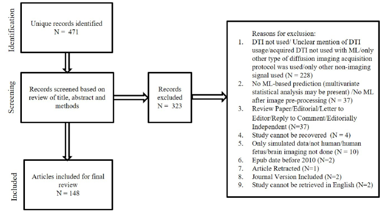
We searched PubMed for ML related applications of DTI published from January 1, 2010 to December 31, 2019. The time-range was selected to review the prior decade of ML related publications using DTI. Three search queries (detailed description of the queries can be found in our supplementary material) having combination of terms “diffusion tensor imaging” and several DTI related terms and “machine learning”, “deep learning” and other ML-related terms were executed in PubMed to retrieve unique articles in the past decade. As shown in Figure 1, we included 148 articles in this review after screening and excluding 323 articles. Though, ours is a literature review, we followed the steps of PRISMA for inclusion of studies in our review. Studies included applied ML to real (in contrast to only simulated data) DTI of human brain. Our study excluded articles if usage of DTI was not confirmed and this included all the studies involving application of several other diffusion imaging acquisition techniques including high-angular diffusion imaging (HARDI). As there is inconsistency in the description of exact criteria for classifying a diffusion MRI acquisition as a HARDI acquisition [39] as opposed to other diffusion MRI acquisitions such as diffusion kurtosis imaging, diffusion spectrum imaging; we excluded works involving only HARDI acquisition and focused on DTI acquisition. From the studies reviewed, we noted the (a) first author and year of publication, (b) unmet need addressed by the study, (c) datasets and ML tasks performed, (d) reference standard, (d) usage of DTI in the study, (e) ML model used and; (f) performance of the models with accomplishments and limitations of the studies. We categorized the studies based on the cohorts that they use for the design of their ML problems. Figure 1 shows a graph of the number of studies published per year.
The following categories were found from the studies included in our work:
-
A.
Studies (n = 15) with fully healthy cohorts
-
B.
Studies (n = 121) that included individual cohorts that may have an addition of healthy controls/relevant control along with the following types of disorders or conditions
-
1.
mental health disorders (n = 25)
-
2.
tumor (n = 19)
-
3.
trauma (n = 5)
-
4.
dementia (n =24)
-
5.
developmental disorders (n = 5)
-
6.
movement disorders (n = 9)
-
7.
other neurological disorders (n = 27)
-
8.
miscellaneous non-neurological disorders or did not state the disease of focus (n = 7)
-
1.
-
C.
Studies (n = 12) that had multiple cohorts with characteristics of A or B or mixed cohorts involving more than one disease type in B
We discuss the specifics of these studies in the next section. Further details of these studies can be found in our supplementary material (sTables 1- 10)
5 Results
5.1 Studies with Fully Healthy Cohorts
Of the 15 articles that involved healthy subjects, three focused on parcellation of specific type of brain areas such as cortex [40], left-dorsal pre-motor cortex [41] and supplementary motor cortex and pre-supplementary motor cortex [42]. Tissue segmentation was done in two studies [43, 44] where the first one focused on the robustness of the technique for noise and field inhomogeneities. Of the four studies working on brain aging, one study [45] classified the patients into age groups and the other three works [46, 47, 48] studied the regression of healthy human brain with age. Additionally, the regression of cognitive performance scores was performed in two studies to predict cognitive ability [49] and memory assessments [50]. Other applications include: denoising [51], network analysis of brain connectivity [52], white matter patterns in infants [53], and zero-shot learning of fMRI task using DTIs [54]. The usage of DTI for all the applications mentioned in this section lied in extracting DTI-parameters, performing DTI based tractography, and estimation of connectivity matrices. Only six of these studies [41, 46, 47, 48, 50, 53] used a dataset involving more than 100 participants and highest number of subjects was 323 used in the study by Ullman et al. [50].
The ML tasks of the studies varied, and various study-specific reference standards were collected. The ML models designed/used for the applications varied accordingly. Nine studies focused on the novelty of the overall ML approach used such as innovations on convolutional filters used in artificial neural networks (ANN) [46], combination of ANN with hybrid genetic algorithm [47], novel modeling of noise distribution [51], multigraph min-max cut [52], and modified Bayesian modeling [50], and incorporation of DTI-based registration in a zero-shot support vector machine (SVM)-based prediction of fMRI task [54]. Existing supervised ML techniques such as SVM, maximum uncertainty linear discriminant analysis (LDA), relevance vector machine, linear regression, partial least squares, Multilayer perceptron (MLP) were used by four studies [44, 45, 48, 49]. Unsupervised ML approaches such as K-means clustering, force directed graph layout, fuzzy C-means clustering with spatial constraints were used by three studies [41, 42, 43] respectively. The study by Gorbach et al. [40] validated pipelines for connectivity-based cortical parcellation. Critsis et al. [44] evaluated the performance of their model in an validation set with an accuracy of 82.1%. Wen et al. [43] validated the clustering methodology for segmentation with a volume overlap over 0.84 WM, gray matter (GM), and CSF.
5.2 Mental Health Disorders
We included 25 studies which focused on various mental health disorders: Autism Spectrum Disorder (ASD) (N=4), first episode of schizophrenia-spectrum disorder (FES) or schizophrenia (SZ) (N=5), depression disorders (N=10), First Episode Psychosis (FEP) (N=2), attention deficit hyperactivity disorder (ADHD) (N=1), obsessive compulsive disorder (OCD) (N=2), and anorexia nervosa (AN) (N=1). Studies on depression disorders developed classification systems for bipolar disorder (BD), major depressive disorder (MDD), and healthy controls (HC) [55], classification of BD patients and controls to data driven phenotypes [56] and classification of pediatric BD patients and controls [57]. Classification of MDD and controls was performed in four studies [58, 59, 60, 61]. Classification among acute, remitted MDD and healthy controls by Qin et al. [62], between treated and untreated MDD by Chu et al. [63], and between treatment responses by in a multi-site study by Wang et al. [64] were performed. Studies on SZ focused on classification of FES and controls [65, 66] and of drug naïve FES and controls [67], and clustering of SZ and controls [68, 69]. The study by Ingalhalikar et al. [69] also performed the clustering in a cohort of ASD and controls. Other studies involving ASD include classification of ASD risk in infants [70], classification of ASD and typically developing controls [71] and regression of ASD-related interview scores [72]. Two studies included classification of FEP patients and controls [73, 74]. Other studies performed classification between healthy controls and other mental health conditions like AN [75], OCD [76, 77], and ADHD [78]. Apart from the studies by Foque et al. [72], regression was performed in two studies for separate purposes: chlorpromazine dose and symptom levels in FES patients [66] and general psychopathology levels and age [79] on publicly available neurodevelopmental cohort. DTI was used to extract study-specific DTI-parameters (sometimes from WM/TBSS skeletons), perform DTI-based tractography, and estimate connectivity matrices. Connectivity matrices were often used for further feature extraction used in ML models. Eight of these studies [55, 56, 64, 65, 66, 75, 78, 79] used a dataset size of more than 100, with the largest dataset (729 studies) used in the study by Alnæs et al. [79].
SVMs and their variations were used for classification in 21 of the 25 mental health-related studies and Support Vector Regression (SVR) was used for regression in one of the studies [66]. The remaining studies focused on Random Forest (RF) [65] and least absolute shrinkage and selection operator (LASSO) and Elastic Net [56] for classification. For clustering, variant of Non-negative matrix factorization (NMF), spectral clustering with modifications [68, 69], K-Means clustering [56, 75] were used. Of the three studies which involved regression [66, 72, 79] , two studies used Kernel Ridge Regression [66, 72] and one study used shrinkage linear regression [79]. One study by Deng et al.[65] validated their model in a hold-out set with an accuracy of 76%.
5.3 Tumor
Of the 19 studies involving tumors, 18 were related to brain tumors, and one was related to nasopharyngeal carcinomas [80] which had a prospective design for the classification of different stages of post-therapy time after the patients were treated for radiotherapy. Fourteen of the studies focused on the following aspects of glioma brain tumors specifically: multimodal segmentation of glioma [81], classification related to tumor biology (i.e. tumor grade) [82, 83, 84], IDH1 or/and MGMT status prediction [85, 86, 87], nuclei-content classification [88], EGFRvIII status prediction [89], tumor cell density regression [90], and tissue type classification for causing recurrence [91], and prognostication by classifying true versus pseudo-progression [92, 93], classification of survival period as good or bad [94]. Segmentation of different types of brain tumors and classification of high grade glioma versus solitary metastasis was investigated in the study by Yang et al. [95]. Classification of different types of intracranial lesions was performed by Svolos et al. [96]. Differentiation of atypical meningiomas from glioblastoma and metastases was performed by Svolos et al. [97]. Shrot et al. [98] performed a study for classification of four types of brain tumors. Six of the 19 studies [80, 82, 89, 93, 96, 97] had dataset sizes of more than 100, with the study by Leng et al. [80] having the largest sample size of 147.
The usage of DTI included derivation of DTI-parameters for feature representation [80, 82, 83, 88, 89, 90, 91, 92, 93, 96, 97, 98], extraction of features using structural connectome [80, 86, 87, 94] and deriving isotropic and anisotropic components for segmentation [81, 88, 95]. Since several studies require tumor and/or its surroundings as the regions-of-interest, (ROI), manual ROIs were used in some applications for identifying tumor or specific regions of tumor and its surroundings [82, 88, 91, 96, 97] and manual seed-points were used in [95]. Atlas-based ROIs were used by [80, 87, 94]. All studies performed classifications and SVMs were used in 13 of 17 studies that performed classification tasks. RF, quadratic discriminant analysis (QDA), LDA, naïve Bayes (NB), and artificial neural networks (ANNs) were used in some of the studies. Variants of matrix completion techniques were used as clustering approaches in one study [87] performing unsupervised prediction of IDH1 and MGMT status. Akbari et al. [89] validated their radiomic signature for glioma in a replication cohort with an AUC of 0.86. Eichinger et al. [85] reported an AUC of 0.91 in validation cohort for IDH classification. Akbari et al. [91] reported an AUC of 0.84 in replication cohort for predicting early recurrence and Hu et al. [88] reported an accuracy of 81.8% in the validation set.
5.4 Trauma
Five studies involved the classification of traumatic brain patients (TBI) injury versus controls. Three of the studies [99, 100, 101] involved patients with the presence of mTBI (mild TBI) and used varying reference standards. The remaining two studies involved patients with history of mTBI whose MRI was acquired on average around 38 months after injury [102] or after retirement from sports [103]. DTI was used for DTI parameter extraction from white matter, brain voxels, performing tractography and for generating connectivity matrices. SVM was used in all studies. Along with classification, two studies performed regression: for neuro-physical outcome scores using SVR [101] and regression for cognitive impairment scores using Elastic Net [102]. Other than one study [99] which had 100 subjects, other studies had a dataset sizes less than 100. The study by Fagerholm et al. [102] reported a correlation of 0.44 in the validation set.
5.5 Dementia
Of the 24 dementia-related studies, one study [104] classified presymptomatic FTD mutation carriers versus controls whereas the majority (N = 12) of studies included Alzheimer’s disease (AD) patients in their cohort. The reference standards for these studies included clinically probable AD based on NINCDS-ADRDA criteria and/or clinical diagnosis of AD [105, 106, 107, 108, 109, 110]. Some studies [111, 112] included additional criteria such as CSF AD markers and one study used MMSE scores and the ADNI protocol [113]. While classification of AD versus healthy controls was the focus of several studies, Bron et al. [107] developed classification systems for discriminating between AD, FTD, and controls. Maggipinto et al. [106] and Eldeeb et al. [114] studied application of DTI measurements for classification between healthy controls, mild cognitive impairment (MCI), and AD. MCI versus healthy classification was performed by several other studies [115, 116, 117, 118, 119, 120, 121, 122] with two studies involving vascular MCI [115, 116] and one study involving amnestic MCI [123]. Classification of various subtypes of MCI was studied by Haller et al. [124]. A multicenter study on the classification of two of types of MCI and healthy controls was performed by Dyrba et al. [125]. A study by O’Dwyer [126] classified apolipoprotein-E4 (a risk-factor for AD) carriers versus non-carriers in healthy young patients. In these studies DTI was used to extract the parametric values, features from white matter, white matter tracts, gray matter tracts and identified ROI, and generate connectivity matrices. Seven of these studies [104, 106, 109, 111, 119, 123, 127] had a dataset size of more than 100 with the highest dataset size of 915 subjects (combination of several cohorts) used in the study by Bouts et al. [127].
All but one of the 24 studies performed classification and SVM and its variations (binary, multi-class) were used in 18 studies for classification. Among these 18 studies, SVM as well as other classifiers including LDA, KNN, Functional Trees, NB were used by three studies [109, 116, 117]. CNN, Adaboost, RF, logistic regression (LR), elastic net regression classifier, sparse group LASSO were used by the other six studies. Two studies- Maggipinto et al. [106] and Lee et al. [122] reported an AUC of 0.86 (AD vs. control classification) and accuracy of 100% (MCI vs. control classification) respectively in corresponding validation sets.
5.6 Developmental Disorders
Of the five studies found regarding developmental disorders, one study focused on genetic variability in PPARG and brain connectivity [128] in preterm infants. Future behavioral profile in neonates was studied by Wee et al. [129]. Classification of dyslexia versus controls in children was performed in two studies [130, 131]. In a set of adult patients, classification of chromosome 22q11.2 deletion syndrome versus controls was studied in the remaining study [132]. Usage of DTI included metric extraction, tractography, and connectivity matrix generation with study-specific variations. Two studies [128, 129] had a data size more than 100 and Krishnan et al. used a dataset of 272 pre-term infants [128]. SVM was used in four of the five studies. One study used Sparse reduced rank regression. Studies did not use an external validation set or were unclear about the same.
5.7 Movement Disorders
All the nine studies related to movement disorders involved patients with Parkinson’s disease (PD). Classification of Parkinson’s patients versus healthy controls was performed in three studies [133, 134, 135] with some differences in the reference standard for the disease (sTable 7 in our supplementary document). One study by Liu et al. [136] involved the identification of a biomarker through feature selection using the United Kingdom brain bank criteria for diagnosis. Six studies focused [133, 137, 138, 139, 140, 141] on several forms of Parkinsonism. Du et al. [139] reported some patients in their cohort to have post-mortem confirmed diagnosis of PD and progressive nuclear palsy (PSP). Two studies [133, 139] used datasets of more than 100 samples and Lei et al. [133] used a dataset of 238 subjects.
Of the eight studies that performed classification, SVMs were used in seven [133, 134, 137, 138, 139, 140, 141] and LR was used in one study [135]. One study [136] performed feature selection and used Folded concave penalized model for the same. The studies reported accuracy or AUC above 0.90 for the tasks they addressed though none of these had external validation sets.
5.8 Other Neurological Disorders
Of the 27 studies related to other neurological disorders, majority [142, 143, 144, 145, 146, 147, 148, 149, 150, 151, 152, 153, 154, 155, 156, 157] of the studies involve epilepsy patients. Various classification tasks within temporal lobe epilepsy (TLE) patients (N=9) were performed including determination of laterality [149, 151, 154]; prediction of seizure frequency [143]; detection of GM and WM abnormalities of focal cortical dysplasia in extratemporal TLE [156]; discrimination between surgical outcomes [145, 146, 150], and distinguishing treatment (vagus nerve stimulation) outcomes [144]. Among these studies, four [143, 148, 149, 150] performed classification between healthy controls and TLE patients as well. A similar classification task in adult cohorts was performed by Del Gaizo et al. [147] whereas children and adolescents were classified into active TLE, remitting TLE and healthy in the study by Amarreh et al. [153]. Classification of mTLE and controls using DTI and T2-relaxometry was performed by Cantor-Rivera et al. [152]. Regression of language performance score of TLE patients was performed by Munsell et al. [142]. Classification of language phenotypes was performed in paediatric epilepsy patients by Paldino et al. [157] and patients of mTLE with hippocampal sclerosis (HS) were found to be different from controls based on white matter abnormalities. MR image voxel-based classification of mTLE having HS and controls was performed by Focke et al. [155]. Classification of amylotropic lateral sclerosis (ALS) versus controls using neuroimaging profile of the corticospinal tract was performed by Sarica et al. [158]. In another study [159], the usage of multimodal MRI was explored for detection and comparison of ALS, predominantly upper motor neuron disease (PUMN), ALS-mimicking conditions and controls. In two separate tasks, classification of controls and relapsing-remitting multiple sclerosis (MS) patients and classification of MS with different degree of disability was performed by Zurita et al. [160]. Wen et al. [161] performed a study on classification of Tourette Syndrome (TS) patients and controls. Imaging and questionnaire data were both used in the classification of migraine versus control patients in [162]. Thalamus segmentation for cerebellar ataxia patients was performed by Stough et al. [163] using multimodal MRI. Clustering of HIV-infected and controls for cognitive impairment [164], classification of presence and absence hepatic minimal encephalopathy [165], classification of normal versus abnormal outcomes in neo-natal encephalopathy [166], classification of stroke outcomes using acute DTI [167], and detection of sensory processing disorder using DTI tractography [168] are other applications. Four of the studies [150, 159, 160, 164] had dataset size more than 100 with the study by Ferraro et al. having the largest dataset size of 265 patients [159].
SVMs were used by 17 of the 23 studies for classification. Other ML models considered were RFs and their variants, multiple kernel learning, Adaboost, naïve Bayes, and gaussian process for ML. Two studies reported results on external validation cohorts. Mithani et al. [144] validated their results on multi-institutional independent dataset with an accuracy of accuracy of 83.3 for predicting treatment outcome. Ferraro et al. [159] validated their model with an accuracy of 95% for classifying PUMN vs. ALS mimicking disorders.
5.9 Miscellaneous Non-neurological Disorders/Not-stated
The diseases/conditions assigned to the studies (n = 7) in this section were related to the classification of venous erectile dysfunction patients and healthy controls [169], exploring amygdala modulation in adolescent representatives of families facing more economic and social challenges [170], classification of world-class gymnasts and healthy controls using neuroplasticity in white matter [171], classification of self-reported drinking histories of adolescents [172]. Three studies did not reveal the state (disease conditions/healthy) of the participants considered and developed methodologies for the segmentation of tissues [173] and WM tracts [174, 175]. Three studies [170, 172, 175] used a dataset size of more than 100; and largest dataset (7000 samples had DTI) was used by Mu et al. [175].
SVMs were used for classification in two studies [169, 171]. Sparse logistic classification model and LR were also used for classification in two studies [171, 172]. For segmentation and regression, fully convolutional networks (FCN), Bayesian decision theory, and combination of maximum a-posteriori, Markov random field, gradient descent, and kernel regression were used. These studies did not use an external validation set or were unclear about the same.
5.10 Multiple or Mixed Cohorts
We found 12 studies belonging to this category. FA and MD maps were reconstructed using ANNs by Aliotta et al. [176] using a training set of healthy patients and the trained model was tested in a cohort of GBM patients. Classification of AD and Late Onset BD was performed by Besga et al. [177] and classification of AD, late onset BD and healthy controls were performed in another study [178]. Classification of AD, non-AD in the context of corticobasal syndrome, and healthy controls were performed by Megdalia et al. [179]. A regression study of age in healthy humans and prediction of age gap in HIV+ and HIV- patients were performed by Kunh et al. [180]. Pathway reconstruction using neuroimaging in a public cohort of healthy and schizophrenic patients was performed in the study [181]. Another study [182] on reconstruction of white matter pathways using longitudinal diffusion MRI was tested separately in healthy control and patients of Huntington disease for different types of analysis. Cortical ROIs were segmented in healthy and MCI patients by Zhang et al. [183]. Clustering of brain sub-networks in healthy and ASD patients was performed [184]. Clustering using DTI connectivity matrices and analyzing children with ASD and TDs and gender differences in different cohorts was performed by Ghanbari et al. [185]. Classification of ASD and typically developing controls and regression of responsiveness scale scores of meditation practitioners was performed by Adluru et al. [186]. Lin et al. [187] created a classification of MCI and severe dizziness in presymptomatic carotid stenosis. Four studies [180, 184, 185, 186] used datasets of size more than 100 and Kuhn et al. [180] used a dataset size of 869 including their independent test set.
SVMs were used for the classification of five studies [177, 178, 179, 186, 187] and Support vector regression was used by two studies [180, 186]. Clustering was performed using NMF and automatic relevance determination based projective NMF in two studies [184, 185]. Generalized Multiple Kernel Learning was used for ROI localization by Zhang et al. [183] and a new technique for fiber reconstruction involving agglomerative hierarchical clustering was used for pathway reconstruction [181]. In the testing set, Kuhn et al. [180] predicted brain age gap which was significantly correlated (r = 0.26) with the cognitive function. Yendiki et al. [182] executed test-retest analysis (average error was 5%) on a separate cohort of 9 healthy patients and sensitivity analysis (22.3% of positions in 18 WM pathways showed significant changes) on 46 Huntington disease patients. Chu et al. [181] demonstrated a better average results (spatial metric 2.72 mm) compared to an existing technique. Zhang et al. [183] validated their method and found its performance (prediction error: 8.08 mm) to be better than four existing techniques.
6 Discussion
Our study found articles relating to DTI applications for several medical disorders and conditions (including healthy). The majority of applications involved the classification of patients of various medical conditions. Many studies also included the clustering of patients, regression of clinical test scores using imaging, segmentation of tissues (normal and abnormal), and novel methods of DTI reconstruction. Classification was performed in 114 studies and SVM was used in the majority (N = 93) of the studies as the preferred classifier. A significant portion (31/44) of publications in the recent years (2018-2019) continued to use traditional ML methods such as SVM, support vector regression and random forest despite phenomenal development in deep learning.
While performing an ML task, DTI was sometimes used in conjunction with other imaging and non-imaging data. Application of human brain DTI-based ML was not confined to subjects for having neurological disease; non-neurological disorders and healthy patients were also considered for designing several studies. Classification of various diseases and controls, clustering of diseased (with various levels of the disease) and healthy cohorts, regression of clinical test scores were major application in studies involving cohorts having abnormal health conditions with classification as the predominant topic of interest. For healthy cohorts, classification, regression, and image segmentation received balanced interest from the researchers.
The majority of the studies had datasets of less than 100 samples. Though most of the studies conducted internal validation through cross-validation, few studies conducted validation on an independent test set. Studies often stated small sample size, study design (such as retrospective, cohort studies), inconsistencies or variabilities in reference standards (often unique to the problem studied), side effects of medications, limited number of directions of diffusion, errors introduced during pre-processing of DTI, crossing-fibers, restriction of extracting imaging features in particular regions, and imbalances in the distribution of samples, as study limitations. Since validating on an independent test set is one of the tests of generalizability of the models, this needs to be addressed by majority of the studies for clinical translation.
While the studies reviewed show promise, before ML techniques can really be translated to the daily healthcare setting to improve the diagnosis, treatment, and prognostication of patients with the neurological conditions reviewed, future work must include larger datasets to validate their accuracy and efficacy. Future work also has to more carefully consider reference standards for diagnosis as these vary within conditions, and the training and evaluation of the ML methods rely heavily on these. Moreover, since several technically varying ML solutions can be developed to solve the same task, researchers and organizations will need to collaborate to develop large scale, diverse, task-specific benchmark datasets to ensure that robust and well-performing sets of ML solutions can be developed to advance healthcare solutions. Ideally, these datasets should be made open-access to allow critical evaluation of these techniques by multiple research teams so that more rapid translation of these techniques to patient care can be achieved with confidence.
7 Limitations
Our study selected papers indexed in PubMed only and as such, does not include articles indexed in other research databases. However, since PubMed is representative of biomedical literature across the world, our study captures a significant view of the research involving ML application related to DTI globally. It is likely that we have excluded some articles from 2019 that were not indexed in PubMed when we executed our search. Due to non-inclusion of HARDI, some connectome related ML applications are excluded from this review. Nevertheless, various ML applications using connectome were considered in our work.
8 Acknowledgements
Funding support from the Canadian Institute for Military and Veteran Research (CIMVHR) is gratefully acknowledged.
References
- Litjens et al. [2017] G. Litjens, T. Kooi, B. E. Bejnordi, A. A. A. Setio, F. Ciompi, M. Ghafoorian, J. A. W. M. van der Laak, B. van Ginneken, and C. I. Sánchez. A Survey on Deep Learning in Medical Image Analysis. Medical Image Analysis, 42:60–88, feb 2017. doi:10.1016/j.media.2017.07.005. URL http://arxiv.org/abs/1702.05747http://dx.doi.org/10.1016/j.media.2017.07.005.
- Mazurowski et al. [2019] M. Mazurowski, M. Buda, A. Saha, and M. Bashir. Deep learning in radiology: An overview of the concepts and a survey of the state of the art with focus on MRI. Journal of Magnetic Resonance Imaging, 49(4), 2019. ISSN 15222586. doi:10.1002/jmri.26534.
- Shickel et al. [2018] B. Shickel, P. J. Tighe, A. Bihorac, and P. Rashidi. Deep EHR: A Survey of Recent Advances in Deep Learning Techniques for Electronic Health Record (EHR) Analysis. IEEE Journal of Biomedical and Health Informatics, 22(5):1589–1604, sep 2018. ISSN 2168-2194. doi:10.1109/JBHI.2017.2767063. URL https://ieeexplore.ieee.org/document/8086133/.
- Wang et al. [2018a] Y. Wang, L. Wang, M. Rastegar-Mojarad, S. Moon, F. Shen, N. Afzal, S. Liu, Y. Zeng, S. Mehrabi, S. Sohn, and H. Liu. Clinical information extraction applications: A literature review. Journal of Biomedical Informatics, 77:34–49, jan 2018a. ISSN 15320464. doi:10.1016/j.jbi.2017.11.011. URL https://linkinghub.elsevier.com/retrieve/pii/S1532046417302563.
- Kim and Mackinnon [2017] D. H. Kim and T. Mackinnon. Artificial intelligence in fracture detection: transfer learning from deep convolutional neural networks. Clinical Radiology, 2017. ISSN 1365229X. doi:10.1016/j.crad.2017.11.015. URL https://www.sciencedirect.com/science/article/pii/S0009926017305354.
- Baker et al. [2017] S. B. Baker, W. Xiang, and I. Atkinson. Internet of Things for Smart Healthcare: Technologies, Challenges, and Opportunities, nov 2017. ISSN 21693536.
- Emanuel and Wachter [2019] E. J. Emanuel and R. M. Wachter. Artificial Intelligence in Health Care: Will the Value Match the Hype?, jun 2019. ISSN 15383598.
- Toh et al. [2019] T. S. Toh, F. Dondelinger, and D. Wang. Looking beyond the hype: Applied AI and machine learning in translational medicine, sep 2019. ISSN 23523964.
- Kantarci et al. [2017] K. Kantarci, M. E. Murray, C. G. Schwarz, R. I. Reid, S. A. Przybelski, T. Lesnick, S. M. Zuk, M. R. Raman, M. L. Senjem, J. L. Gunter, B. F. Boeve, D. S. Knopman, J. E. Parisi, R. C. Petersen, C. R. Jack, and D. W. Dickson. White-matter integrity on DTI and the pathologic staging of Alzheimer’s disease. Neurobiology of Aging, 56:172–179, aug 2017. ISSN 15581497. doi:10.1016/j.neurobiolaging.2017.04.024.
- Brueggen et al. [2017] K. Brueggen, M. J. Grothe, M. Dyrba, A. Fellgiebel, F. Fischer, M. Filippi, F. Agosta, P. Nestor, E. Meisenzahl, J. Blautzik, L. Frölich, L. Hausner, A. L. Bokde, G. Frisoni, M. Pievani, S. Klöppel, D. Prvulovic, F. Barkhof, P. J. Pouwels, J. Schröder, H. Hampel, K. Hauenstein, and S. Teipel. The European DTI Study on Dementia — A multicenter DTI and MRI study on Alzheimer’s disease and Mild Cognitive Impairment. NeuroImage, 144:305–308, jan 2017. ISSN 10959572. doi:10.1016/j.neuroimage.2016.03.067.
- Wallace et al. [2018] E. J. Wallace, J. L. Mathias, and L. Ward. Diffusion tensor imaging changes following mild, moderate and severe adult traumatic brain injury: a meta-analysis. Brain Imaging and Behavior, 12(6):1607–1621, dec 2018. ISSN 19317565. doi:10.1007/s11682-018-9823-2.
- Tymofiyeva et al. [2017] O. Tymofiyeva, C. G. Connolly, T. C. Ho, M. D. Sacchet, E. Henje Blom, K. Z. LeWinn, D. Xu, and T. T. Yang. DTI-based connectome analysis of adolescents with major depressive disorder reveals hypoconnectivity of the right caudate. Journal of Affective Disorders, 207:18–25, jan 2017. ISSN 15732517. doi:10.1016/j.jad.2016.09.013.
- Ugwu et al. [2015] I. D. Ugwu, F. Amico, A. Carballedo, A. J. Fagan, and T. Frodl. Childhood adversity, depression, age and gender effects on white matter microstructure: a DTI study. Brain Structure and Function, 220(4):1997–2009, jul 2015. ISSN 18632661. doi:10.1007/s00429-014-0769-x.
- Ranzenberger and Snyder [2018] L. R. Ranzenberger and T. Snyder. Diffusion Tensor Imaging. In StatPearls [Internet]. StatPearls Publishing, Treasure Island (FL), feb 2018. URL http://www.ncbi.nlm.nih.gov/pubmed/30726046.
- Douglas et al. [2015] D. B. Douglas, M. Iv, P. K. Douglas, A. Anderson, S. B. Vos, R. Bammer, M. Zeineh, and M. Wintermark. Diffusion Tensor Imaging of TBI: Potentials and Challenges. Topics in magnetic resonance imaging : TMRI, 24(5):241–51, oct 2015. ISSN 1536-1004. doi:10.1097/RMR.0000000000000062.
- Blamire [2018] A. M. Blamire. MR approaches in neurodegenerative disorders, oct 2018. ISSN 00796565.
- Rathore et al. [2017] S. Rathore, M. Habes, M. A. Iftikhar, A. Shacklett, and C. Davatzikos. A review on neuroimaging-based classification studies and associated feature extraction methods for Alzheimer’s disease and its prodromal stages. NeuroImage, 155:530–548, jul 2017. ISSN 1095-9572. doi:10.1016/j.neuroimage.2017.03.057.
- Davatzikos [2019] C. Davatzikos. Machine learning in neuroimaging: Progress and challenges, aug 2019. ISSN 10959572.
- Mateos-Pérez et al. [2018] J. M. Mateos-Pérez, M. Dadar, M. Lacalle-Aurioles, Y. Iturria-Medina, Y. Zeighami, and A. C. Evans. Structural neuroimaging as clinical predictor: A review of machine learning applications, jan 2018. ISSN 22131582.
- Pellegrini et al. [2018] E. Pellegrini, L. Ballerini, M. d. C. Hernandez, F. M. Chappell, V. González-Castro, D. Anblagan, S. Danso, S. Muñoz-Maniega, D. Job, C. Pernet, G. Mair, T. J. MacGillivray, E. Trucco, and J. M. Wardlaw. Machine learning of neuroimaging for assisted diagnosis of cognitive impairment and dementia: A systematic review. Alzheimer’s and Dementia: Diagnosis, Assessment and Disease Monitoring, 10:519–535, 2018. ISSN 23528729. doi:10.1016/j.dadm.2018.07.004.
- Jo et al. [2019] T. Jo, K. Nho, and A. J. Saykin. Deep Learning in Alzheimer’s Disease: Diagnostic Classification and Prognostic Prediction Using Neuroimaging Data. Frontiers in Aging Neuroscience, 11:220, aug 2019. ISSN 1663-4365. doi:10.3389/fnagi.2019.00220.
- Lotan et al. [2019] E. Lotan, R. Jain, N. Razavian, G. M. Fatterpekar, and Y. W. Lui. State of the Art: Machine Learning Applications in Glioma Imaging. American Journal of Roentgenology, 212(1):26–37, jan 2019. ISSN 0361-803X. doi:10.2214/AJR.18.20218.
- Farquharson et al. [2013] S. Farquharson, J. D. Tournier, F. Calamante, G. Fabinyi, M. Schneider-Kolsky, G. D. Jackson, and A. Connelly. White matter fiber tractography: Why we need to move beyond DTI. Journal of Neurosurgery, 118(6):1367–1377, jun 2013. ISSN 00223085. doi:10.3171/2013.2.JNS121294.
- Chilamkurthy et al. [2018] S. Chilamkurthy, R. Ghosh, S. Tanamala, M. Biviji, N. G. Campeau, V. K. Venugopal, V. Mahajan, P. Rao, and P. Warier. Deep learning algorithms for detection of critical findings in head CT scans: a retrospective study. The Lancet, 392(10162):2388–2396, 2018. ISSN 1474547X. doi:10.1016/S0140-6736(18)31645-3.
- Yang et al. [2018] H. Yang, J. Zhang, Q. Liu, and Y. Wang. Multimodal MRI-based classification of migraine: Using deep learning convolutional neural network 08 Information and Computing Sciences 0801 Artificial Intelligence and Image Processing. BioMedical Engineering Online, 17(1):1–14, 2018. ISSN 1475925X. doi:10.1186/s12938-018-0587-0.
- Xie et al. [2015a] Y. Xie, F. Xing, X. Kong, H. Su, and L. Yang. Beyond classification: Structured regression for robust cell detection using convolutional neural network. In Lecture Notes in Computer Science (including subseries Lecture Notes in Artificial Intelligence and Lecture Notes in Bioinformatics), volume 9351, pages 358–365. Springer Verlag, 2015a. ISBN 9783319245737. doi:10.1007/978-3-319-24574-4_43.
- Liu et al. [2019] Y. Liu, P. H. C. Chen, J. Krause, and L. Peng. How to Read Articles That Use Machine Learning: Users’ Guides to the Medical Literature. JAMA - Journal of the American Medical Association, 322(18):1806–1816, 2019. ISSN 15383598. doi:10.1001/jama.2019.16489.
- Saha et al. [2020] A. Saha, S. Tso, J. Rabski, A. Sadeghian, and M. D. Cusimano. Machine learning applications in imaging analysis for patients with pituitary tumors: a review of the current literature and future directions, jan 2020. ISSN 15737403.
- Belić et al. [2019] M. Belić, V. Bobić, M. Badža, N. Šolaja, M. Đurić Jovičić, and V. S. Kostić. Artificial intelligence for assisting diagnostics and assessment of Parkinson’s disease—A review, sep 2019. ISSN 18726968.
- Noor et al. [2019] M. B. T. Noor, N. Z. Zenia, M. S. Kaiser, M. Mahmud, and S. Al Mamun. Detecting Neurodegenerative Disease from MRI: A Brief Review on a Deep Learning Perspective. In Lecture Notes in Computer Science (including subseries Lecture Notes in Artificial Intelligence and Lecture Notes in Bioinformatics), volume 11976 LNAI, pages 115–125. Springer, dec 2019. ISBN 9783030370770. doi:10.1007/978-3-030-37078-7_12.
- De Filippis et al. [2019] R. De Filippis, E. A. Carbone, R. Gaetano, A. Bruni, V. Pugliese, C. Segura-Garcia, and P. De Fazio. Machine learning techniques in a structural and functional MRI diagnostic approach in schizophrenia: A systematic review, 2019. ISSN 11782021.
- Poulin et al. [2019] P. Poulin, D. Jörgens, P. M. Jodoin, and M. Descoteaux. Tractography and machine learning: Current state and open challenges. Magnetic Resonance Imaging, 64:37–48, dec 2019. ISSN 18735894. doi:10.1016/j.mri.2019.04.013.
- Alexander et al. [2007] A. L. Alexander, J. E. Lee, M. Lazar, and A. S. Field. Diffusion Tensor Imaging of the Brain. Neurotherapeutics, 4(3):316–329, jul 2007. ISSN 19337213. doi:10.1016/j.nurt.2007.05.011.
- Soares et al. [2013] J. M. Soares, P. Marques, V. Alves, and N. Sousa. A hitchhiker’s guide to diffusion tensor imaging. Frontiers in Neuroscience, 7(7 MAR):31, mar 2013. ISSN 16624548. doi:10.3389/fnins.2013.00031.
- Le Bihan et al. [2001] D. Le Bihan, J.-F. Mangin, C. Poupon, C. A. Clark, S. Pappata, N. Molko, and H. Chabriat. Diffusion tensor imaging: Concepts and applications. Journal of Magnetic Resonance Imaging, 13(4):534–546, apr 2001. ISSN 1053-1807. doi:10.1002/jmri.1076.
- Haddad et al. [2019] S. M. H. Haddad, C. J. M. Scott, M. Ozzoude, M. F. Holmes, S. R. Arnott, N. D. Nanayakkara, J. Ramirez, S. E. Black, D. Dowlatshahi, S. C. Strother, R. H. Swartz, S. Symons, M. Montero-Odasso, and R. Bartha. Comparison of quality control methods for automated diffusion tensor imaging analysis pipelines. PLOS ONE, 14(12):e0226715, dec 2019. ISSN 1932-6203. doi:10.1371/journal.pone.0226715.
- Hasan et al. [2011] K. M. Hasan, I. S. Walimuni, H. Abid, and K. R. Hahn. A review of diffusion tensor magnetic resonance imaging computational methods and software tools. Computers in Biology and Medicine, 41(12):1062–1072, dec 2011. ISSN 18790534. doi:10.1016/j.compbiomed.2010.10.008.
- Liu et al. [2015] B. Liu, T. Zhu, and J. Zhong. Comparison of quality control software tools for diffusion tensor imaging. Magnetic Resonance Imaging, 33(3):276–285, apr 2015. ISSN 18735894. doi:10.1016/j.mri.2014.10.011.
- Mueller et al. [2015] B. A. Mueller, K. O. Lim, L. Hemmy, and J. Camchong. Diffusion MRI and its Role in Neuropsychology, sep 2015. ISSN 15736660.
- Gorbach et al. [2018] N. S. Gorbach, M. Tittgemeyer, and J. M. Buhmann. Pipeline validation for connectivity-based cortex parcellation. NeuroImage, 181:219–234, 2018. doi:10.1016/j.neuroimage.2018.06.066. URL https://doi.org/10.1016/j.neuroimage.2018.06.066.
- Genon et al. [2018] S. Genon, A. Reid, H. Li, L. Fan, V. I. Müller, E. C. Cieslik, F. Hoffstaedter, R. Langner, C. Grefkes, A. R. Laird, P. T. Fox, T. Jiang, K. Amunts, and S. B. Eickhoff. The heterogeneity of the left dorsal premotor cortex evidenced by multimodal connectivity-based parcellation and functional characterization. NeuroImage, 2018. ISSN 10959572. doi:10.1016/j.neuroimage.2017.02.034.
- Crippa et al. [2011] A. Crippa, L. Cerliani, L. Nanetti, and J. B. Roerdink. Heuristics for connectivity-based brain parcellation of SMA/pre-SMA through force-directed graph layout. NeuroImage, 2011. ISSN 10538119. doi:10.1016/j.neuroimage.2010.09.075.
- Wen et al. [2013] Y. Wen, L. He, K. M. von Deneen, and Y. Lu. Brain tissue classification based on DTI using an improved fuzzy C-means algorithm with spatial constraints. Magnetic resonance imaging, 31(9):1623–30, nov 2013. ISSN 1873-5894. doi:10.1016/j.mri.2013.05.007.
- Ciritsis et al. [2018] A. Ciritsis, A. Boss, and C. Rossi. Automated pixel-wise brain tissue segmentation of diffusion-weighted images via machine learning. NMR in Biomedicine, 31(7):e3931, jul 2018. ISSN 09523480. doi:10.1002/nbm.3931.
- Robinson et al. [2010] E. C. Robinson, A. Hammers, A. Ericsson, A. David Edwards, and D. Rueckert. Identifying population differences in whole-brain structural networks: A machine learning approach. NeuroImage, 50:910–919, 2010. doi:10.1016/j.neuroimage.2010.01.019.
- Kawahara et al. [2017] J. Kawahara, C. J. Brown, S. P. Miller, B. G. Booth, V. Chau, R. E. Grunau, J. G. Zwicker, and G. Hamarneh. BrainNetCNN: Convolutional neural networks for brain networks; towards predicting neurodevelopment. NeuroImage, 146:1038–1049, feb 2017. ISSN 10538119. doi:10.1016/j.neuroimage.2016.09.046. URL http://www.ncbi.nlm.nih.gov/pubmed/27693612%****␣DTI_review_mauscript.bbl␣Line␣425␣****https://linkinghub.elsevier.com/retrieve/pii/S1053811916305237.
- Lin et al. [2016] L. Lin, C. Jin, Z. Fu, B. Zhang, G. Bin, and S. Wu. Predicting healthy older adult’s brain age based on structural connectivity networks using artificial neural networks. Computer methods and programs in biomedicine, 125:8–17, mar 2016. ISSN 1872-7565. doi:10.1016/j.cmpb.2015.11.012.
- Mwangi et al. [2013] B. Mwangi, K. M. Hasan, and J. C. Soares. Prediction of individual subject’s age across the human lifespan using diffusion tensor imaging: a machine learning approach. NeuroImage, 75:58–67, jul 2013. ISSN 1095-9572. doi:10.1016/j.neuroimage.2013.02.055.
- Yun et al. [2013] R. Yun, C. C. Lin, S. Wu, C. C. Huang, C. P. Lin, and Y. P. Chao. A prediction model for cognitive performance in health ageing using diffusion tensor imaging with graph theory. In Proceedings of the Annual International Conference of the IEEE Engineering in Medicine and Biology Society, EMBS, 2013. ISBN 9781457702167. doi:10.1109/EMBC.2013.6609553.
- Ullman et al. [2014] H. Ullman, R. Almeida, and T. Klingberg. Structural maturation and brain activity predict future working memory capacity during childhood development. The Journal of neuroscience : the official journal of the Society for Neuroscience, 34(5):1592–8, jan 2014. ISSN 1529-2401. doi:10.1523/JNEUROSCI.0842-13.2014.
- Maximov et al. [2012] I. I. Maximov, E. Farrher, F. Grinberg, and N. J. Shah. Spatially variable Rician noise in magnetic resonance imaging. Medical image analysis, 16(2):536–48, feb 2012. ISSN 1361-8423. doi:10.1016/j.media.2011.12.002.
- Wang et al. [2014] D. Wang, Y. Wang, F. Nie, J. Yan, W. Cai, A. J. Saykin, L. Shen, and H. Huang. Human connectome module pattern detection using a new multi-graph MinMax cut model. In Lecture Notes in Computer Science (including subseries Lecture Notes in Artificial Intelligence and Lecture Notes in Bioinformatics), 2014. ISBN 9783319104423. doi:10.1007/978-3-319-10443-0_40.
- Hua et al. [2012] Z. Hua, D. B. Dunson, J. H. Gilmore, M. A. Styner, and H. Zhu. Semiparametric Bayesian local functional models for diffusion tensor tract statistics. NeuroImage, 63(1):460–74, oct 2012. ISSN 1095-9572. doi:10.1016/j.neuroimage.2012.06.027.
- Fuchigami et al. [2018] T. Fuchigami, Y. Shikauchi, K. Nakae, M. Shikauchi, T. Ogawa, and S. Ishii. Zero-shot fMRI decoding with three-dimensional registration based on diffusion tensor imaging. Scientific Reports, 8(1):1–11, dec 2018. ISSN 20452322. doi:10.1038/s41598-018-30676-3.
- Deng et al. [2018a] F. Deng, Y. Wang, H. Huang, M. Niu, S. Zhong, L. Zhao, Z. Qi, X. Wu, Y. Sun, C. Niu, Y. He, L. Huang, and R. Huang. Abnormal segments of right uncinate fasciculus and left anterior thalamic radiation in major and bipolar depression. Progress in Neuro-Psychopharmacology and Biological Psychiatry, 2018a. ISSN 18784216. doi:10.1016/j.pnpbp.2017.09.006.
- Wu et al. [2017] M.-J. Wu, B. Mwangi, I. E. Bauer, I. C. Passos, M. Sanches, G. B. Zunta-Soares, T. D. Meyer, K. M. Hasan, and J. C. Soares. Identification and individualized prediction of clinical phenotypes in bipolar disorders using neurocognitive data, neuroimaging scans and machine learning. NeuroImage, 145(Pt B):254–264, jan 2017. ISSN 1095-9572. doi:10.1016/j.neuroimage.2016.02.016.
- Mwangi et al. [2015] B. Mwangi, M.-J. Wu, I. E. Bauer, H. Modi, C. P. Zeni, G. B. Zunta-Soares, K. M. Hasan, and J. C. Soares. Predictive classification of pediatric bipolar disorder using atlas-based diffusion weighted imaging and support vector machines. Psychiatry research, 234(2):265–271, nov 2015. ISSN 1872-7123. doi:10.1016/j.pscychresns.2015.10.002.
- Chu et al. [2018a] S. H. Chu, C. Lenglet, M. W. Schreiner, B. Klimes-Dougan, K. Cullen, and K. K. Parhi. Anatomical Biomarkers for Adolescent Major Depressive Disorder from Diffusion Weighted Imaging using SVM Classifier. In Proceedings of the Annual International Conference of the IEEE Engineering in Medicine and Biology Society, EMBS, volume 2018-July, pages 2740–2743. Institute of Electrical and Electronics Engineers Inc., oct 2018a. ISBN 9781538636466. doi:10.1109/EMBC.2018.8512852.
- Qin et al. [2014] J. Qin, M. Wei, H. Liu, J. Chen, R. Yan, L. Hua, K. Zhao, Z. Yao, and Q. Lu. Abnormal hubs of white matter networks in the frontal-parieto circuit contribute to depression discrimination via pattern classification. Magnetic Resonance Imaging, 2014. ISSN 18735894. doi:10.1016/j.mri.2014.08.037.
- Fang et al. [2012] P. Fang, L.-L. Zeng, H. Shen, L. Wang, B. Li, L. Liu, and D. Hu. Increased cortical-limbic anatomical network connectivity in major depression revealed by diffusion tensor imaging. PloS one, 7(9):e45972, sep 2012. ISSN 1932-6203. doi:10.1371/journal.pone.0045972.
- Bi et al. [2016] K. Bi, L. Hua, M. Wei, J. Qin, Z. Yao, and Q. Lu. Dynamic functional-structural coupling within acute functional state change phases: Evidence from a depression recognition study. Journal of Affective Disorders, 191:145–155, 2016. ISSN 15732517. doi:10.1016/j.jad.2015.11.041.
- Qin et al. [2015] J. Qin, M. Wei, H. Liu, J. Chen, R. Yan, Z. Yao, and Q. Lu. Altered anatomical patterns of depression in relation to antidepressant treatment: Evidence from a pattern recognition analysis on the topological organization of brain networks. Journal of Affective Disorders, 180:129–137, 2015. ISSN 15732517. doi:10.1016/j.jad.2015.03.059.
- Chu et al. [2018b] S. H. Chu, C. Lenglet, M. W. Schreiner, B. Klimes-Dougan, K. Cullen, and K. K. Parhi. Classifying Treated vs. Untreated MDD Adolescents from Anatomical Connectivity using Nonlinear SVM. In Proceedings of the Annual International Conference of the IEEE Engineering in Medicine and Biology Society, EMBS, volume 2018-July, pages 1–4. Institute of Electrical and Electronics Engineers Inc., oct 2018b. ISBN 9781538636466. doi:10.1109/EMBC.2018.8513168.
- Wang et al. [2019] X. Wang, J. Qin, J. Zhu, K. Bi, S. Zhang, R. Yan, P. Zhao, Z. Yao, and Q. Lu. Rehabilitative compensatory mechanism of hierarchical subnetworks in major depressive disorder: A longitudinal study across multi-sites. European Psychiatry, 58:54–62, 2019. ISSN 17783585. doi:10.1016/j.eurpsy.2019.02.004.
- Deng et al. [2019] Y. Deng, K. S. Hung, S. S. Lui, W. W. Chui, J. C. Lee, Y. Wang, Z. Li, H. K. Mak, P. C. Sham, R. C. Chan, and E. F. Cheung. Tractography-based classification in distinguishing patients with first-episode schizophrenia from healthy individuals. Progress in Neuro-Psychopharmacology and Biological Psychiatry, 88:66–73, jan 2019. ISSN 18784216. doi:10.1016/j.pnpbp.2018.06.010.
- Mikolas et al. [2018] P. Mikolas, J. Hlinka, A. Skoch, Z. Pitra, T. Frodl, F. Spaniel, and T. Hajek. Machine learning classification of first-episode schizophrenia spectrum disorders and controls using whole brain white matter fractional anisotropy. BMC Psychiatry, 2018. ISSN 1471244X. doi:10.1186/s12888-018-1678-y.
- Zhuang et al. [2019] H. Zhuang, R. Liu, C. Wu, Z. Meng, D. Wang, D. Liu, M. Liu, and Y. Li. Multimodal classification of drug-naïve first-episode schizophrenia combining anatomical, diffusion and resting state functional resonance imaging. Neuroscience Letters, 705:87–93, 2019. ISSN 18727972. doi:10.1016/j.neulet.2019.04.039.
- Zwir et al. [2015] I. Zwir, D. Mamah, J. Arnedo, D. M. Svrakic, D. A. Baranger, D. M. Barch, G. A. de Erausquin, C. R. Cloninger, and M. P. Harms. Decomposition of brain diffusion imaging data uncovers latent schizophrenias with distinct patterns of white matter anisotropy. NeuroImage, 2015. ISSN 10538119. doi:10.1016/j.neuroimage.2015.06.083.
- Ingalhalikar et al. [2012] M. Ingalhalikar, R. Verma, T. P. L. Roberts, R. Gur, A. R. Smith, and L. Bloy. Identifying Sub-Populations via Unsupervised Cluster Analysis on Multi-Edge Similarity Graphs. 2012. doi:10.1007/978-3-642-33418-4_32.
- Jin et al. [2015] Y. Jin, C.-Y. Wee, F. Shi, K.-H. Thung, D. Ni, P.-T. Yap, and D. Shen. Identification of infants at high-risk for autism spectrum disorder using multiparameter multiscale white matter connectivity networks. Human brain mapping, 36(12):4880–96, dec 2015. ISSN 1097-0193. doi:10.1002/hbm.22957.
- Ingalhalikar et al. [2011] M. Ingalhalikar, D. Parker, L. Bloy, T. P. L. Roberts, and R. Verma. Diffusion based abnormality markers of pathology: toward learned diagnostic prediction of ASD. NeuroImage, 57(3):918–27, aug 2011. ISSN 1095-9572. doi:10.1016/j.neuroimage.2011.05.023.
- Fouque et al. [2011] A. L. Fouque, P. Fillard, A. Bargiacchi, A. Cachia, M. Zilbovicius, B. Thyreau, E. Le Floch, P. Ciuciu, and E. Duchesnay. Voxelwise multivariate statistics and brain-wide machine learning using the full diffusion tensor. In Lecture Notes in Computer Science (including subseries Lecture Notes in Artificial Intelligence and Lecture Notes in Bioinformatics), 2011. doi:10.1007/978-3-642-23629-7_2.
- Peruzzo et al. [2015] D. Peruzzo, U. Castellani, C. Perlini, M. Bellani, V. Marinelli, G. Rambaldelli, A. Lasalvia, S. Tosato, K. De Santi, V. Murino, M. Ruggeri, P. Brambilla, P.-V. Group, D. Peruzzo, Á. U. Castellani, V. Murino, M. Bellani, Á. V. Marinelli, Á. G. Rambaldelli, Á. M. Ruggeri, A. Lasalvia, Á. S. Tosato, Á. K. De Santi, and P. Brambilla. Classification of first-episode psychosis: a multi-modal multi-feature approach integrating structural and diffusion imaging. Journal of Neural Transmission, 122:897–905, 2015. doi:10.1007/s00702-014-1324-x.
- Pettersson-Yeo et al. [2013] W. Pettersson-Yeo, S. Benetti, A. F. Marquand, F. Dell’Acqua, S. C. Williams, P. Allen, D. Prata, P. McGuire, and A. Mechelli. Using genetic, cognitive and multi-modal neuroimaging data to identify ultra-high-risk and first-episode psychosis at the individual level. Psychological Medicine, 2013. ISSN 00332917. doi:10.1017/S003329171300024X.
- Pfuhl et al. [2016] G. Pfuhl, J. A. King, D. Geisler, B. Roschinski, F. Ritschel, M. Seidel, F. Bernardoni, D. K. Müller, T. White, V. Roessner, and S. Ehrlich. Preserved white matter microstructure in young patients with anorexia nervosa? Human brain mapping, 37(11):4069–4083, nov 2016. ISSN 1097-0193. doi:10.1002/hbm.23296.
- Li et al. [2014a] F. Li, X. Huang, W. Tang, Y. Yang, B. Li, G. J. Kemp, A. Mechelli, and Q. Gong. Multivariate pattern analysis of DTI reveals differential white matter in individuals with obsessive-compulsive disorder. Human brain mapping, 35(6):2643–51, jun 2014a. ISSN 1097-0193. doi:10.1002/hbm.22357.
- Mas et al. [2016] S. Mas, P. Gassó, A. Morer, A. Calvo, N. Bargalló, A. Lafuente, and L. Lázaro. Integrating genetic, neuropsychological and neuroimaging data to model early-onset obsessive compulsive disorder severity. PLoS ONE, 11(4), 2016. ISSN 19326203. doi:10.1371/journal.pone.0153846.
- Chaim-Avancini et al. [2017] T. M. Chaim-Avancini, J. Doshi, M. V. Zanetti, G. Erus, M. A. Silva, F. L. S. Duran, M. Cavallet, M. H. Serpa, S. C. Caetano, M. R. Louza, C. Davatzikos, and G. F. Busatto. Neurobiological support to the diagnosis of ADHD in stimulant-naïve adults: pattern recognition analyses of MRI data. Acta psychiatrica Scandinavica, 136(6):623–636, dec 2017. ISSN 1600-0447. doi:10.1111/acps.12824.
- Alnæs et al. [2018] D. Alnæs, T. Kaufmann, N. T. Doan, A. Córdova-Palomera, Y. Wang, F. Bettella, T. Moberget, O. A. Andreassen, and L. T. Westlye. Association of heritable cognitive ability and psychopathology with white matter properties in children and adolescents. JAMA Psychiatry, 75(3):287–295, mar 2018. ISSN 2168622X. doi:10.1001/jamapsychiatry.2017.4277.
- Leng et al. [2019] X. Leng, P. Fang, H. Lin, C. Qin, X. Tan, Y. Liang, C. Zhang, H. Wang, J. An, D. Wu, Q. Liu, and S. Qiu. Application of a machine learning method to whole brain white matter injury after radiotherapy for nasopharyngeal carcinoma. Cancer Imaging, 19(1), mar 2019. ISSN 14707330. doi:10.1186/s40644-019-0203-y.
- Soltaninejad et al. [2018] M. Soltaninejad, G. Yang, T. Lambrou, N. Allinson, T. L. Jones, T. R. Barrick, F. A. Howe, and X. Ye. Supervised learning based multimodal MRI brain tumour segmentation using texture features from supervoxels. Computer methods and programs in biomedicine, 157:69–84, apr 2018. ISSN 1872-7565. doi:10.1016/j.cmpb.2018.01.003.
- Shibata et al. [2014] S. Shibata, Y. Yamao, N. Oishi, R. Inano, T. Kunieda, S. Miyamoto, H. Fukuyama, T. Kikuchi, and Y. Arakawa. Voxel-based clustered imaging by multiparameter diffusion tensor images for glioma grading. NeuroImage: Clinical, 2014. ISSN 22131582. doi:10.1016/j.nicl.2014.08.001.
- Vamvakas et al. [2019] A. Vamvakas, S. C. Williams, K. Theodorou, E. Kapsalaki, K. Fountas, C. Kappas, K. Vassiou, and I. Tsougos. Imaging biomarker analysis of advanced multiparametric MRI for glioma grading. Physica Medica, 60:188–198, 2019. ISSN 1724191X. doi:10.1016/j.ejmp.2019.03.014.
- Citak-Er et al. [2018] F. Citak-Er, Z. Firat, I. Kovanlikaya, U. Ture, and E. Ozturk-Isik. Machine-learning in grading of gliomas based on multi-parametric magnetic resonance imaging at 3T. Computers in Biology and Medicine, 99(8):154–160, 2018. ISSN 18790534. doi:10.1016/j.compbiomed.2018.06.009.
- Eichinger et al. [2017] P. Eichinger, E. Alberts, C. Delbridge, S. Trebeschi, A. Valentinitsch, S. Bette, T. Huber, J. Gempt, B. Meyer, J. Schlegel, C. Zimmer, J. S. Kirschke, B. H. Menze, and B. Wiestler. Diffusion tensor image features predict IDH genotype in newly diagnosed WHO grade II/III gliomas. Scientific Reports, 7(1), 2017. ISSN 20452322. doi:10.1038/s41598-017-13679-4.
- Chen et al. [2017a] L. Chen, H. Zhang, K.-H. Thung, L. Liu, J. Lu, J. Wu, Q. Wang, and D. Shen. Multi-label Inductive Matrix Completion for Joint MGMT and IDH1 Status Prediction for Glioma Patients. Medical image computing and computer-assisted intervention : MICCAI … International Conference on Medical Image Computing and Computer-Assisted Intervention, 10434:450–458, sep 2017a. doi:10.1007/978-3-319-66185-8_51.
- Chen et al. [2018] L. Chen, H. Zhang, J. Lu, K. Thung, A. Aibaidula, L. Liu, S. Chen, L. Jin, J. Wu, Q. Wang, L. Zhou, and D. Shen. Multi-Label Nonlinear Matrix Completion With Transductive Multi-Task Feature Selection for Joint MGMT and IDH1 Status Prediction of Patient With High-Grade Gliomas. IEEE transactions on medical imaging, 37(8):1775–1787, 2018. ISSN 1558-254X. doi:10.1109/TMI.2018.2807590.
- Hu et al. [2015] L. S. Hu, S. Ning, J. M. Eschbacher, N. Gaw, A. C. Dueck, K. A. Smith, P. Nakaji, J. Plasencia, S. Ranjbar, S. J. Price, N. Tran, J. Loftus, R. Jenkins, B. P. O’Neill, W. Elmquist, L. C. Baxter, F. Gao, D. Frakes, J. P. Karis, C. Zwart, K. R. Swanson, J. Sarkaria, T. Wu, J. R. Mitchell, and J. Li. Multi-Parametric MRI and Texture Analysis to Visualize Spatial Histologic Heterogeneity and Tumor Extent in Glioblastoma. PloS one, 10(11):e0141506, nov 2015. ISSN 1932-6203. doi:10.1371/journal.pone.0141506.
- Akbari et al. [2018] H. Akbari, S. Bakas, J. M. Pisapia, M. P. Nasrallah, M. Rozycki, M. Martinez-Lage, J. J. Morrissette, N. Dahmane, D. M. O’Rourke, and C. Davatzikos. In vivo evaluation of EGFRvIII mutation in primary glioblastoma patients via complex multiparametric MRI signature. Neuro-Oncology, 20(8):1068–1079, 2018. ISSN 15235866. doi:10.1093/neuonc/noy033.
- Hu et al. [2019] L. S. Hu, H. Yoon, J. M. Eschbacher, L. C. Baxter, A. C. Dueck, A. Nespodzany, K. A. Smith, P. Nakaji, Y. Xu, L. Wang, J. P. Karis, A. Hawkins-Daarud, K. Singleton, and P. R. Jackson. Accurate Patient-specific Machine Learning Models Of Glioblastoma Invasion Using Transfer Learning HHS Public Access. AJNR Am J Neuroradiol, 40(3):418–425, 2019. doi:10.3174/ajnr.A5981.
- Akbari et al. [2016] H. Akbari, L. Macyszyn, X. Da, M. Bilello, R. L. Wolf, M. Martinez-Lage, G. Biros, M. Alonso-Basanta, D. M. O Rourke, and C. Davatzikos. Imaging Surrogates of Infiltration Obtained Via Multiparametric Imaging Pattern Analysis Predict Subsequent Location of Recurrence of Glioblastoma. Neurosurgery, 78(4):572–80, apr 2016. ISSN 1524-4040. doi:10.1227/NEU.0000000000001202.
- Qian et al. [2016] X. Qian, H. Tan, J. Zhang, W. Zhao, M. D. Chan, and X. Zhou. Stratification of pseudoprogression and true progression of glioblastoma multiform based on longitudinal diffusion tensor imaging without segmentation. Medical Physics, 43(11):5889–5902, nov 2016. ISSN 00942405. doi:10.1118/1.4963812.
- Zhang et al. [2016] J. Zhang, H. Yu, X. Qian, K. Liu, H. Tan, T. Yang, M. Wang, K. C. Li, M. D. Chan, W. Debinski, A. Paulsson, G. Wang, and X. Zhou. Pseudo progression identification of glioblastoma with dictionary learning. Computers in biology and medicine, 73:94–101, jun 2016. ISSN 1879-0534. doi:10.1016/j.compbiomed.2016.03.027.
- Liu et al. [2016a] L. Liu, H. Zhang, I. Rekik, X. Chen, Q. Wang, and D. Shen. Outcome prediction for patient with high-grade gliomas from brain functional and structural networks. In Lecture Notes in Computer Science (including subseries Lecture Notes in Artificial Intelligence and Lecture Notes in Bioinformatics), 2016a. ISBN 9783319467221. doi:10.1007/978-3-319-46723-8_4.
- Yang et al. [2014] G. Yang, T. L. Jones, T. R. Barrick, and F. A. Howe. Discrimination between glioblastoma multiforme and solitary metastasis using morphological features derived from the p: Q tensor decomposition of diffusion tensor imaging. NMR in Biomedicine, 2014. ISSN 10991492. doi:10.1002/nbm.3163.
- Svolos et al. [2013a] P. Svolos, E. Tsolaki, E. Kapsalaki, K. Theodorou, K. Fountas, I. Fezoulidis, and I. Tsougos. Investigating brain tumor differentiation with diffusion and perfusion metrics at 3T MRI using pattern recognition techniques. Magnetic Resonance Imaging, 31(9):1567–1577, 2013a. ISSN 0730725X. doi:10.1016/j.mri.2013.06.010.
- Svolos et al. [2013b] P. Svolos, E. Tsolaki, K. Theodorou, K. Fountas, E. Kapsalaki, I. Fezoulidis, and I. Tsougos. Classification methods for the differentiation of atypical meningiomas using diffusion and perfusion techniques at 3-T MRI. Clinical imaging, 37(5):856–64, sep 2013b. ISSN 1873-4499. doi:10.1016/j.clinimag.2013.03.006.
- Shrot et al. [2019] S. Shrot, M. Salhov, N. Dvorski, E. Konen, A. Averbuch, and C. Hoffmann. Application of MR morphologic, diffusion tensor, and perfusion imaging in the classification of brain tumors using machine learning scheme. Neuroradiology, 61(7):757–765, 2019. ISSN 14321920. doi:10.1007/s00234-019-02195-z.
- Vergara et al. [2016] V. M. Vergara, A. R. Mayer, E. Damaraju, K. A. Kiehl, and V. Calhoun. Detection of Mild Traumatic Brain Injury by Machine Learning Classification Using Resting State Functional Network Connectivity and Fractional Anisotropy. Journal of Neurotrauma, 34(5):1045–1053, sep 2016. ISSN 0897-7151. doi:10.1089/neu.2016.4526.
- Zheng et al. [2017] Z. S. Zheng, N. Reggente, E. Lutkenhoff, A. M. Owen, and M. M. Monti. Disentangling disorders of consciousness: Insights from diffusion tensor imaging and machine learning. Human brain mapping, 38(1):431–443, jan 2017. ISSN 1097-0193. doi:10.1002/hbm.23370.
- Hellyer et al. [2013] P. J. Hellyer, R. Leech, T. E. Ham, V. Bonnelle, and D. J. Sharp. Individual prediction of white matter injury following traumatic brain injury. Annals of Neurology, 73(4):489–499, 2013. ISSN 03645134. doi:10.1002/ana.23824.
- Fagerholm et al. [2015] E. D. Fagerholm, P. J. Hellyer, G. Scott, R. Leech, and D. J. Sharp. Disconnection of network hubs and cognitive impairment after traumatic brain injury. Brain : a journal of neurology, 138(Pt 6):1696–709, jun 2015. ISSN 1460-2156. doi:10.1093/brain/awv075.
- Goswami et al. [2016] R. Goswami, P. Dufort, M. C. Tartaglia, R. E. Green, A. Crawley, C. H. Tator, R. Wennberg, D. J. Mikulis, M. Keightley, and K. D. Davis. Frontotemporal correlates of impulsivity and machine learning in retired professional athletes with a history of multiple concussions. Brain Structure and Function, 221(4):1911–1925, 2016. ISSN 18632661. doi:10.1007/s00429-015-1012-0.
- Feis et al. [2018] R. A. Feis, M. J. Bouts, J. L. Panman, L. C. Jiskoot, E. G. Dopper, T. M. Schouten, F. de Vos, J. van der Grond, J. C. van Swieten, and S. A. Rombouts. Single-subject classification of presymptomatic frontotemporal dementia mutation carriers using multimodal MRI. NeuroImage: Clinical, 20:188–196, 2018. ISSN 22131582. doi:10.1016/j.nicl.2018.07.014.
- Wada et al. [2019] A. Wada, K. Tsuruta, R. Irie, K. Kamagata, T. Maekawa, S. Fujita, S. Koshino, K. Kumamaru, M. Suzuki, A. Nakanishi, M. Hori, and S. Aoki. Differentiating Alzheimer’s disease from dementia with lewy bodies using a deep learning technique based on structural brain connectivity. Magnetic Resonance in Medical Sciences, 18(3):219–224, 2019. ISSN 18802206. doi:10.2463/mrms.mp.2018-0091.
- Maggipinto et al. [2017] T. Maggipinto, R. Bellotti, N. Amoroso, D. Diacono, G. Donvito, E. Lella, A. Monaco, M. Antonella Scelsi, and S. Tangaro. DTI measurements for Alzheimer’s classification. Physics in medicine and biology, 62(6):2361–2375, mar 2017. ISSN 1361-6560. doi:10.1088/1361-6560/aa5dbe.
- Bron et al. [2017] E. E. Bron, M. Smits, J. M. Papma, R. M. E. Steketee, R. Meijboom, M. de Groot, J. C. van Swieten, W. J. Niessen, and S. Klein. Multiparametric computer-aided differential diagnosis of Alzheimer’s disease and frontotemporal dementia using structural and advanced MRI. European radiology, 27(8):3372–3382, aug 2017. ISSN 1432-1084. doi:10.1007/s00330-016-4691-x.
- Li et al. [2014b] M. Li, Y. Qin, F. Gao, W. Zhu, and X. He. Discriminative analysis of multivariate features from structural MRI and diffusion tensor images. Magnetic resonance imaging, 32(8):1043–51, oct 2014b. ISSN 1873-5894. doi:10.1016/j.mri.2014.05.008.
- Dyrba et al. [2013] M. Dyrba, M. Ewers, M. Wegrzyn, I. Kilimann, and C. Plant. Robust Automated Detection of Microstructural White Matter Degeneration in Alzheimer’s Disease Using Machine Learning Classification of Multicenter DTI Data. PLoS ONE, 8(5):64925, 2013. doi:10.1371/journal.pone.0064925.
- Chen et al. [2017b] Y. Chen, M. Sha, X. Zhao, J. Ma, H. Ni, W. Gao, and D. Ming. Automated detection of pathologic white matter alterations in Alzheimer’s disease using combined diffusivity and kurtosis method. Psychiatry research. Neuroimaging, 264:35–45, jun 2017b. ISSN 1872-7506. doi:10.1016/j.pscychresns.2017.04.004.
- Schouten et al. [2017] T. M. Schouten, M. Koini, F. de Vos, S. Seiler, M. de Rooij, A. Lechner, R. Schmidt, M. van den Heuvel, J. van der Grond, and S. A. Rombouts. Individual classification of Alzheimer’s disease with diffusion magnetic resonance imaging. NeuroImage, 152:476–481, 2017. ISSN 10959572. doi:10.1016/j.neuroimage.2017.03.025.
- Dyr [2015] Multimodal analysis of functional and structural disconnection in Alzheimer’s disease using multiple kernel SVM. Human Brain Mapping, 36(6):2118–2131, 2015. ISSN 10970193. doi:10.1002/hbm.22759.
- Patil et al. [2013] R. B. Patil, R. Piyush, and S. Ramakrishnan. Identification of brain white matter regions for diagnosis of Alzheimer using Diffusion Tensor Imaging. In Proceedings of the Annual International Conference of the IEEE Engineering in Medicine and Biology Society, EMBS, pages 6535–6538, 2013. ISBN 9781457702167. doi:10.1109/EMBC.2013.6611052.
- Eldeeb et al. [2018] G. W. Eldeeb, N. Zayed, and I. A. Yassine. Alzheimer’S Disease Classification Using Bag-Of-Words Based on Visual Pattern of Diffusion Anisotropy for DTI Imaging. In Proceedings of the Annual International Conference of the IEEE Engineering in Medicine and Biology Society, EMBS, volume 2018-July, pages 57–60. Institute of Electrical and Electronics Engineers Inc., oct 2018. ISBN 9781538636466. doi:10.1109/EMBC.2018.8512203.
- Ciulli et al. [2016] S. Ciulli, L. Citi, E. Salvadori, R. Valenti, A. Poggesi, D. Inzitari, M. Mascalchi, N. Toschi, L. Pantoni, and S. Diciotti. Prediction of Impaired Performance in Trail Making Test in MCI Patients with Small Vessel Disease Using DTI Data. IEEE Journal of Biomedical and Health Informatics, 20(4):1026–1033, 2016. ISSN 21682194. doi:10.1109/JBHI.2016.2537808.
- Diciotti et al. [2015] S. Diciotti, S. Ciulli, A. Ginestroni, E. Salvadori, A. Poggesi, L. Pantoni, D. Inzitari, M. Mascalchi, and N. Toschi. Multimodal MRI classification in vascular mild cognitive impairment. In Proceedings of the Annual International Conference of the IEEE Engineering in Medicine and Biology Society, EMBS, volume 2015-Novem, pages 4278–4281. Institute of Electrical and Electronics Engineers Inc., nov 2015. ISBN 9781424492718. doi:10.1109/EMBC.2015.7319340.
- Pineda-Pardo et al. [2014] J. A. Pineda-Pardo, R. Bruña, M. Woolrich, A. Marcos, A. C. Nobre, F. Maestú, and D. Vidaurre. Guiding functional connectivity estimation by structural connectivity in MEG: An application to discrimination of conditions of mild cognitive impairment. NeuroImage, 101:765–777, 2014. ISSN 10959572. doi:10.1016/j.neuroimage.2014.08.002.
- Zhu et al. [2014] D. Zhu, K. Li, D. P. Terry, A. N. Puente, L. Wang, D. Shen, L. S. Miller, and T. Liu. Connectome-scale assessments of structural and functional connectivity in MCI. Human brain mapping, 35(7):2911–23, jul 2014. ISSN 1097-0193. doi:10.1002/hbm.22373.
- Xie et al. [2015b] Y. Xie, Z. Cui, Z. Zhang, Y. Sun, C. Sheng, K. Lid, G. Gong, Y. Han, and J. Jia. Identification of Amnestic Mild Cognitive Impairment Using Multi-Modal Brain Features: A Combined Structural MRI and Diffusion Tensor Imaging Study. Journal of Alzheimer’s Disease, 47(2):509–522, jul 2015b. ISSN 18758908. doi:10.3233/JAD-150184.
- Wee et al. [2012] C.-Y. Wee, P.-T. Yap, D. Zhang, K. Denny, J. N. Browndyke, G. G. Potter, K. A. Welsh-Bohmer, L. Wang, and D. Shen. Identification of MCI individuals using structural and functional connectivity networks. NeuroImage, 59(3):2045–56, feb 2012. ISSN 1095-9572. doi:10.1016/j.neuroimage.2011.10.015.
- O’Dwyer et al. [2012a] L. O’Dwyer, F. Lamberton, A. L. Bokde, M. Ewers, Y. O. Faluyi, C. Tanner, B. Mazoyer, D. O’Neill, M. Bartley, D. R. Collins, T. Coughlan, D. Prvulovic, and H. Hampel. Using support vector machines with multiple indices of diffusion for automated classification of mild cognitive impairment. PLoS ONE, 7(2):32441, 2012a. ISSN 19326203. doi:10.1371/journal.pone.0032441.
- Lee et al. [2013] W. Lee, B. Park, and K. Han. Classification of diffusion tensor images for the early detection of Alzheimer’s disease. Computers in Biology and Medicine, 43(10):1313–1320, 2013. ISSN 00104825. doi:10.1016/j.compbiomed.2013.07.004.
- Knight et al. [2019] M. J. Knight, A. Wearn, E. Coulthard, and R. A. Kauppinen. T2 Relaxometry and Diffusion Tensor Indices of the Hippocampus and Entorhinal Cortex Improve Sensitivity and Specificity of MRI to Detect Amnestic Mild Cognitive Impairment and Alzheimer’s Disease Dementia. Journal of Magnetic Resonance Imaging, 49(2):445–455, feb 2019. ISSN 15222586. doi:10.1002/jmri.26195.
- Haller et al. [2013] S. Haller, P. Missonnier, F. R. Herrmann, C. Rodriguez, M. P. Deiber, D. Nguyen, G. Gold, K. O. Lovblad, and P. Giannakopoulos. Individual classification of mild cognitive impairment subtypes by support vector machine analysis of white matter DTI. American Journal of Neuroradiology, 34(2):283–291, 2013. ISSN 01956108. doi:10.3174/ajnr.A3223.
- Dyrba et al. [2015] M. Dyrba, F. Barkhof, A. Fellgiebel, M. Filippi, L. Hausner, K. Hauenstein, T. Kirste, S. J. Teipel, F. Agosta, J. Blautzik, A. L. Bokde, M. Ewers, F. Fischer, G. B. Frisoni, L. Frolich, H. Hampel, F. Hentschel, M. Hüll, F. Jessen, V. Kljajevic, S. Klöppel, T. Meindl, L. O’Dwyer, M. Pievani, and P. J. Pouwels. Predicting Prodromal Alzheimer’s Disease in Subjects with Mild Cognitive Impairment Using Machine Learning Classification of Multimodal Multicenter Diffusion-Tensor and Magnetic Resonance Imaging Data. Journal of Neuroimaging, 25(5):738–747, 2015. ISSN 15526569. doi:10.1111/jon.12214.
- O’Dwyer et al. [2012b] L. O’Dwyer, F. Lamberton, S. Matura, M. Scheibe, J. Miller, D. Rujescu, D. Prvulovic, and H. Hampel. White matter differences between healthy young ApoE4 carriers and non-carriers identified with tractography and support vector machines. PloS one, 7(4):e36024, apr 2012b. ISSN 1932-6203. doi:10.1371/journal.pone.0036024.
- Bouts et al. [2019] M. J. Bouts, J. van der Grond, M. W. Vernooij, M. Koini, T. M. Schouten, F. de Vos, R. A. Feis, L. G. Cremers, A. Lechner, R. Schmidt, M. de Rooij, W. J. Niessen, M. A. Ikram, and S. A. Rombouts. Detection of mild cognitive impairment in a community-dwelling population using quantitative, multiparametric MRI-based classification. Human Brain Mapping, 40(9):2711–2722, jun 2019. ISSN 10970193. doi:10.1002/hbm.24554.
- Krishnan et al. [2017] M. L. Krishnan, Z. Wang, P. Aljabar, G. Ball, G. Mirza, A. Saxena, S. J. Counsell, J. V. Hajnal, G. Montana, and A. D. Edwards. Machine learning shows association between genetic variability in PPARG and cerebral connectivity in preterm infants. Proceedings of the National Academy of Sciences of the United States of America, 114(52):13744–13749, dec 2017. ISSN 1091-6490. doi:10.1073/pnas.1704907114.
- Wee et al. [2017] C.-Y. Wee, T. A. Tuan, B. F. P. Broekman, M. Y. Ong, Y.-S. Chong, K. Kwek, L. P.-C. Shek, S.-M. Saw, P. D. Gluckman, M. V. Fortier, M. J. Meaney, and A. Qiu. Neonatal neural networks predict children behavioral profiles later in life. Human brain mapping, 38(3):1362–1373, mar 2017. ISSN 1097-0193. doi:10.1002/hbm.23459.
- Chimeno et al. [2014] Y. G. Chimeno, B. G. Zapirain, I. S. Prieto, and B. Fernandez-Ruanova. Automatic classification of dyslexic children by applying machine learning to fMRI images. In Bio-Medical Materials and Engineering, volume 24, pages 2995–3002, 2014. doi:10.3233/BME-141120.
- Cui et al. [2016] Z. Cui, Z. Xia, M. Su, H. Shu, and G. Gong. Disrupted white matter connectivity underlying developmental dyslexia: A machine learning approach. Human Brain Mapping, 37(4):1443–1458, 2016. ISSN 10970193. doi:10.1002/hbm.23112.
- Tylee et al. [2017] D. S. Tylee, Z. Kikinis, T. P. Quinn, K. M. Antshel, W. Fremont, M. A. Tahir, A. Zhu, X. Gong, S. J. Glatt, I. L. Coman, M. E. Shenton, W. R. Kates, and N. Makris. Machine-learning classification of 22q11.2 deletion syndrome: A diffusion tensor imaging study. NeuroImage: Clinical, 15:832–842, 2017. ISSN 22131582. doi:10.1016/j.nicl.2017.04.029.
- Lei et al. [2019] H. Lei, Z. Huang, F. Zhou, A. Elazab, E. L. Tan, H. Li, J. Qin, and B. Lei. Parkinson’s Disease Diagnosis via Joint Learning from Multiple Modalities and Relations. IEEE Journal of Biomedical and Health Informatics, 23(4):1437–1449, 2019. ISSN 21682208. doi:10.1109/JBHI.2018.2868420.
- Haller et al. [2012] S. Haller, S. Badoud, D. Nguyen, V. Garibotto, K. O. Lovblad, and P. R. Burkhard. Individual detection of patients with Parkinson Disease using support vector machine analysis of diffusion tensor imaging data: Initial results. American Journal of Neuroradiology, 33(11):2123–2128, dec 2012. ISSN 01956108. doi:10.3174/ajnr.A3126.
- Liu et al. [2017] Y. Liu, Y. Wang, C. Huang, and D. Zeng. Estimating personalized diagnostic rules depending on individualized characteristics. Statistics in Medicine, 36(7):1099–1117, mar 2017. ISSN 10970258. doi:10.1002/sim.7182.
- Liu et al. [2016b] H. Liu, G. Du, L. Zhang, M. M. Lewis, X. Wang, T. Yao, R. Li, and X. Huang. Folded concave penalized learning in identifying multimodal MRI marker for Parkinson’s disease. Journal of Neuroscience Methods, 268:1–6, 2016b. ISSN 1872678X. doi:10.1016/j.jneumeth.2016.04.016.
- Jin et al. [2019] L. Jin, Q. Zeng, J. He, Y. Feng, S. Zhou, and Y. Wu. A ReliefF-SVM-based method for marking dopamine-based disease characteristics: A study on SWEDD and Parkinson’s disease. Behavioural Brain Research, 356:400–407, jan 2019. ISSN 18727549.
- Morisi et al. [2018] R. Morisi, D. N. Manners, G. Gnecco, N. Lanconelli, C. Testa, S. Evangelisti, L. Talozzi, L. L. Gramegna, C. Bianchini, G. Calandra-Buonaura, L. Sambati, G. Giannini, P. Cortelli, C. Tonon, and R. Lodi. Multi-class parkinsonian disorders classification with quantitative MR markers and graph-based features using support vector machines. Parkinsonism and Related Disorders, 47:64–70, 2018. ISSN 18735126. doi:10.1016/j.parkreldis.2017.11.343.
- Du et al. [2017] G. Du, M. M. Lewis, S. Kanekar, N. W. Sterling, L. He, L. Kong, R. Li, and X. Huang. Combined diffusion tensor imaging and apparent transverse relaxation rate differentiate Parkinson disease and atypical parkinsonism. American Journal of Neuroradiology, 38(5):966–972, may 2017. ISSN 1936959X. doi:10.3174/ajnr.A5136.
- Cherubini et al. [2014a] A. Cherubini, M. Morelli, R. Nisticó, M. Salsone, G. Arabia, R. Vasta, A. Augimeri, M. E. Caligiuri, and A. Quattrone. Magnetic resonance support vector machine discriminates between Parkinson disease and progressive supranuclear palsy. Movement Disorders, 29(2):266–269, 2014a. ISSN 08853185. doi:10.1002/mds.25737.
- Cherubini et al. [2014b] A. Cherubini, R. Nisticó, F. Novellino, M. Salsone, S. Nigro, G. Donzuso, and A. Quattrone. Magnetic resonance support vector machine discriminates essential tremor with rest tremor from tremor-dominant Parkinson disease. Movement Disorders, 29(9):1216–1219, 2014b. ISSN 15318257. doi:10.1002/mds.25869.
- Munsell et al. [2019] B. C. Munsell, G. Wu, J. Fridriksson, K. Thayer, N. Mofrad, N. Desisto, D. Shen, and L. Bonilha. Relationship between neuronal network architecture and naming performance in temporal lobe epilepsy: A connectome based approach using machine learning. Brain and Language, 193:45–57, jun 2019. ISSN 10902155. doi:10.1016/j.bandl.2017.08.006.
- hyun Park and Ohn [2019] C. hyun Park and S. H. Ohn. A challenge of predicting seizure frequency in temporal lobe epilepsy using neuroanatomical features. Neuroscience Letters, 692:115–121, 2019. ISSN 18727972. doi:10.1016/j.neulet.2018.11.005.
- Mithani et al. [2019] K. Mithani, M. Mikhail, B. R. Morgan, S. Wong, A. G. Weil, S. Deschenes, S. Wang, B. Bernal, M. R. Guillen, A. Ochi, H. Otsubo, I. Yau, W. Lo, E. Pang, S. Holowka, O. C. Snead, E. Donner, J. T. Rutka, C. Go, E. Widjaja, and G. M. Ibrahim. Connectomic Profiling Identifies Responders to Vagus Nerve Stimulation. Annals of Neurology, 86(5):743–753, nov 2019. ISSN 0364-5134. doi:10.1002/ana.25574.
- Gleichgerrcht et al. [2018] E. Gleichgerrcht, B. Munsell, S. Bhatia, W. A. Vandergrift, C. Rorden, C. McDonald, J. Edwards, R. Kuzniecky, and L. Bonilha. Deep learning applied to whole-brain connectome to determine seizure control after epilepsy surgery. Epilepsia, 59(9):1643–1654, sep 2018. ISSN 1528-1167. doi:10.1111/epi.14528.
- Taylor et al. [2018] P. N. Taylor, N. Sinha, Y. Wang, S. B. Vos, J. de Tisi, A. Miserocchi, A. W. McEvoy, G. P. Winston, and J. S. Duncan. The impact of epilepsy surgery on the structural connectome and its relation to outcome. NeuroImage: Clinical, 18:202–214, 2018. ISSN 22131582. doi:10.1016/j.nicl.2018.01.028. URL https://doi.org/10.1016/j.nicl.2018.01.028.
- Del Gaizo et al. [2017] J. Del Gaizo, N. Mofrad, J. H. Jensen, D. Clark, R. Glenn, J. Helpern, and L. Bonilha. Using machine learning to classify temporal lobe epilepsy based on diffusion MRI. Brain and Behavior, 7(10), 2017. ISSN 21623279. doi:10.1002/brb3.801.
- Fang et al. [2017] P. Fang, J. An, L. L. Zeng, H. Shen, S. Qiu, and D. Hu. Mapping the convergent temporal epileptic network in left and right temporal lobe epilepsy. Neuroscience Letters, 639:179–184, 2017. ISSN 18727972. doi:10.1016/j.neulet.2016.12.029.
- Kamiya et al. [2016] K. Kamiya, S. Amemiya, Y. Suzuki, N. Kunii, K. Kawai, H. Mori, A. Kunimatsu, N. Saito, S. Aoki, and K. Ohtomo. Machine learning of dti structural brain connectomes for lateralization of temporal lobe epilepsy. Magnetic Resonance in Medical Sciences, 15(1):121–129, 2016. ISSN 18802206. doi:10.2463/mrms.2015-0027.
- Munsell et al. [2015] B. C. Munsell, C. Y. Wee, S. S. Keller, B. Weber, C. Elger, L. A. T. da Silva, T. Nesland, M. Styner, D. Shen, and L. Bonilha. Evaluation of machine learning algorithms for treatment outcome prediction in patients with epilepsy based on structural connectome data. NeuroImage, 118:219–230, sep 2015. ISSN 10959572. doi:10.1016/j.neuroimage.2015.06.008.
- Pustina et al. [2015] D. Pustina, B. Avants, M. Sperling, R. Gorniak, X. He, G. Doucet, P. Barnett, S. Mintzer, A. Sharan, and J. Tracy. Predicting the laterality of temporal lobe epilepsy from PET, MRI, and DTI: A multimodal study: Predicting temporal lobe epilepsy laterality. NeuroImage: Clinical, 9:20–31, 2015. ISSN 22131582. doi:10.1016/j.nicl.2015.07.010.
- Cantor-Rivera et al. [2015] D. Cantor-Rivera, A. R. Khan, M. Goubran, S. M. Mirsattari, and T. M. Peters. Detection of temporal lobe epilepsy using support vector machines in multi-parametric quantitative MR imaging. Computerized Medical Imaging and Graphics, 41:14–28, 2015. ISSN 18790771. doi:10.1016/j.compmedimag.2014.07.002.
- Amarreh et al. [2014] I. Amarreh, M. E. Meyerand, C. Stafstrom, B. P. Hermann, and R. M. Birn. Individual classification of children with epilepsy using support vector machine with multiple indices of diffusion tensor imaging. NeuroImage: Clinical, 4:757–764, 2014. ISSN 22131582. doi:10.1016/j.nicl.2014.02.006.
- An et al. [2014] J. An, P. Fang, W. Wang, Z. Liu, D. Hu, and S. Qiu. Decreased white matter integrity in mesial temporal lobe epilepsy: A machine learning approach. NeuroReport, 25(10):788–794, 2014. ISSN 1473558X. doi:10.1097/WNR.0000000000000178.
- Focke et al. [2012] N. K. Focke, M. Yogarajah, M. R. Symms, O. Gruber, W. Paulus, and J. S. Duncan. Automated MR image classification in temporal lobe epilepsy. NeuroImage, 59(1):356–362, 2012. ISSN 10538119. doi:10.1016/j.neuroimage.2011.07.068.
- Wang et al. [2018b] Y. Wang, Y. Zhou, H. Wang, J. Cui, B. A. Nguchu, X. Zhang, B. Qiu, X. Wang, and M. Zhu. Voxel-based automated detection of focal cortical dysplasia lesions using diffusion tensor imaging and T2-weighted MRI data. Epilepsy and Behavior, 84:127–134, 2018b. ISSN 15255069. doi:10.1016/j.yebeh.2018.04.005.
- Paldino et al. [2014] M. J. Paldino, K. Hedges, and W. Zhang. Independent contribution of individual white matter pathways to language function in pediatric epilepsy patients. NeuroImage: Clinical, 6:327–332, 2014. ISSN 22131582. doi:10.1016/j.nicl.2014.09.017.
- Sarica et al. [2017] A. Sarica, A. Cerasa, P. Valentino, J. Yeatman, M. Trotta, S. Barone, A. Granata, R. Nisticò, P. Perrotta, F. Pucci, and A. Quattrone. The corticospinal tract profile in amyotrophic lateral sclerosis. Human Brain Mapping, 38(2):727–739, 2017. ISSN 10970193. doi:10.1002/hbm.23412.
- Ferraro et al. [2017] P. M. Ferraro, F. Agosta, N. Riva, M. Copetti, E. G. Spinelli, Y. Falzone, G. Sorarù, G. Comi, A. Chiò, and M. Filippi. Multimodal structural MRI in the diagnosis of motor neuron diseases. NeuroImage: Clinical, 16:240–247, 2017. ISSN 22131582. doi:10.1016/j.nicl.2017.08.002.
- Zurita et al. [2018] M. Zurita, C. Montalba, T. Labbé, J. P. Cruz, J. Dalboni da Rocha, C. Tejos, E. Ciampi, C. Cárcamo, R. Sitaram, and S. Uribe. Characterization of relapsing-remitting multiple sclerosis patients using support vector machine classifications of functional and diffusion MRI data. NeuroImage: Clinical, 20(September):724–730, 2018. ISSN 22131582. doi:10.1016/j.nicl.2018.09.002. URL https://doi.org/10.1016/j.nicl.2018.09.002.
- Wen et al. [2017] H. Wen, Y. Liu, I. Rekik, S. Wang, J. Zhang, Y. Zhang, Y. Peng, and H. He. Disrupted topological organization of structural networks revealed by probabilistic diffusion tractography in Tourette syndrome children. Human Brain Mapping, 38(8):3988–4008, aug 2017. ISSN 10970193. doi:10.1002/hbm.23643.
- Garcia-Chimeno et al. [2017] Y. Garcia-Chimeno, B. Garcia-Zapirain, M. Gomez-Beldarrain, B. Fernandez-Ruanova, and J. C. Garcia-Monco. Automatic migraine classification via feature selection committee and machine learning techniques over imaging and questionnaire data. BMC Medical Informatics and Decision Making, 17(1):38, apr 2017. ISSN 14726947. doi:10.1186/s12911-017-0434-4.
- Stough et al. [2014] J. V. Stough, J. Glaister, C. Ye, S. H. Ying, J. L. Prince, and A. Carass. Automatic method for thalamus parcellation using multi-modal feature classification. In Lecture Notes in Computer Science (including subseries Lecture Notes in Artificial Intelligence and Lecture Notes in Bioinformatics), volume 8675 LNCS, pages 169–176, 2014. ISBN 9783319104423. doi:10.1007/978-3-319-10443-0_22.
- Underwood et al. [2017] J. Underwood, J. H. Cole, M. Caan, D. De Francesco, R. Leech, R. A. Van Zoest, T. Su, G. J. Geurtsen, B. A. Schmand, P. Portegies, M. Prins, F. W. Wit, C. A. Sabin, C. Majoie, P. Reiss, A. Winston, and D. J. Sharp. Gray and White Matter Abnormalities in Treated Human Immunodeficiency Virus Disease and Their Relationship to Cognitive Function. Clinical Infectious Diseases, 65(3):422–432, 2017. ISSN 15376591. doi:10.1093/cid/cix301.
- Chen et al. [2015] H. J. Chen, R. Chen, M. Yang, G. J. Teng, and E. H. Herskovits. Identification of minimal hepatic encephalopathy in patients with cirrhosis based on white matter imaging and Bayesian data mining. American Journal of Neuroradiology, 36(3):481–487, 2015. ISSN 1936959X. doi:10.3174/ajnr.A4146.
- Ziv et al. [2013] E. Ziv, O. Tymofiyeva, D. M. Ferriero, A. J. Barkovich, C. P. Hess, and D. Xu. A machine learning approach to automated structural network analysis: Application to neonatal encephalopathy. PLoS ONE, 8(11), 2013. ISSN 19326203. doi:10.1371/journal.pone.0078824.
- Moulton et al. [2019] E. Moulton, R. Valabregue, S. Lehéricy, Y. Samson, and C. Rosso. Multivariate prediction of functional outcome using lesion topography characterized by acute diffusion tensor imaging. NeuroImage: Clinical, 23:101821, jan 2019. ISSN 22131582. doi:10.1016/j.nicl.2019.101821.
- Payabvash et al. [2019] S. Payabvash, E. M. Palacios, J. P. Owen, M. B. Wang, T. Tavassoli, M. Gerdes, A. Brandes-Aitken, E. J. Marco, and P. Mukherjee. Diffusion tensor tractography in children with sensory processing disorder: Potentials for devising machine learning classifiers. NeuroImage: Clinical, 23, jan 2019. ISSN 22131582. doi:10.1016/j.nicl.2019.101831.
- Li et al. [2018] L. Li, W. Fan, J. Li, Q. Li, J. Wang, Y. Fan, T. Ye, J. Guo, S. Li, Y. Zhang, Y. Cheng, Y. Tang, H. Zeng, L. Yang, and Z. Zhu. Abnormal brain structure as a potential biomarker for venous erectile dysfunction: evidence from multimodal MRI and machine learning. European Radiology, 28(9):3789–3800, 2018. ISSN 14321084. doi:10.1007/s00330-018-5365-7.
- Goetschius et al. [2019] L. G. Goetschius, T. C. Hein, W. I. Mattson, N. Lopez-Duran, H. L. Dotterer, R. C. Welsh, C. Mitchell, L. W. Hyde, and C. S. Monk. Amygdala-prefrontal cortex white matter tracts are widespread, variable and implicated in amygdala modulation in adolescents. NeuroImage, 191:278–291, 2019. ISSN 10959572. doi:10.1016/j.neuroimage.2019.02.009.
- Deng et al. [2018b] F. Deng, L. Zhao, C. Liu, M. Lu, S. Zhang, H. Huang, L. Chen, X. Wu, C. Niu, Y. He, J. Wang, and R. Huang. Plasticity in deep and superficial white matter: a DTI study in world class gymnasts. Brain Structure and Function, 223(4):1849–1862, 2018b. ISSN 18632661. doi:10.1007/s00429-017-1594-9.
- Park et al. [2018] S. H. Park, Y. Zhang, D. Kwon, Q. Zhao, N. M. Zahr, A. Pfefferbaum, E. V. Sullivan, and K. M. Pohl. Alcohol use effects on adolescent brain development revealed by simultaneously removing confounding factors, identifying morphometric patterns, and classifying individuals. Scientific Reports, 8(1):1–14, dec 2018. ISSN 20452322. doi:10.1038/s41598-018-26627-7.
- Wen and He [2014] Y. Wen and L. He. A statistical approach to segmentation of diffusion tensor imaging. In Bio-Medical Materials and Engineering, volume 24, pages 1253–1259, 2014. doi:10.3233/BME-130927.
- Hao and Fletcher [2013] X. Hao and P. T. Fletcher. Joint fractional segmentation and multi-tensor estimation in diffusion MRI. In Lecture Notes in Computer Science (including subseries Lecture Notes in Artificial Intelligence and Lecture Notes in Bioinformatics), volume 7917 LNCS, pages 340–351, 2013. ISBN 9783642388675. doi:10.1007/978-3-642-38868-2_29.
- Mu et al. [2019] Y. Mu, Q. Li, and Y. Zhang. White Matter Segmentation Algorithm for DTI Images Based on Super-Pixel Full Convolutional Network. Journal of Medical Systems, 43(9), 2019. ISSN 1573689X. doi:10.1007/s10916-019-1431-1.
- Aliotta et al. [2019] E. Aliotta, H. Nourzadeh, J. Sanders, D. Muller, and D. B. Ennis. Highly accelerated, model-free diffusion tensor MRI reconstruction using neural networks. Medical Physics, 46(4):1581–1591, apr 2019. ISSN 00942405. doi:10.1002/mp.13400.
- Besga et al. [2016] A. Besga, D. Chyzhyk, I. González-Ortega, A. Savio, B. Ayerdi, J. Echeveste, M. Graña, and A. González-Pinto. Eigenanatomy on Fractional Anisotropy Imaging Provides White Matter Anatomical Features Discriminating Between Alzheimer’s Disease and Late Onset Bipolar Disorder. Current Alzheimer Research, 13(5):557–565, 2016. ISSN 15672050. doi:10.2174/1567205013666151116125349.
- Besga et al. [2012] A. Besga, M. Termenon, M. Graña, J. Echeveste, J. M. Pérez, and A. Gonzalez-Pinto. Discovering Alzheimer’s disease and bipolar disorder white matter effects building computer aided diagnostic systems on brain diffusion tensor imaging features. Neuroscience Letters, 520(1):71–76, jun 2012. ISSN 03043940. doi:10.1016/j.neulet.2012.05.033.
- Medaglia et al. [2017] J. D. Medaglia, W. Huang, S. Segarra, C. Olm, J. Gee, M. Grossman, A. Ribeiro, C. T. McMillan, and D. S. Bassett. Brain network efficiency is influenced by the pathologic source of corticobasal syndrome. Neurology, 89(13):1373–1381, sep 2017. ISSN 1526632X. doi:10.1212/WNL.0000000000004324.
- Kuhn et al. [2018] T. Kuhn, T. Kaufmann, N. T. Doan, L. T. Westlye, J. Jones, R. A. Nunez, S. Y. Bookheimer, E. J. Singer, C. H. Hinkin, and A. D. Thames. An augmented aging process in brain white matter in HIV. Human Brain Mapping, 39(6):2532–2540, 2018. ISSN 10970193. doi:10.1002/hbm.24019.
- Chu et al. [2015] C. Y. Chu, J. P. Huang, C. Y. Sun, Y. L. Zhang, W. Y. Liu, and Y. M. Zhu. Multifiber pathway reconstruction using bundle constrained streamline. Computerized Medical Imaging and Graphics, 46:291–299, dec 2015. ISSN 18790771. doi:10.1016/j.compmedimag.2015.07.010.
- Yendiki et al. [2016] A. Yendiki, M. Reuter, P. Wilkens, H. D. Rosas, and B. Fischl. Joint reconstruction of white-matter pathways from longitudinal diffusion MRI data with anatomical priors. NeuroImage, 127:277–286, feb 2016. ISSN 1095-9572. doi:10.1016/j.neuroimage.2015.12.003.
- Zhang et al. [2013] T. Zhang, D. Zhu, X. Jiang, B. Ge, X. Hu, J. Han, L. Guo, and T. Liu. Predicting cortical ROIs via joint modeling of anatomical and connectional profiles. Medical Image Analysis, 17(6):601–615, aug 2013. ISSN 13618415. doi:10.1016/j.media.2013.03.007.
- Ball et al. [2017] G. Ball, R. Beare, and M. L. Seal. Network component analysis reveals developmental trajectories of structural connectivity and specific alterations in autism spectrum disorder. Human Brain Mapping, 38(8):4169–4184, aug 2017. ISSN 10970193. doi:10.1002/hbm.23656.
- Ghanbari et al. [2013] Y. Ghanbari, J. Herrington, R. C. Gur, R. T. Schultz, and R. Verma. Locality preserving non-negative basis learning with graph embedding. In Lecture Notes in Computer Science (including subseries Lecture Notes in Artificial Intelligence and Lecture Notes in Bioinformatics), volume 7917 LNCS, pages 316–327, 2013. ISBN 9783642388675. doi:10.1007/978-3-642-38868-2_27.
- Adluru et al. [2013] N. Adluru, B. M. Hanlon, A. Lutz, J. E. Lainhart, A. L. Alexander, and R. J. Davidson. Penalized likelihood phenotyping: Unifying voxelwise analyses and multi-voxel pattern analyses in neuroimaging. Neuroinformatics, 11(2):227–247, 2013. ISSN 15392791. doi:10.1007/s12021-012-9175-9.
- Lin et al. [2014] C. J. Lin, P. C. Tu, C. M. Chern, F. J. Hsiao, F. C. Chang, H. L. Cheng, C. W. Tang, Y. C. Lee, W. T. Chen, and I. H. Lee. Connectivity features for identifying cognitive impairment in presymptomatic carotid stenosis. PLoS ONE, 9(1):e85441, 2014. ISSN 19326203. doi:10.1371/journal.pone.0085441.