Cellular and Developmental Basis of Avian Structural Coloration
Abstract
Vivid structural colors in birds are a conspicuous and vital part of
their phenotype. They are produced by a rich diversity of integumentary
photonic nanostructures in skin and feathers. Unlike pigmentary
coloration, whose molecular genetic basis is being elucidated, little is
known regarding the pathways underpinning organismal structural
coloration. Here, we review available data on the development of avian
structural colors. In particular, feather photonic nanostructures are
understood to be intracellularly self-assembled by physicochemical
forces typically seen in soft colloidal systems. We identify promising
avenues for future research that can address current knowledge gaps,
which is also highly relevant for the sustainable engineering of
advanced bioinspired and biomimetic materials.
Keywords: structural colors, biophotonic nanostructures, self-assembly, skin coloration, plumage coloration
Introduction
While pigmentary colors result from wavelength-selective molecular absorption and re-emission of light, vivid saturated colors are produced via physical or structural means, or sometimes a combination of both Vukusic2003 ; 2* . Organismal structural colors arise from light scattering by biophotonic nanostructures with compositional variation (i.e., refractive index contrast) on the order of visible light wavelengths Vukusic2003 ; PCbook . They can be further classified based on whether the scattering is incoherent (e.g., white color), arising from uncorrelated or spatially independent scatterers (in Rayleigh, Tyndall or Mie regimes depending on particle size) or coherent, as a result of constructive interference of light due to periodic or quasi-periodic spatial material variation with characteristic length scales of about 100-350 nm Vukusic2003 ; PCbook . The latter class of interference colors, especially (ultra)violet, blue and green hues are quite conspicuous in animals and produced by a stunning diversity of underlying epidermal or integumentary photonic nanostructures Vukusic2003 ; Hill2006a ; Saranathan2015 . They constitute a very important aspect of the appearance of animals including birds, as they are often used in aposematism, crypsis or in inter- and intra-sexual signaling 2* Hill2006b .
By contrast to pigment-based coloration and pattern formation, there is a dearth of developmental studies on organismal structural coloration in general, and their underlying genetic basis is only just unraveling 7** ; 8* Morse2020 . This is in part, because most model species lack structural coloration or remain uninvestigated, even if present. In this review, we describe the progress to date in understanding the genetics and development of structural color production in birds, a cosmopolitan group of over 10,000 species with vibrant and diverse coloration Hill2006a ; Hill2006b . We identify promising avenues for future research that can address current knowledge gaps.
Structural coloration in avian skin
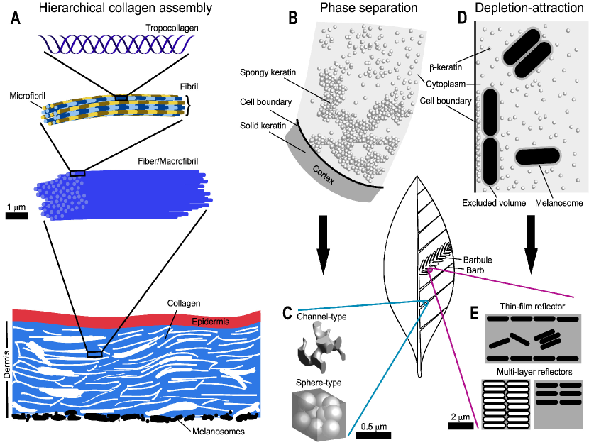
Non-iridescent structural colors prominently occur in bare skin (especially around the eye), bill (ramphotheca) and feet (podotheca) and has convergently evolved in over 50 bird families, likely driven by sexual selection Prum2003 ; 11** (Figs. 1A-B). They are produced by constructive light interference from 2D quasi-periodic arrays of parallel collagen (Refractive Index 1.42) fibrils in the mucopolysaccharide matrix (RI 1.35) of the dermis and underlain by a layer of melanin granules called melanosomes Prum2003 ; 11** (Fig. 2A). The collagen fibrils are in turn bundled into larger macrofibrils (fibers), tens of microns in diameter and several hundred microns in length that are apparently produced by a single collagenocyte in such a way that their longitudinal axis is aligned nearly parallel to the skin surface Prum2003 . However, the Velvet Asity (Philepitta castanea) uniquely among birds has evolved a 2D photonic crystal (PC) PCbook analog with ordered hexagonal arrangement of collagen fibrils that is derived from ancestral quasi-periodic state found in the sister sunbird asities (Neodrepanis spp.) Prum2003 ; Prum1999 . This transition is apparently driven by female preference for highly saturated hues in this lek polygynous species rather than for directional optical properties, as the overall papillose geometry of the facial caruncle and the polycrystalline nature of macrofibrils attenuate iridescence Prum2003 ; Prum1999 .
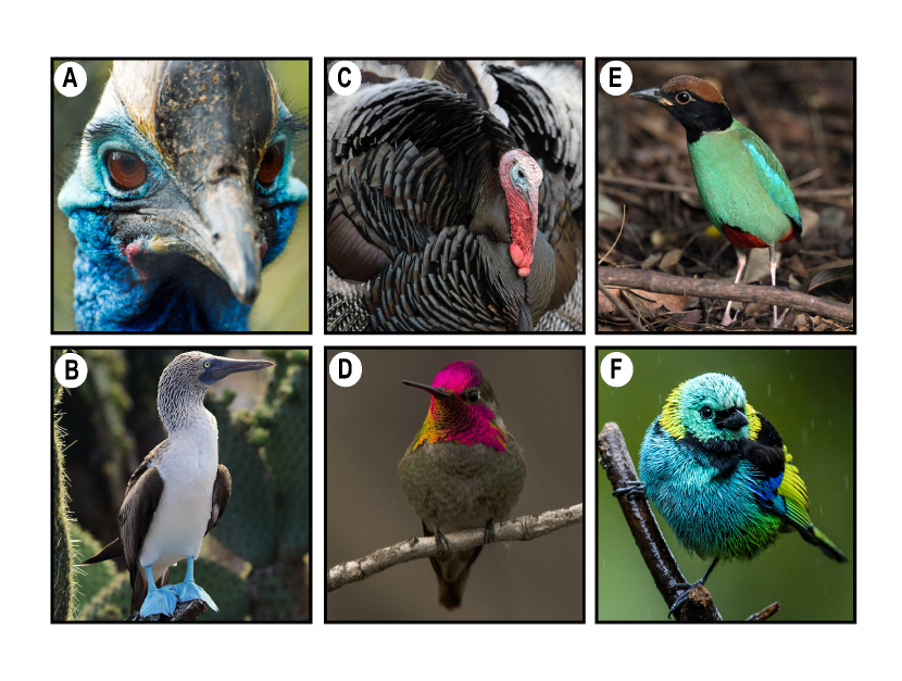
Photo Credits: A - Lucy Bridges, B - Rene Leubert, D - Daniel Arndt, F - Nick Athanas, C and E - Vinodkumar Saranathan (All images are cropped and reproduced under CC-BY-NC-SA 2.0)
The genetic and developmental basis of structural color producing dermal collagen arrays remains unstudied. Nevertheless, Prum and coworkers Prum2003 have suggested some prerequisites for the evolution of photonic dermal collagen arrays. In a plausible sequence, these include – loss of feathers exposing bare skin (apteria), thickening of dermis with a concomitant proliferation of collagen arrays to increase scattering efficiency given their low refractive index contrast, dermal melanization to absorb any unscattered light that would otherwise wash out structural hues, and near-uniform specification of larger than usual fibrils ( 100 nm) to make avian visible hues. It is conceivable that evolution of bare skin in birds is accompanied by dermal thickening for mechanical reasons and melanization as protection against UV damage.
Here, we focus on the molecular basis of collagen fibrillogenesis, which ultimately determines the photonic aspect of dermal collagen arrays (Fig. 2A). The hierarchical assembly of triple-helical collagen proteins that occurs in the dermal extracellular matrix (ECM) into collagen fibres is intrinsic and integral to the structure and function of vertebrate connective tissues, including dermis 13* . Collagens, specified by the diverse COL gene family, are ubiquitous and largely conserved across vertebrates, although birds have a slightly reduced diversity Haq2019 . The fact that collagen synthesis and fibrillogenesis in vertebrates are tightly regulated 13* , with large irregularly sized fibrils seen pathologically Liu1997 , suggests that photonic organization of fibrillar bundles likely stems from molecular regulation of fibril diameter, inter-fibril spacing and arrangement. One promising regulator is Tenascin-X (TNX; Tenascin-Y in birds Hagios1996 ) with epidermal growth factor (EGF) and fibronectin (FN) domains, which is implicated in determining inter-fibril spacing as well as accelerating rate of fibril formation by interacting with ECM proteoglycans such as decorin 13* . Interestingly, loss of either decorin Zhang2009 or TNX Minamitani2004 results in irregularly arranged, large-diameter fibrils. The ETS family transcription factor FLI1 (with known avian homolog) is also of interest as it represses fibrillar collagen genes, while upregulating the production of small leucine-rich proteoglycan (including decorin), during fibrillogenesis Asano2009 . However, spatio-temporal changes in fibrillar and fibril-associated collagen expression could result in similar changes in phenotype Liu1997 , suggesting that family-specific differences in collagen fibril composition could also be responsible for the repeated evolution of this trait in birds Haq2019 .
Structural coloration in avian plumages
Iridescent Feather Barbule Coloration
Structural coloration in feather barbules is generally iridescent (Figs. 1C-D) and produced by interference from biological analogs of 1DPCs PCbook – thin-film or multi-layer (spaced or close-packed) arrays of melanosomes (RI 2.0) embedded in a -keratin medium (RI 1.58) 11** (Fig. 2E). However, considerable systematic variation exists in the morphology and arrangement of melanosomes within iridescent barbules (See Fig. 3 of Durrer1986, )Gruson2019 ; Maia2013 . The melanosomes can be spherical, lozenge to rod-shaped about 1-2 m in length with various aspect ratios, or pancake-shaped. The melanosomes involved in barbule iridescence are usually comprised of eumelanin, although some pigeons (Columba trocaz) utilize phaeomelanins 11** . Some taxa (starlings, hummingbirds and quetzals) have evolved arrays of novel hollow/air-filled (RI 1.0) rod- and pancake-shaped melanosomes from ancestral solid types, leading to an increased refractive index contrast and thereby, extravagant interference colors Durrer1986 . Some have evolved close-packed 2DPC-like PCbook square (peafowl) or hexagonal (ducks and trogons) arrangement of melanosomes 11** Durrer1986 , although optically they seem to function as multilayers Stavenga2017 ; Freyer2019 . These diverse melanosomal arrays have convergently evolved numerous times across birds and show complex evolutionary history 11** , with both solid and hollow melanosomes in different plumage patches in some species Gruson2019 .
Published more than 50 years ago, Durrer and Villiger’s 25** description of hollow melanosome assembly in barbules of a starling (Lamprotornis) still remains an authoritative source on the ontogeny of iridescent barbule coloration. During feather development, melanosomes produced within specialized melanocytes are dendritically transferred to the barbule plate keratinocytes via endocytosis-like process Durrer1986 ; 25** . Melanosomes in developing non-iridescent barbules have an exclusion zone around them and seem randomly embedded in a matrix of rapidly polymerizing keratin that physically prevents the migration of melanosomes to the cell boundary 25** . Whereas, in iridescent barbules, -keratin is proliferating and polymerizing into small fibrils that do not fuse and remain confined to the center of the cell, with the cytoplasmic melanosomes free to diffuse to the cell membrane. The melanosomes are eventually mechanically confined by the keratin mass as it grows to fill the cell volume, and ordered into a marginal monolayer as the cell flattens upon death and dehydration 25** . A relatively recent developmental study in Blue-black Grassquit (Volatinia jacarina) observed a greater density of larger, more uniformly-sized melanosomes in barbules of iridescent males relative to non-iridescent females Maia2012 . Based on their observations that the organization of the melanosomes into a flat monolayer occurs late in barbule development, when the cell is dying, they proposed that depletion-attraction forces re-organize melanosomes into a monolayer, as opposed to cellular or molecular mechanisms Maia2012 ; Ghosh2015 . When melanosomes aggregate, the volume that keratins are normally excluded from occupying is reduced, increasing entropy and lowering the free-energy of the system (Fig. 2D).
In Lamprotornis sp. 25** , hollow melanosomes are already formed within melanocytes before being transferred to keratinocytes. Premelanosomes, large vesicles (m diameter) of Golgi origin filled with fine granular tyrosinase are rapidly generated within melanocytes, and later incorporate many small vesicles from the cytoplasm. A zigzag 5-6 layer lamellae forms centrally around which the smaller vesicles are organized like beads on a string. As the premelanosomes flatten, melanin is rapidly synthesized around the foam-like central matrix, which eventually becomes the air-filled internal structure of hollow melanosomes. By contrast, in regular solid melanosomes, melanin synthesis occurs at the zigzag lamellae and completely fills the premelanosome Birbeck1962 . Another recent study on the ontogeny of hollow melanosomes in iridescent barbules of Wild Turkey (Meleagris gallopavo) indicated that melanosomes are mostly solid and randomly oriented during transport Shawkey2015 . Once inside the barbule keratinocyte, however, they are mostly oriented in the same direction, and electron-dense material from the core is lost, prior to being close-packed into a hexagonal lattice. Shawkey et al.Shawkey2015 suggest that in turkeys, melanosomes could be a composite with a phaeomelanin core and eumelanin mantle, and that the chemically unstable phaeomelanin core can degrade upon changes in local environment (e.g., pH). That the mechanisms for hollow melanosome ontogeny are convergent is not surprising given iridescent barbule coloration using hollow melanosomes has evolved numerous independent times across birds 11** Gruson2019 ; Maia2013 .
Several outstanding questions remain. Although it is becoming clear that melanosomes late in development assemble via a ” crowding mechanism” Ghosh2015 , this needs to be reconciled with spatiotemporal changes in keratin synthesis and polymerization in barbule keratinocytes 25** , which can affect the position of the melanosomes relative to the cell boundary. Furthermore, depletion-attraction alone cannot explain how spaced multi-layers form (Fig. 2E), let alone more complex arrays with double-layered melanosomes in Birds of Paradise 11** Durrer1986 . Some authors have argued that the high aspect ratios of melanosomes in iridescent barbules relative to non-iridescent ones is enough for the self-emergence of layering Shawkey2015 ; Norden2019 . However, the observed melanosome aspect ratios seem far from the optimum under granular packing considerations of spherocylinders Baule2013 . Moreover, melanosome morphology can vary in a single species more than previously appreciated 11** Durrer1986 ; Gruson2019 , further confounding these analyses.
Given the key role of melanosomes in iridescent color generation, it is conceivable that convergent regulatory changes in melanin synthesis pathway have led to the repeated evolution of barbule iridescence in birds. The genetics of melanin-based coloration is well-studied in animals 32** , and recent progress in butterfly coloration suggest changes to single master regulatory genes can pleiotropically induce structural coloration 7** , while loss of melanin pathway genes can alter the gross morphology of scales themselves Matsuoka2018 . We wonder if similar changes could pleiotropically affect keratin expression and feather morphology in birds. The role of Melanocortin-1 Receptor (MC1R) in determining melanin patterning is inconsistent, but non-coding and coding differences in its repressor, agouti signaling protein (ASIP), is functionally significant across organisms 32** Funk2019 . A recent comparative genomic study on birds of paradise with extravagant iridescent barbule coloration Prost2019 suggests other promising candidates under putative positive selection – ADAMTS20, implicated in melanocyte development through KIT ligand functioning, and ATP7B, involved in copper transport, an element essential for melanogenesis. A similar study in galliforms Gao2018 recovered functional changes in four melanogenesis genes, two of which are KIT and ASIP, but these were not specific to iridescence. More interestingly, they found difference in -keratin gene expression between white and iridescent green feathers. Recently, melanocytes themselves have been found to autonomously determine color patterning and ASIP expression in adjacent dermal tissue, and this could be investigated via melanocyte transplantation from iridescent to non-iridescent barbule plates Funk2019 .
Non-iridescent Feather Barb Coloration
By contrast to iridescent barbule colors (Figs. 1C-D), structural colors in feather barbs are usually non-iridescent (Figs. 1E-F) and have evolved in over 45 families across 12 bird orders 11** ; 37* . Two main classes of 3D glassy or quasi-periodic photonic nanostructures are recognized in spongy barb medullary cells (Fig. 2C) – interconnected networks with anastomosing air channels and -keratin rods (channel-type), and random close-packed arrays of spherical air voids in a -keratin matrix (sphere-type). Some species with slate to blue-gray plumages have evolved rudimentary barb structural coloration produced by highly disordered versions of channel- and sphere-type nanostructures 37* . However, in Little Penguin (Eudyptula minor), non-iridescent blue barb colors are uniquely produced by 2D quasi-periodic bundles of parallel -keratin fibres in air Dalba2011 . Recently, Saranathan et al.Saranathan2020 discovered at least 3 parallel transitions in barb nanostructures of leafbirds, tanagers, and manakins from ancestral 3D photonic glasses to derived 3DPCs, apparently driven by female preference for highly saturated colors Prum1999 , rather than directional signaling, as the random Pointillist presentation of the crystal domains reduces iridescence.
The channel- and sphere-type barb nanostructures (Fig. 2C) are remarkably similar to highly stereotypical morphologies seen during phase separation of binary mixtures via spinodal decomposition and nucleation-and-growth, respectively 37* Dufresne2009 . Beyond structural analogies, there is growing evidence that barb photonic nanostructures are self-assembled within spongy medullary cells by visco-elastic phase separation of -keratin from cytoplasm (Fig. 2B), followed by a dynamic self-arrest as a result of the competition between coarsening and cross-polymerization of neighboring keratin fibrils 37* Saranathan2020 ; Dufresne2009 . The lone ground-breaking study on the development of spongy barb nanostructures along maturation gradients in a growing feather germ of Blue-and-Yellow Macaw (Ara Ararauna) is consistent with the phase separation hypothesis 41** . Around mid-development, medullary barb cells develop small electron-lucent, fluid-filled droplets in the center that coalesce and grow in a manner reminiscent of capillary transport seen in drying coffee-ring stains 41** . At this stage, electron-dense granules (likely keratohyalin) are seen near the edges of barb keratinocytes 41** Matulionis1970 . Once the droplets grow to fill most of the cell volume pushing all electron-dense materials (e.g., nucleus) to the periphery, -keratin synthesis and polymerization commences from cell edges. The first -keratin structures to form is a reticulate matrix of solid keratin fibres with a distinct ” crown-of-thorns” appearance, very similar to those seen at the periphery of hollow medullary cells in white feathers, but they are too large to make a visible interference color. Interestingly, the development of non-iridescent feather barbs seems identical up until this stage, when cells normally die leaving behind the characteristic foam-like, pneumatic medulla Matulionis1970 . In photonic barbs, however, development proceeds further and in just over a few hours, the characteristic channel-type network spontaneously appears at the cell boundaries from a cytoplasmic background filled with granular material whose sizes corresponds well with RNPs Matulionis1970 ; 43** . As this polymerizing spongy network grows, the volume occupied by the electron-lucent droplet shrinks. During this process, the barb cells have not yet apoptosed since nuceli remain visible 41** . When the cells finally die, the cytoplasm, nuclei and other cellular machinery are replaced by air. During this entire process, neither membrane nor cytoskeletal templates or prepatterns were observed directing the assembly of -keratin into spongy networks, consistent with a phase separation process 41** . Turing-type patterning, which produces (quasi-)periodic stripes and spots, is another unlikely alternative, as this process usually occurs in 2D (not 3D), and often breaks down with growth over time Saranathan2020 ; Maini2012 . Whereas, liquid-liquid phase separation within cells is a growing paradigm to explain the fundamental organization and functioning of cells, including how RNPs can lead to the development of fibrous, self-organized pathologies 43** . It is plausible that birds have co-opted such innate cellular processes for photonic self-assembly, and future studies will have to investigate the identity and function of RNPs during barb development 41** Matulionis1970 .
Feather development and genetic basis of keratin expression are typically studied in Chicken (Gallus gallus), Japanese Quail (Coturnix japonica) and Zebra Finch (Taenopygia guttata) none of which have barb structural coloration, while homology relationships between and among - and -keratins are only just being uncovered Greenwold2014 ; 46* . Across birds, there is extreme variation in copy numbers of -keratin genes (6 in owls - 149 in Zebra Finch; average 34). A complex pattern of differential expression of different types of keratin genes (scale and claw, feather, keratinocyte) from multiple chromosomal loci in different feather tissues has been documented Greenwold2014 ; 46* , with many of the feather -keratin genes evolving their own chromosome-wise transcription factors Bhattacharjee2016 . Nevertheless, barb ridge specific (e.g., barb vs. barbule) expression profiles are unavailable 46* . We believe the key to unlocking the molecular basis for -keratin self-assembly lies in generating tissue-specific, time-resolved expression of feather and feather-associated keratins, and identifying copy number variation unique to each family/genus Gao2018 46* Bruders2020 . Different family-specific combinations of -keratins might confer different macromolecular properties (e.g., hydropathy, charge) that can aid or hinder self-assembly, while tuning the stoichiometry of expressed keratins may predictably determine the length scale at which the phase separation arrests, which is crucial for color production. Comparatively studying the tissue-specific molecular structure of feather keratins or looking for differences in the macro-molecular packing of -keratin filaments, for instance, in spongy photonic barbs vs. hollow white barbs or barbules could also help illuminate the structure-function relationships underpinning keratin self-assembly.
Conclusions and Future Directions
Major aspects of organismal structural color pathways remain currently opaque. Nevertheless, we have highlighted how birds appear to have co-opted developmental programs behind collagen fibrillogenesis in dermis, melanosome synthesis and inclusion in feather barbules, and keratin polymerization in feathers barbs, to produce structural coloration. The redundant regulatory control of fibrillogenesis, melanosynthesis and cornification provides alternative pathways that can be modified by selection. This could explain the repeated convergence of avian structural coloration. We have also discussed the important role played by short-ranged attractive and long-ranged repulsive forces typically seen in soft colloidal systems Morse2020 Ghosh2015 43** Ji2015 , in feather nanostructure development.
Interrogating molecular regulation of collagen fibril spacing by manipulating collagen fibrillogenesis 13* is one future challenge that is tractable in Silky Chicken (artificially-selected variant of model Gallus gallus), with its hypermelanized dermis and unique blue earlobes. Another direction is to determine the exact role played by high melanosome densities in self-assembly during iridescent barbule development Maia2012 . The putative function(s) of cytoplasmic RNPs in flocculation and/or self-arrest also needs to be investigated 43** Ji2015 Comparative transcriptomics and genome-wide association studies might represent promising complementary approaches to cell and developmental biology. Two of 50 bird species with published complete genomes have barb structural coloration, while at least a quarter have barbule iridescence Zhang2014 . As successfully demonstrated for pigmentary coloration Gao2018 , these methods could help identify candidate genes, whose functions can be tested using latest genome-editing technologies (e.g., CRISPR-Cas9) in existing model species with iridescence – Silky, Domestic Chicken, and Turkey.
A burgeoning number of studies in physics and engineering are looking to organismal structural coloration, which have been evolutionarily optimized over millions of years of selection, as a rich reservoir for the bioinspired design and synthesis of functional materials, given current challenges in sustainable manufacture and synthetic self-assembly at visible optical lengthscales Morse2020 McDougal2019 . Genetic and developmental knowledge of biophotonic nanostructures may lead to next-generation technologies that directly biomimic in vivo self-assembly using bio-similar and biodegradable materials in vitro.
Acknowledgements.
VS acknowledges support from Yale-NUS start-up funds (R-607-261-182-121) and a NRF CRP Award (CRP20-2017-0004), and is grateful to Eric Dufresne, Antónia Monteiro, Dan Morse, and Rick Prum for stimulating discussions over the years.Declaration of Interests: The authors declare no conflict of interests.
References
- (1) Vukusic P, Sambles JR: Photonic structures in biology, Nature 2003, 424:852–855. doi: 10.1038/nature01941
- (2) Cuthill IC, Allen WL, Arbuckle K, Caspers B, Chaplin G, Hauber ME, Hill GE, Jablonski NG, Jiggins CD, Kelber A, et al.: The biology of color. Science 2017, 357:eaan0221. doi: 10.1126/science.aan0221 This review provides a nice overview of the biological aspects of animal coloration, ranging from their origin and evolution, diversity of visual reception and cognition, to its multiple functions, including aposematism, crypsis and inter- and intra-sexual signalling.
- (3) Joannopoulos JD, Johnson SG, Winn JN, Meade RD: Photonic crystals: Molding the flow of light edn 2nd. New Jersey: Princeton University Press; 2008.
- (4) Hill GE, McGraw KJ (Ed): Bird Coloration, Volume 1, Mechanisms and measurements Cambridge, MA: Harvard University Press; 2006.
- (5) Saranathan V, Seago AE, Narayanan S, Sandy A, Mochrie SGJ, Dufresne ER, Cao H, Osuji CO, Prum RO: Structural Diversity of Arthropod Biophotonic Nanostructures Spans Amphiphilic Phase-Space. Nano Letters 2015, 15:3735–3742. doi: 10.1021/acs.nanolett.5b00201
- (6) Hill GE, McGraw KJ (Ed): Bird Coloration, Volume 2, Function and Evolution Cambridge, MA: Harvard University Press; 2006.
- (7) Zhang L, Mazo-Vargas A, Reed RD: Single master regulatory gene coordinates the evolution and development of butterfly color and iridescence. Proceedings of the National Academy of Sciences 2017, 114:10707–10712. doi: 10.1073/pnas.1709058114 This path-breaking study uses genome-editing tools to uncover the deeply conserved dual role of the optix gene in regulating pigmentary but also structural coloration in nymphalid butterflies. This important work suggests that the genetic link between structural and pigmentary colors is closer than previously appreciated.
- (8) Airoldi CA, Ferria J, Glover BJ: The cellular and genetic basis of structural colour in plants. Current Opinion in Plant Biology 2019, 47:81–87. doi: 10.1016/j.pbi.2018.10.002 Analogous to the present study, this review summarizes the current knowledge about the production of structural coloration in plants, a very rare trait. An emerging idea is the co-option of core developmental pathways to produce derived structural colors.
- (9) Morse DE, Taxon E: Reflectin needs its intensity amplifier: Realizing the potential of tunable structural biophotonics. Applied Physics Letters 2020, 117:220501. doi: 10.1063/5.0026546
- (10) Prum RO, Torres RH: Structural colouration of avian skin: convergent evolution of coherently scattering dermal collagen arrays. Journal of Experimental Biology 2003, 206:2409–2429.
- (11) Prum RO: Anatomy, physics, and evolution of avian structural colors. In Bird Coloration, Volume 1 Mechanisms and Measurements. Edited by Hill GE, McGraw KJ: Harvard University Press; 2006:295–353. vol 1. This wide-ranging book chapter comprehensively surveys the evolution, morphological diversity and optics of avian structural coloration, with a level of detail outside the scope of this review.
- (12) Prum RO, Torres R, Kovach C, Williamson S, Goodman SM: Coherent light scattering by nanostructured collagen arrays in the caruncles of the Malagasy Asities (Eurylaimidae: Aves). The Journal of Experimental Biology 1999, 202:3507–3522.
- (13) Kadler KE, Hill A, Canty-Laird EG: Collagen fibrillogenesis: fibronectin, integrins, and minor collagens as organizers and nucleators. Current Opinion in Cell Biology 2008, 20:495–501. doi: 10.1016/j.ceb.2008.06.008 This comprehensive review provides a modern perspective on the hierarchical assembly of the ubiqituous, triply-helical collagen proteins into increasingly complex fibrillar morphologies. Interestingly, they point out that while collagen can self-assemble into fibrils in vitro, their biological assembly is under tight molecular control, the disruption of which leads to various pathophysiologies. Another important perspective is that collagen microfibrils are a biomaterial composite made up of different types of collagens that can tune their assembly and mechanical properties.
- (14) Haq F, Ahmed N, Qasim M: Comparative genomic analysis of collagen gene diversity. 3 Biotech 2019, 9:83–83. doi: 10.1007/s13205-019-1616-9
- (15) Liu X, Wu H, Byrne M, Krane S, Jaenisch R: Type III collagen is crucial for collagen I fibrillogenesis and for normal cardiovascular development. Proceedings of the National Academy of Sciences 1997, 94:1852–1856. doi: 10.1073/pnas.94.5.1852
- (16) Hagios C, Koch M, Spring J, Chiquet M, Chiquet-Ehrismann R: Tenascin-Y: a protein of novel domain structure is secreted by differentiated fibroblasts of muscle connective tissue. Journal of Cell Biology 1996, 134:1499–1512. doi: 10.1083/jcb.134.6.1499
- (17) Zhang G, Chen S, Goldoni S, Calder BW, Simpson HC, Owens RT, McQuillan DJ, Young MF, Iozzo RV, Birk DE: Genetic Evidence for the Coordinated Regulation of Collagen Fibrillogenesis in the Cornea by Decorin and Biglycan. Journal of Biological Chemistry 2009, 284:8888–8897. doi: 10.1074/jbc.M806590200
- (18) Minamitani T, Ikuta T, Saito Y, Takebe G, Sato M, Sawa H, Nishimura T, Nakamura F, Takahashi K, Ariga H, et al.: Modulation of collagen fibrillogenesis by tenascin-X and type VI collagen. Experimental Cell Research 2004, 298:305–315. doi: 10.1016/j.yexcr.2004.04.030
- (19) Asano Y, Markiewicz M, Kubo M, Szalai G, Watson DK, Trojanowska M: Transcription Factor Fli1 Regulates Collagen Fibrillogenesis in Mouse Skin. Molecular and Cellular Biology 2009, 29:425–434. doi: 10.1128/MCB.01278-08
- (20) Durrer H: The Skin of Birds: Colouration. In Biology of the Integument 2: Vertebrates. Edited by: Springer-Verlag; 1986:239–247. vol 2.
- (21) Gruson H, Elias M, Andraud C, Djediat C, Berthier S, Doutrelant C, Gomez D: Hummingbird iridescence: an unsuspected structural diversity influences colouration at multiple scales. bioRxiv 2019:699744. doi: 10.1101/699744
- (22) Maia R, Rubenstein DR, Shawkey MD: Key ornamental innovations facilitate diversification in an avian radiation. Proceedings of the National Academy of Sciences 2013, 110:10687–10692. doi: 10.1073/pnas.1220784110
- (23) Stavenga DG, Kooi CJvd, Wilts BD: Structural coloured feathers of mallards act by simple multilayer photonics. Journal of The Royal Society Interface 2017, 14:20170407. doi: 10.1098/rsif.2017.0407
- (24) Freyer P, Wilts BD, Stavenga DG: Reflections on iridescent neck and breast feathers of the peacock, Pavo cristatus. Interface Focus 2019, 9:20180043. doi: 10.1098/rsfs.2018.0043
- (25) Durrer H, Villiger W: Bildung der schillerstruktur beim glanzstar. Zeitschift fur Zellforschungen 1967, 81:445–456. doi: 10.1007/BF00342767 Although written in German more than a half-century ago, this pioneering study on the development of hollow air-filled melanosomes and their organization leading to iridescence in feather barbules remains authoritative. The beautiful sequence of TEM images are easy to follow and are indeed more eloquent than our descriptions can do justice.
- (26) Maia R, Macedo RH, Shawkey MD: Nanostructural self-assembly of iridescent feather barbules through depletion attraction of melanosomes during keratinization. Journal of The Royal Society Interface 2012, 9:734–743. doi: 10.1098/rsif.2011.0456
- (27) Ghosh P, Mondal J, Ben-Jacob E, Levine H: Mechanically-driven phase separation in a growing bacterial colony. Proceedings of the National Academy of Sciences 2015, 112:E2166–E2173. doi: 10.1073/pnas.1504948112
- (28) Birbeck MSC: Electron Micsroscopy of Melanocytes. British Medical Bulletin 1962, 18:220–222. doi: 10.1093/oxfordjournals.bmb.a069982
- (29) Shawkey MD, D’Alba L, Xiao M, Schutte M, Buchholz R: Ontogeny of an iridescent nanostructure composed of hollow melanosomes. Journal of Morphology 2015, 276:378–384. doi: 10.1002/jmor.20347
- (30) Nordén KK, Faber JW, Babarović F, Stubbs TL, Selly T, Schiffbauer JD, Peharec Štefanić P, Mayr G, Smithwick FM, Vinther J: Melanosome diversity and convergence in the evolution of iridescent avian feathers-Implications for paleocolor reconstruction. Evolution 2019, 73:15–27. doi: 10.1111/evo.13641
- (31) Baule A, Mari R, Bo L, Portal L, Makse HA: Mean-field theory of random close packings of axisymmetric particles. Nature Communications 2013, 4:2194. doi: 10.1038/ncomms3194}
- (32) . Orteu A, Jiggins CD: The genomics of coloration provides insights into adaptive evolution. Nature Reviews Genetics 2020, 21:461–475. doi: 10.1038/s41576-020-0234-z This thorough recent review discusses the successful use of cutting-edge genomic approaches to study the genetic basis of pigmentary coloration and its evolution in animals, that we advocate for the study of animal structural coloration.
- (33) Matsuoka Y, Monteiro A: Melanin Pathway Genes Regulate Color and Morphology of Butterfly Wing Scales. Cell Rep 2018, 24:56–65. doi: 10.1016/j.celrep.2018.05.092
- (34) Funk ER, Taylor SA: High-throughput sequencing is revealing genetic associations with avian plumage color. The Auk 2019, 136:ukz048. doi: 10.1093/auk/ukz048
- (35) Prost S, Armstrong EE, Nylander J, Thomas GWC, Suh A, Petersen B, Dalen L, Benz BW, Blom MPK, Palkopoulou E, et al.: Comparative analyses identify genomic features potentially involved in the evolution of birds-of-paradise. GigaScience 2019, 8:giz003. doi: 10.1093/gigascience/giz003
- (36) Gao G, Xu M, Bai C, Yang Y, Li G, Xu J, Wei Z, Min J, Su G, Zhou X, et al.: Comparative genomics and transcriptomics of Chrysolophus provide insights into the evolution of complex plumage coloration. GigaScience 2018, 7:giy113. doi: 10.1093/gigascience/giy113
- (37) Saranathan V, Forster JD, Noh H, Liew SF, Mochrie SGJ, Cao H, Dufresne ER, Prum RO: Structure and Optical Function of Amorphous Photonic Nanostructures from Avian Feather Barbs: A Comparative Small Angle X-ray Scattering (SAXS) Analysis of 230 Bird Species. Journal of The Royal Society Interface 2012, 9:2563–2580. doi: 10.1098/rsif.2012.0191 This study uses synchrotron Small Angle X-ray Scattering to comparatively characterize the nanostructure and optical function of non-iridescent feather barb nanostructures from an unprecedented 230 species of birds, covering nearly every genus with such colors. The authors demonstrate barb morphologies across birds are highly stereotyped (channel- or sphere-type) just like under binary phase separation. They further provide the first comparisons of experimental structure factors from barb photonic glasses with those from polymer blends undergoing spinodal decomposition or colloidally self-assembled quasi-periodic arrays of dielectric spheres as a test of the phase separation hypothesis.
- (38) D’Alba L, Saranathan V, Clarke JA, Vinther JA, Prum RO, Shawkey MD: Colour-producing -keratin nanofibres in blue penguin (Eudyptula minor) feathers. Biology Letters 2011, 7:543–546. doi: 10.1098/rsbl.2010.1163
- (39) Saranathan V, Narayanan S, Sandy AR, Dufresne ER, Prum RO: Single Gyroid and Inverse b.c.c. Photonic Crystals in Bird Feathers. bioRxiv 2020.08.27.271213. doi: 10.1101/2020.08.27.271213
- (40) Dufresne ER, Noh H, Saranathan V, Mochrie SGJ, Cao H, Prum RO: Self-assembly of amorphous biophotonic nanostructures by phase separation. Soft Matter 2009, 5:1792–1795. doi: 10.1039/B902775K
- (41) Prum RO, Dufresne ER, Quinn T, Waters K: Development of colour-producing -keratin nanostructures in avian feather barbs. Journal of The Royal Society Interface 2009, 6:S253–S265. doi: 10.1098/rsif.2008.0466.focus This seminal study describes the intracellular development of channel-type photonic nanostructures in medullary barb cells of the Blue-and-Yellow Macaw. Their results unambiguously show the absence of any cytoskeletal or membrane patterning in guiding the characteristic dendritic pattern formation in the late stages of barb cell cornification and provide in our opinion, incontrovertible proof for the self-assembly of biophotonic nanostructures, likely via phase separation.
- (42) Matulionis DH: Morphology of the developing down feathers of chick embryos. Zeitschrift für Anatomie und Entwicklungsgeschichte 1970, 132:107–157. doi: 10.1007/BF00523275
- (43) Shin Y, Brangwynne CP: Liquid phase condensation in cell physiology and disease. Science 2017, 357:eaaf4382. doi: 10.1126/science.aaf4382 This fairly recent review compiles examples of phase transitions that are integral to the structure and function of cells, the impeding of which leads to pathological aggregation. The authors especially develop the paradigm of intracellular liquid-liquid phase separation to explain the formation and assembly of membrane-less intracellular condensates (biomolecular droplet phases).
- (44) Maini PK, Woolley TE, Baker RE, Gaffney EA, Lee SS: Turing’s model for biological pattern formation and the robustness problem. Interface Focus 2012, 2:487–496. doi: 10.1098/rsfs.2011.0113
- (45) Greenwold MJ, Bao W, Jarvis ED, Hu H, Li C, Gilbert MTP, Zhang G, Sawyer RH: Dynamic evolution of the alpha () and beta () keratins has accompanied integument diversification and the adaptation of birds into novel lifestyles. BMC Evolutionary Biology 2014, 14:249. doi: 10.1186/s12862-014-0249-1
- (46) Ng CS, Li W-H: Genetic and Molecular Basis of Feather Diversity in Birds. Genome Biology and Evolution 2018, 10:2572–2586. doi: 10.1093/gbe/evy180 This important review addresses available molecular genomic and developmental data that underlie the diversity of bird feathers, in term of morphology and pigmentation, both within individual in model species and among avian species.
- (47) Bhattacharjee MJ, Yu C-P, Lin J-J, Ng CS, Wang T-Y, Lin H-H, Li W-H: Regulatory Divergence among Beta-Keratin Genes during Bird Evolution. Molecular Biology and Evolution 2016, 33:2769–2780. doi: 10.1093/molbev/msw165
- (48) Bruders R, Van Hollebeke H, Osborne EJ, Kronenberg Z, Maclary E, Yandell M, Shapiro MD: A copy number variant is associated with a spectrum of pigmentation patterns in the rock pigeon (Columba livia). PLoS Genet 2020, 16:e1008274. doi: 10.1371/journal.pgen.1008274
- (49) Ji S, Walz JY: Depletion forces and flocculation with surfactants, polymers and particles - Synergistic effects. Current Opinion in Colloid & Interface Science 2015, 20:39-45. doi: 10.1016/j.cocis.2014.11.006
- (50) Zhang G, Jarvis ED, Gilbert MTP: A flock of genomes. Science 2014, 346:1308-1309. doi: 10.1126/science.346.6215.1308
- (51) McDougal A, Miller B, Singh M, Kolle M: Biological growth and synthetic fabrication of structurally colored materials. Journal of Optics 2019, 21:073001. doi: 10.1088/2040-8986/aaff39
Box 1: Glossary
Refractive Index – this dimensionless metric describes the
amount of retardation of light in a dense medium relative to vacuum. The
RI determines the extent to which light rays are bent (refraction) when
entering a dense material from air. See PCbook .
Biophotonic Nanostructure – a nanoscale feature in the animal
integument with random, quasi-periodic or periodic variation in material
composition that leads to a refractive index contrast (e.g., -keratin and air). See Vukusic2003 11** .
Photonic Crystal – A concept borrowed from solid-state
physics, a photonic crystal describes periodic variations in material
composition that interferes with the propagation of light through the
material. See Vukusic2003 ; PCbook .
1D, 2D, 3D (photonic materials) – The designation 1D, 2D or
3D describes whether the material varies in composition or refractive
index along one (e.g., a thin-film or multilayer lamellae),
two (e.g., square or honeycomb lattices of cylindrical
holes), or three (e.g., gem opals and cubic crystals)
principal orthogonal directions (e.g., Cartesian axes). For
instance, in a 2D columnar or fibrillar nanostructure, there is no
variation along column/fibril axis (say z-axis), but there
is in the cross-sectional plane (along x and y). See Vukusic2003 ; PCbook 11** .
Iridescence – a change in hue with angle of illumination or
angle of observation.
Self-assembly – a ubiquitous phenomenon that describes the
spontaneous or emergent organization of materials at macro-molecular
length scales, usually driven by thermal fluctuations and interactions
among molecules.
Phase separation – a process that describes the spontaneous
unmixing of immiscible binary or ternary mixtures, under unfavorable
conditions. See 43** .
Depletion-attraction – a process that describes the
attractive force that brings together larger colloidal particles that
reduces the excluded volume around the large particles that a competing
smaller solute (depletant) cannot normally occupy, thereby increasing
the positional entropy for the depletants and lowering the overall
free-energy of the system. Depletion-attraction can also lead to phase
separation. See Ji2015 .