Robustness Investigation on Deep Learning CT Reconstruction for Real-Time Dose Optimization
Abstract
In computed tomography (CT), automatic exposure control (AEC) is frequently used to reduce radiation dose exposure to patients. For organ-specific AEC, a preliminary CT reconstruction is necessary to estimate organ shapes for dose optimization, where only a few projections are allowed for real-time reconstruction. In this work, we investigate the performance of automated transform by manifold approximation (AUTOMAP) in such applications. For proof of concept, we investigate its performance on the MNIST dataset first, where the dataset containing all the 10 digits are randomly split into a training set and a test set. We train the AUTOMAP model for image reconstruction from 2 projections or 4 projections directly. The test results demonstrate that AUTOMAP is able to reconstruct most digits well with a false rate of 1.6% and 6.8% respectively. In our subsequent experiment, the MNIST dataset is split in a way that the training set contains 9 digits only while the test set contains the excluded digit only, for instance “2”. In the test results, the digit “2”s are falsely predicted as “3” or “5” when using 2 projections for reconstruction, reaching a false rate of 94.4%. For the application in medical images, AUTOMAP is also trained on patients’ CT images. The test images reach an average root-mean-square error of 290 HU. Although the coarse body outlines are well reconstructed, some organs are misshaped.
Index Terms:
Sparse-view reconstruction, deep learning, automatic exposure control, computed tomographyI Introduction
Computed tomography (CT) is widely used for disease diagnosis and interventions in modern medicine. To reduce health risks caused by X-rays, automatic exposure control (AEC) is frequently used in contemporary CT scanners to control the effective dose as low as reasonably achievable while keeping the image quality. For accurate organ-specific AEC, a preliminary reconstruction of patient organs is necessary for dose optimization, which needs to be fast enough for real-time AEC. Therefore, a reconstruction from extreme sparse-view projections, e.g. 4 projections or 2 projections, is preferred.
For image reconstruction from sparse-view data, conventional filtered back-projection (FBP) based reconstruction algorithms perform poorly. Even for compressed sensing technologies, no satisfactory images are reconstructed from such extreme sparse views. However, the emerging deep learning methods offer a possible solution. Some achievements have been reported to reconstruct images directly from projection data by deep learning [1, 2, 3, 4]. Automated transform by manifold approximation (AUTOMAP) [2] is a generic deep learning reconstruction framework for multiple imaging modalities including CT. It learns a joint manifold linking projections and reconstructed images in a supervised manner. As the robustness of deep learning is a concern for clinical applications [5], in this paper, we investigate the performance of AUTOMAP for such extreme sparse-view reconstruction.
II Methods
According to [2], the AUTOMAP neural network is implemented for parallel-beam CT reconstruction from 4 projections or 2 two projections. As a proof of concept, the performance of AUTOMAP is evaluated on the MNIST dataset first. Afterwards, its performance is evaluated on patients’ CT images.
II-A Neural network architecture
The AUTOMAP used in our experiments consists of 3 cascaded fully-connected layers, each with a tanh activation function, and 3 convolutional layers, each with a ReLu activation function. Batch normalization is included to smooth the training process.
II-B Training and test
For the experiments on the MNIST dataset, 48000 images from MNIST are used for training and test. In our first investigation, we randomly split the 48000 images containing all the 10 digits into 43000 images for training and 5000 for test. The images are first resized to and encoded using the parallel-beam Radon transform into sinograms with a detector size of 64. The detector pixel size is the same as the image pixel size, simply assuming 5 mm per pixel. The projections from , , and are used as the first sparse-view condition, and those from and are used as the second sparse-view condition. In the second investigation, the dataset is split based on the digits. We exclude the images of digit “2” from the dataset and train the model using remaining images using the same sparse-view conditions. The images of digit “2” are used for test afterwards.
To demonstrate the performance of AUTOMAP on medical images, we further train the model using 6900 sinograms from 12 patients’ CT volumes. Sinograms from 11 CT volumes are used for training, while those from the excluded one are used for test. The other experimental settings are the same as the first experiment.
For each training, 50 epochs are applied. The loss function used is mean square error (MSE) and the optimizer used is RMSProp with a learning rate of 0.00002.
II-C Evaluation
For the experiment on the MINIST images, the image quality of reconstructed images is evaluated by root-mean-square error (RMSE) and false digit rate. A false digit means that the reconstructed image is observed as a different digit rather than the digit “2”, or it cannot be recognized as a digit at all. For the experiment on CT images, RMSE in Hounsfield unit (HU) is calculated.
III Results And Discussion
|
|
|
|
|||||||||
|---|---|---|---|---|---|---|---|---|---|---|---|---|
|
0.123 | 0.169 | 1.6% | 6.8% | ||||||||
|
0.134 | 0.244 | 8.4% | 94.4% |
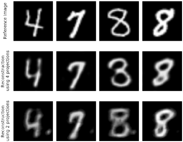
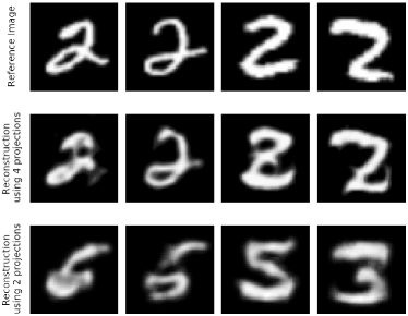
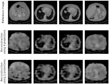
Fig. 1 exhibits that AUTOMAP is able to reconstruct most digits well when the training dataset contains all the 10 digits. In Fig. 2 where the digit “2” is excluded for training, most digits are recognized as “2” well for reconstructions from 4 projections, although one result appears like the letter “”. However, most reconstructions appear as false digits, e.g., “3”, “5” or “6”, when only 2 projections are used. As shown in TAB. I, the false digit rate is 94.4% when training on the MNIST dataset excluding “2” using 2 projections. The reconstructions on the CT images are displayed in Fig. 3. The coarse body outlines are reconstructed by AUTOMAP. However, some organs are misshaped. These reconstructions have an average RMSE value of 290 HU. Whether such images are sufficient for dose optimization in clinical applications needs further investigation.
IV Conclusion
As a proof of concept, AUTOMAP shows its robustness for sparse-view reconstruction when using 4 projections on the MINIST dataset. However, it tends to reconstruct false digits from 2 projections when one digit is excluded for training. The experiment on the patients’ CT images demonstrates that AUTOMAP is able to reconstruct the coarse body outlines well. However, some organs are misshaped.
References
- [1] T. Würfl, F. C. Ghesu, V. Christlein, and A. Maier, “Deep learning computed tomography,” in Proc. MICCAI, 2016, pp. 432–440.
- [2] B. Zhu, J. Z. Liu, S. F. Cauley, B. R. Rosen, and M. S. Rosen, “Image reconstruction by domain-transform manifold learning,” Nature, vol. 555, no. 7697, pp. 487–492, 2018.
- [3] Y. Li, K. Li, C. Zhang, J. Montoya, and G.-H. Chen, “Learning to reconstruct computed tomography images directly from sinogram data under a variety of data acquisition conditions,” IEEE Trans. Med. Imaging, vol. 38, no. 10, pp. 2469–2481, 2019.
- [4] X. Ying, H. Guo, K. Ma, J. Wu, Z. Weng, and Y. Zheng, “X2CT-GAN: reconstructing ct from biplanar x-rays with generative adversarial networks,” in Proc. IEEE CVPR, 2019, pp. 10 619–10 628.
- [5] Y. Huang, T. Würfl, K. Breininger, L. Liu, G. Lauritsch, and A. Maier, “Some investigations on robustness of deep learning in limited angle tomography,” in Proc. MICCAI, 2018, pp. 145–153.