:
\theoremsep
\jmlrvolumeML4H Extended Abstract Arxiv Index
\jmlryear2020
\jmlrsubmitted2020
\jmlrpublished
\jmlrworkshopMachine Learning for Health (ML4H) 2020
Pneumothorax and chest tube classification on chest x-rays for detection of missed pneumothorax
Abstract
Chest x-ray imaging is widely used for the diagnosis of pneumothorax and there has been significant interest in developing automated methods to assist in image interpretation. We present an image classification pipeline which detects pneumothorax as well as the various types of chest tubes that are commonly used to treat pneumothorax. Our multi-stage algorithm is based on lung segmentation followed by pneumothorax classification, including classification of patches that are most likely to contain pneumothorax. This algorithm achieves state of the art performance for pneumothorax classification on an open-source benchmark dataset. Unlike previous work, this algorithm shows comparable performance on data with and without chest tubes and thus has an improved clinical utility. To evaluate these algorithms in a realistic clinical scenario, we demonstrate the ability to identify real cases of missed pneumothorax in a large dataset of chest x-ray studies.
fig:Chest-tubes

fig:diagram

1 Introduction
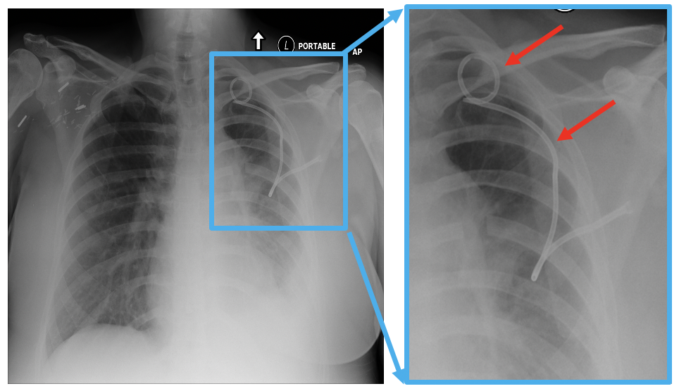
Pneumothorax is a potentially life-threatening condition where air collects in the pleural space between the lung and chest wall. There has been significant interest in developing automated methods for detecting pneumothorax on chest x-rays. One notable limitation of previous work is the lack of associated methods for chest tube recognition. Chest tubes are flexible plastic tubes that are inserted through the chest wall to drain air from the pleural space in order to treat pneumothorax. Significant proportions of pneumothorax cases in chest x-ray datasets contain chest tubes (70% in (Majkowska et al., 2020)). Since chest tubes may appear as prominent features on chest x ray images (see \figurereffig:Chest-tubes), there is a risk that deep learning models will detect the presence of chest tubes rather than the pneumothorax itself. Most prior work does not consider the effect of chest tubes (Irvin et al., 2019; Sze-To and Wang, 2019; Rajpurkar et al., 2017; Wang et al., 2017; Taylor et al., 2018) and the recent work from (Majkowska et al., 2020) demonstrates a 13.2% drop in sensitivity when evaluating data without chest tubes.
tab:results Method AUC (all data) AUC (no tubes) AUC (only tubes) AUC % change with no tubes Rajpurkar et al. (2017) 0.895 0.816 0.914 -8.8% Sze-To and Wang (2019) 0.844 0.787 0.858 -6.8% Majkowska et al. (2020) 0.940 0.890 - -5.3% A - DN full image 0.941 0.941 0.941 0.0% B - DN apical/basilar 0.932 0.878 0.946 -5.8% C - U-Net 0.921 0.927 0.919 0.7% Ensemble A + C 0.952 0.953 0.952 0.1% Ensemble A + B + C 0.958 0.948 0.960 -1.0%
2 Pneumothorax and chest tube classification pipeline
In this work we present an image classification pipeline that detects both pneumothorax and chest tubes. The pipeline is shown in the red box in \figurereffig:diagram. All images are first evaluated by an X-ray view classification deep learning model so that only frontal (AP or PA) images are processed by the rest of the pipeline. Pneumothorax classification consists of first performing lung segmentation with a U-Net (Ronneberger et al., 2015). This is followed by three separate approaches for pneumothorax classification which are ensembled. The first method crops the image to the lung region and then classifies the image with a DenseNet-121 model (Huang et al., 2016). The second method utilizes a U-Net to segment the pneumothorax region on the same cropped and resized image. The third method extracts four patches from the left/right, basilar/apical lung regions. These specific lung regions are chosen as they are the most likely to contain a small pneumothorax. The three different models are ensenbled to create the final output for pneumothorax classification. The chest tube classification algorithm is a DenseNet-121 model. The input to this model is the full chest X-ray image without any cropping.
All deep learning models were trained separately on both open-source data and a private multi-site data set with annotations done by radiologists. For pneumothorax segmentation, we used 8,222 images from the NIH ChestXray14 dataset (Wang et al., 2017) with pixel level annotations that were provided as part of the Kaggle Pneumothorax Challenge (SIIM-ACR, 2019). We also used an additional 6,011 images from the private dataset. The pneumothorax and chest tube classification algorithms were developed using 25,173 images from the private dataset. Training labels for pneumothorax and chest tubes were generated in a semi-automated way from the radiology reports. These sentences were manually categorized as positive or negative to generate verified image level labels. When available, the location of the pneumothorax (left/right and base/apex) was also captured from the report for training the apical/basilar classifier. Additional details about the training data can be found in \appendixrefapd:first.
fig:missed-finding

3 Missed pneumothorax detection
To evaluate these algorithms in a realistic clinical scenario, we combine the image algorithms with natural language processing (NLP) to identify studies with potential missed pneumothorax (pneumothorax detected on images but not mentioned in radiology report). Missed findings that are clearly noticeable on retrospect are a known problem in radiology that can result from a number of factors such as reader fatigue and distractions such as phone calls (van Ginneken et al., 2011; Brady, 2017). The idea of combining image classification algorithms with NLP to detect missed findings was recently demonstrated for intracranial hemorrhage on non-contrast CT scan (Rao et al., 2020).
The NLP (previously described in Guo et al. (2017)) analyzes radiology reports and classifies the report as positive if there is any positive mention of pneumothorax. Any study that has a negative output from the NLP, a negative output for chest tube image classification and a positive output for pneumothorax image classification are identified as potentially having a missed pneumothorax. Studies with a chest tube are not considered for potential missed findings since the presence of the chest tube indicates that there is already a clinical awareness of a pneumothorax or other pleural space abnormality.
4 Results
We evaluated pneumothorax classification performance using the radiologist adjudicated test set made available by Majkowska et al. (2020) (see \appendixrefapd:first). We also compare our results with two other models that have been open sourced (Sze-To and Wang, 2019; Rajpurkar et al., 2017). These results are shown in \tablereftab:results. In addition to having a higher overall AUC, our model maintains similar performance for samples that do not contain chest tubes. In comparison, the other models we evaluated had a significant drop in AUC (5.3%-8.8%).
We also evaluated the chest tube classification algorithm on a test set of 2,977 images with radiologist annotated ground truth. AUC was .977 for the classification of standard chest tubes and .987 for pigtail catheters.
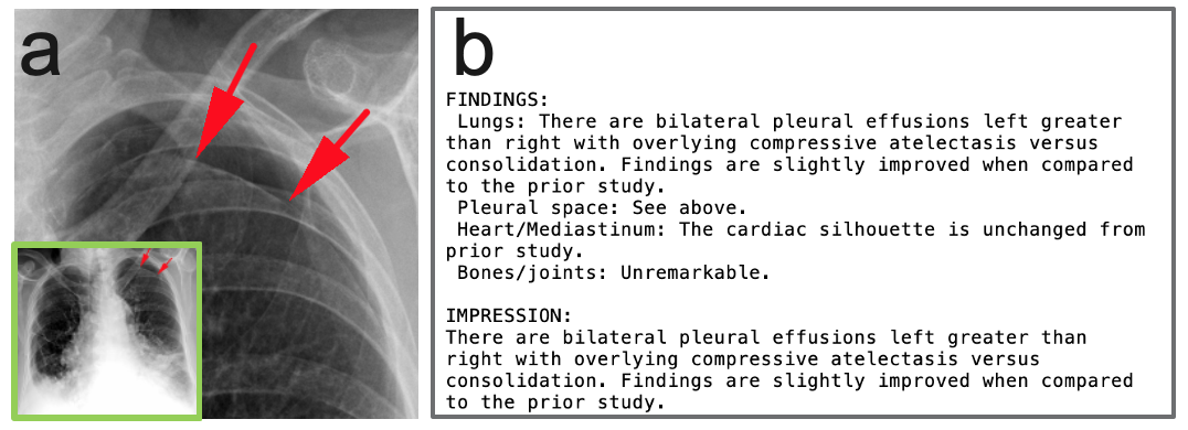
To demonstrate the ability to identify true missed findings in a realistic clinical setting, we analyzed a set of 20,000 chest x-ray studies and their associated radiology reports. Using the combined image and NLP algorithm pipeline, we identified 62 studies as having a potential missed pneumothorax. A board-certified radiologist reviewed both the radiology report text and images and identified nine cases of missed pneumothorax (see example in \figurereffig:missed-finding).
5 Summary of contributions
-
•
We developed an image classification pipeline that achieves state of the art performance for pneumothorax classification and also includes the ability to identify chest tubes.
-
•
We demonstrate that, unlike previous works, this pipeline achieves comparable performance on pneumothorax classification in data with and without the presence of chest tubes which significantly improves the clinical utility.
-
•
We utilize these image classification algorithms in combination with an NLP algorithm to demonstrate the ability to identify real cases of missed pneumothorax in a large and diverse dataset of chest X-ray studies
References
- Brady (2017) Adrian P. Brady. Error and discrepancy in radiology: inevitable or avoidable? Insights into Imaging, 8:171–182, 2 2017. ISSN 1869-4101. 10.1007/s13244-016-0534-1. URL http://link.springer.com/10.1007/s13244-016-0534-1.
- Guo et al. (2017) Yufan Guo, Deepika Kakrania, Tyler Baldwin, and Tanveer Syeda-Mahmood. Efficient clinical concept extraction in electronic medical records. AAAI Conference on Artificial Intelligence, 2017. URL https://aaai.org/ocs/index.php/AAAI/AAAI17/paper/view/14794.
- Huang et al. (2016) Gao Huang, Zhuang Liu, and Kilian Q Weinberger. Densely connected convolutional networks. CoRR, abs/1608.06993, 2016. URL http://arxiv.org/abs/1608.06993.
- Irvin et al. (2019) Jeremy Irvin, Pranav Rajpurkar, Michael Ko, Yifan Yu, Silviana Ciurea-Ilcus, Chris Chute, Henrik Marklund, Behzad Haghgoo, Robyn L Ball, Katie S Shpanskaya, Jayne Seekins, David A Mong, Safwan S Halabi, Jesse K Sandberg, Ricky Jones, David B Larson, Curtis P Langlotz, Bhavik N Patel, Matthew P Lungren, and Andrew Y Ng. Chexpert: A large chest radiograph dataset with uncertainty labels and expert comparison. CoRR, abs/1901.07031, 2019. URL http://arxiv.org/abs/1901.07031.
- Majkowska et al. (2020) Anna Majkowska, Sid Mittal, David F. Steiner, Joshua J. Reicher, Scott Mayer McKinney, Gavin E. Duggan, Krish Eswaran, Po-Hsuan Cameron Chen, Yun Liu, Sreenivasa Raju Kalidindi, Alexander Ding, Greg S. Corrado, Daniel Tse, and Shravya Shetty. Chest radiograph interpretation with deep learning models: Assessment with radiologist-adjudicated reference standards and population-adjusted evaluation. Radiology, 294:421–431, 2 2020. ISSN 0033-8419. 10.1148/radiol.2019191293. URL http://pubs.rsna.org/doi/10.1148/radiol.2019191293.
- Rajpurkar et al. (2017) Pranav Rajpurkar, Jeremy Irvin, Kaylie Zhu, Brandon Yang, Hershel Mehta, Tony Duan, Daisy Yi Ding, Aarti Bagul, Curtis Langlotz, Katie S Shpanskaya, Matthew P Lungren, and Andrew Y Ng. Chexnet: Radiologist-level pneumonia detection on chest x-rays with deep learning. CoRR, abs/1711.05225, 2017. URL http://arxiv.org/abs/1711.05225.
- Rao et al. (2020) Balaji Rao, Vahe Zohrabian, Paul Cedeno, Atin Saha, Jay Pahade, and Melissa A. Davis. Utility of artificial intelligence tool as a prospective radiology peer reviewer - detection of unreported intracranial hemorrhage. Academic Radiology, 2 2020. ISSN 10766332. 10.1016/j.acra.2020.01.035. URL https://linkinghub.elsevier.com/retrieve/pii/S1076633220300842.
- Ronneberger et al. (2015) Olaf Ronneberger, Philipp Fischer, and Thomas Brox. U-net: Convolutional networks for biomedical image segmentation. CoRR, abs/1505.04597, 2015. URL http://arxiv.org/abs/1505.04597.
- SIIM-ACR (2019) SIIM-ACR. Siim-acr pneumothorax segmentation. Kaggle, 2019. URL https://www.kaggle.com/c/siim-acr-pneumothorax-segmentation.
- Sze-To and Wang (2019) Antonio Sze-To and Zihe Wang. tchexnet: Detecting pneumothorax on chest x-ray images using deep transfer learning. Image Analysis and Recognition, pages 325–332, 2019.
- Taylor et al. (2018) Andrew G Taylor, Clinton Mielke, and John Mongan. Automated detection of moderate and large pneumothorax on frontal chest x-rays using deep convolutional neural networks: A retrospective study. PLoS medicine, 15:e1002697–e1002697, 11 2018. ISSN 1549-1676. 10.1371/journal.pmed.1002697. URL https://pubmed.ncbi.nlm.nih.gov/30457991https://www.ncbi.nlm.nih.gov/pmc/articles/PMC6245672/.
- van Ginneken et al. (2011) Bram van Ginneken, Cornelia M. Schaefer-Prokop, and Mathias Prokop. Computer-aided diagnosis: How to move from the laboratory to the clinic. Radiology, 261:719–732, 12 2011. ISSN 0033-8419. 10.1148/radiol.11091710. URL http://pubs.rsna.org/doi/10.1148/radiol.11091710.
- Wang et al. (2017) Xiaosong Wang, Yifan Peng, Le Lu, Zhiyong Lu, Mohammadhadi Bagheri, and Ronald M Summers. Chestx-ray8: Hospital-scale chest x-ray database and benchmarks on weakly-supervised classification and localization of common thorax diseases. CoRR, abs/1705.02315, 2017. URL http://arxiv.org/abs/1705.02315.
Appendix A Data Details
tab:training-datasets summarizes the data used for development of the algorithms and \tablereftab:test-datasets summarizes the data used for testing and evaluation.
tab:training-datasets Dataset description Total # samples # positive samples Ground truth NIH ChestX-ray14 and SIIM-Kaggle 8,222 1,059 Pixel level annotation Pneux segmentation 6,011 1,958 Pixel level annotation Pneux and chest tube classification 25,173 6,597 pneux full image 3,217 pneux apex/base 5,742 chest tube Image level labels from report
tab:test-datasets Dataset description Total # samples # positive samples Chest tube classification 2,977 823 (506 standard, 329 pigtail) Pneumothorax classification 1,962 195 (156 with chest tubes, 39 without) Missed pneumothorax evaluation 20,000 NA