Elasto-capillary network model of inhalation
Abstract
The seemingly simple process of inhalation relies on a complex interplay between muscular contraction in the thorax, elasto-capillary interactions in individual lung branches, propagation of air between different connected branches, and overall air flow into the lungs. These processes occur over considerably different length and time scales; consequently, linking them to the biomechanical properties of the lungs, and quantifying how they together control the spatiotemporal features of inhalation, remains a challenge. We address this challenge by developing a computational model of the lungs as a hierarchical, branched network of connected liquid-lined flexible cylinders coupled to a viscoelastic thoracic cavity. Each branch opens at a rate and a pressure that is determined by input biomechanical parameters, enabling us to test the influence of changes in the mechanical properties of lung tissues and secretions on inhalation dynamics. By summing the dynamics of all the branches, we quantify the evolution of overall lung pressure and volume during inhalation, reproducing the shape of measured breathing curves. Using this model, we demonstrate how changes in lung muscle contraction, mucus viscosity and surface tension, and airway wall stiffness—characteristic of many respiratory diseases, including those arising from COVID-19, cystic fibrosis, chronic obstructive pulmonary disease, asthma, and emphysema—drastically alter inhaled lung capacity and breathing duration. Our work therefore helps to identify the key factors that control breathing dynamics, and provides a way to quantify how disease-induced changes in these factors lead to respiratory distress.
I Introduction
The ongoing COVID-19 crisis highlights the critical importance of lung biomechanics in our everyday lives: COVID-19 patients frequently develop shortness of breath and often, debilitating and possibly fatal respiratory failure [1, 2, 3, 4, 5]. These complications are thought to arise in part from virus-induced alterations in the biomechanical properties of the lungs—specifically, an increase in the surface tension of the airway mucus lining and a decrease in the strength of the thoracic muscles [6, 7]. Such complications also manifest in diverse other disorders arising from cystic fibrosis (CF), chronic obstructive pulmonary disease (COPD), asthma, and emphysema; these are again thought to be linked to changes in airway surface tension or muscular contraction, as well as to other alterations in the mechanics of airway tissues and secretions such as an increase in mucus viscosity and a decrease in airway wall stiffness [8, 9, 10, 11]. As a result, treatments frequently rely on mechanical ventilation and exogenous administration of surfactant and/or mucus-thinning agents [4, 12, 13, 14, 15, 16, 17, 18, 19, 20, 21, 22, 23]. However, these interventions often proceed by trial-and-error due to a limited understanding of how biomechanical factors impact the overall dynamics of breathing.
While experiments provide a wealth of information quantifying muscle strength, mucus surface tension and viscosity, and lung airway wall stiffness, directly connecting alterations in these tissue-scale biomechanical factors to organ-scale alterations in breathing is challenging. In particular, measurements of tissue properties can be invasive and often do not provide a way to assess the larger-scale impact of variations in these factors, while measurements of overall breathing dynamics are non-invasive but do not shed light on the underlying biomechanical factors at play. Computational models provide a promising way to overcome these limitations. For example, computational fluid dynamics approaches are capable of resolving air pressure and flow-induced stresses in the lungs with exquisite detail [24, 25, 26, 27, 28, 29, 30, 31, 32, 33, 34, 35, 36, 37]; however, they are computationally intensive and frequently focus on static lung morphologies for simplicity. Conversely, sophisticated pulmonary mechanics models have been developed to elaborate the competition between capillary, viscous, and elastic stresses in determining how individual lung branches deform [38, 39, 40, 41, 42, 43, 44, 45, 46, 47, 48, 49, 50, 51]; however, these models do not incorporate the complex hierarchical structure of the lungs and thus cannot reproduce the full dynamics of breathing. Models that simplify the representation of the different lung branches as an interconnected network provide a promising way to bridge these extremes; however, previous implementations have not treated dynamic changes in lung structure during breathing or have only been used to specifically investigate the influence of structural heterogeneity on breathing [52, 53, 54, 55]. Thus, an understanding of how lung biomechanics impacts respiration in general remains elusive.
Here, we address this problem by developing a dynamic network model of the lungs that connects the multi-scaled processes underlying inhalation: contraction of the thoracic muscles, opening of the individual lung branches, flow of the mucus lining, propagation of air between different connected branches, and overall air flow into the lungs. We hypothesize that the network representation of these processes resolves the relevant length and time scales, while still providing a simplified and computationally tractable representation of the interconnected and hierarchical geometry of the lungs. In support of this hypothesis, we show that our model can describe the evolution of overall lung pressure and volume, as well as the hierarchical and heterogeneous opening of different lung branches, during inhalation starting from a completely closed respiratory zone as a proof of principle. We use the model to demonstrate how disease-induced weakening of the thoracic muscles, increased mucus viscosity and surface tension, and alterations in lung airway wall elasticity impact inhalation. Thus, our results help elucidate how lung biomechanics control breathing dynamics.
Theory of inhalation dynamics
I.1 Lung network representation
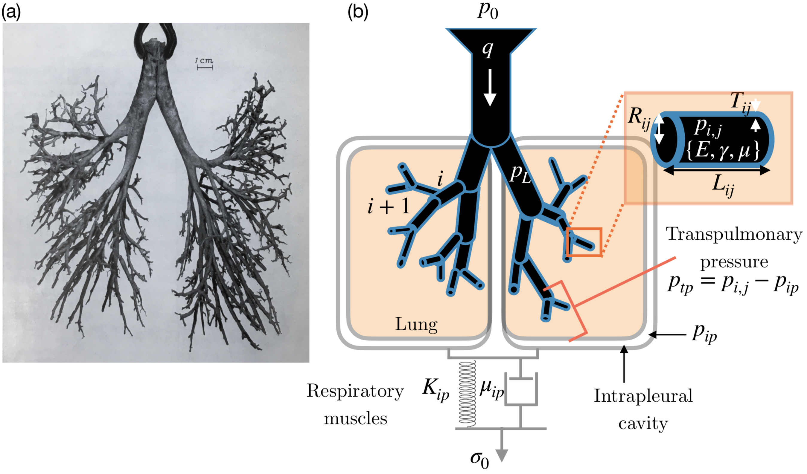
Motivated by morphological data, we computationally represent the lungs as a binary branched network of thin-walled, liquid-lined flexible cylinders coupled to a viscoelastic thoracic cavity (Figure 1). This tree can be classified into two sections leading from the trachea [57, 58], indexed by the generation number : the conducting zone (), which has a constant open volume and does not contribute to oxygen uptake into the bloodstream, and the respiratory zone (), which has branches that can collapse and open during respiration, and is the primary site of oxygen uptake. We therefore represent the conducting zone as one static airway branch, and the respiratory zone as a binary tree spanning generations 17 through 23. We index each individual branch by the labels and , where denotes the generation number and corresponds to the index of the branch within a given generation . The branches are all connected; thus, branches deeper in the lungs are only able to open when branches above them have opened.
Each branch is characterized by an open inner radius , length , and wall thickness , and therefore an open airway volume (Fig. 1b, right inset). For each generation, the mean values of these morphological parameters , , and are given by experimental measurements of the mean branch radius, length, and thickness, respectively (Table 1). To incorporate heterogeneity, a natural feature of the lungs, we then randomly select the individual , , and from a uniform distribution bounded by 25 of , , and , respectively. The results shown in Figs. 2–7 all utilize the same lung architecture parameterized by the same values of , to isolate the influence of biomechanical factors on breathing. However, an advantage of our network representation is that it is generalizable: specific values of the morphological parameters can be incorporated in future extensions of this work. For example, our model could be used to assess the distributions of outcomes across different airways with different , or between different realizations of the same airways having the same , given the importance of structural heterogeneity on breathing [55].
| Generation | Mean radius (mm) | Mean length (mm) | Mean wall thickness (mm) |
|---|---|---|---|
| 17 | 0.270 | 1.41 | 0.0236 |
| 18 | 0.250 | 1.17 | 0.0229 |
| 19 | 0.235 | 0.99 | 0.0226 |
| 20 | 0.225 | 0.83 | 0.0227 |
| 21 | 0.215 | 0.70 | 0.0228 |
| 22 | 0.205 | 0.59 | 0.0231 |
| 23 | 0.204 | 0.50 | 0.0250 |
| Biomechanical parameters | Value | Reference |
|---|---|---|
| Young’s modulus of the lung airway wall | 5 kPa | [60, 61] |
| Poisson’s ratio of the airway wall | 0.5 | [62] |
| Mucus dynamic shear viscosity | 100 mPa-s | [63, 64, 65] |
| Applied muscular stress | 500 Pa | [66, 67, 68, 69] |
| Initial volume of the intrapleural cavity Vip,0 | 20 mL | [70] |
| Maximal open airway volume V0 | 1.675 L | [56] |
| Initial pressure of the intrapleural cavity | – 400 Pa | [71] |
| Effective bulk modulus of the intrapleural cavity | 100 kPa | [60, 61] |
| Mucus surface tension | 15 mN/m | [72, 73] |
Further, to incorporate the biomechanical properties of lung tissues, we make the simplifying assumption that the inner walls of the branches are uniformly coated by a Newtonian fluid of negligible thickness with dynamic shear viscosity and surface tension , and the lung airway wall is a linear elastic solid with Young’s modulus ; we use values of , , and obtained from experimental measurements, as listed in Table 2, and take them to be constant throughout the lungs. This model therefore represents a key first step toward computationally describing the lungs, which in reality have non-Newtonian mucus with a non-negligible, generation-dependent thickness, as well as generation-dependent values of the parameters , , and . However, our network representation enables specific branch-dependent values of these biomechanical parameters to be incorporated, which would be another useful direction for future work.
I.2 Stress exerted by thoracic muscles
As a first step toward modeling the full dynamics of respiration, here we consider the process of inhalation starting from a completely closed respiratory zone—characteristic of newborn infants or patients with severe respiratory distress [74]. We therefore initialize the model with all branches with closed. Building on this model to explore the additional dynamics of exhalation as well as multiple breathing cycles will be an important direction for future research.
Inhalation begins with the contraction of the thoracic muscles that, as a first approximation, we assume pull with a constant stress on the intrapleural cavity. Motivated by previous work [75, 61], we model the viscoelastic behavior of the chest using a Kelvin-Voigt model, which treats the chest as a combination of an elastic spring and a viscous dashpot connected in parallel (Fig. 1b, bottom): , where and are the effective elastic and viscous constants characterizing the intrapleural cavity and represents the volumetric strain in the intrapleural cavity, where represents the difference in the cavity volume compared to its initial value . Thus, the intrapleural space expands over time in a stress-dependent manner:
| (1) |
where is the maximal strain and is the characteristic time scale of inhalation. Based on previous measurements [60, 61], we take kPa and s.
I.3 Elasto-capillary interactions in individual branches
The expansion of the thoracic cavity reduces the intrapleural pressure (Fig. 1, right): given a fixed amount of air within the intrapleural space, , where kPa as determined experimentally [76] and is given by Eq. 1. Thus, the transpulmonary pressure difference across the lung airway wall transiently increases; here is the air pressure in the respiratory zone, which we take to be constant throughout due to the low air flow resistance of the respiratory zone, as justified in the Methods.
As established in many previous studies [77, 78, 79, 55], as increases, it exceeds the threshold pressure required to open a collapsed branch that is in contact with the open region of the lungs [38]. In this case, an air finger propagates into the branch at a speed that is determined by a complex interplay between viscous forces in the airway mucus lining as the branch is pulled apart, capillary forces holding the walls of the branch together, and elastic forces resisting bending of the branch walls. Motivated by the results of previous three-dimensional numerical solutions [38], we estimate this speed using the relation where is the capillary number and is a dimensionless parameter that quantifies the competition between capillary and elastic stresses in the branch, with bending stiffness and is the Poisson’s ratio of the airway wall. This equation highlights three key features of branch opening. First, capillary forces hold the walls of a branch together, and thus, the transpulmonary pressure must overcome the capillary pressure threshold to force a branch to open. Second, the elastic energy penalty associated with deforming a branch also promotes opening, as quantified by . Third, branch opening is not instantaneous, but is limited by viscous dissipation as the mucus lining is pushed apart, as quantified by .
We directly implement this relation into our model, with the condition that a given branch can only open if its proximal end is in contact with the open region of the lungs. For ease of computation we treat the branch as being split into a fully open fraction with time-dependent volume and a remaining fully closed fraction. In non-dimensional form, this relation can then be expressed as
| (2) |
where the hat notation indicates that the variables , , , , and have been normalized by the characteristic branch-scale quantities , , , , and , respectively, where the subscript 17 refers to the mean value at the first generation of the respiratory zone. This non-dimensional form reveals that the biomechanical parameter is a key factor that governs the amount of lung opening during inhalation: when is large, elasticity tends to peel the lung branches open, while when is small, capillarity tends to hold lung branches shut.
I.4 Overall opening of the lungs
The physics described in the previous subsection governs the opening of individual branches; summing over all open regions of the airways then yields the total opened lung volume , where is the constant open volume of the conducting zone and is the time-dependent open volume of the respiratory zone, with given by Eq. 2. Thus, as increases, the open volume of the respiratory zone increases, causing the lung pressure to transiently decrease. This decrease in pressure draws air into the lungs from the atmosphere with a volumetric flow rate of magnitude through the conducting zone (Methods), where kPa is atmospheric pressure.
I.5 Computational implementation of the model
Analysis of these coupled processes enables us to quantitatively model the full dynamics of inhalation. Specifically, as detailed in the Methods, we input values of the morphological parameters and the biomechanical parameters and iteratively solve the equations described in the Theory section at uniform discrete time steps . This scheme thus enables us to determine the full evolution of the pressures , the volumes and the flow rate over time , where the tilde notation indicates lung-scale variables that have been normalized by the atmospheric pressure , maximal open lung volume , characteristic flow rate , and breathing time respectively. Importantly, this network representation reduces computational cost: the complete dynamics of inhalation can be obtained within a matter of minutes on a conventional personal computer. For example, the entirety of the results in Fig. 2 were obtained using a laptop with an 8th Gen Intel Core i7-8750H 6 core processor with 2.2 GHz and 16GB RAM in 20 minutes. Thus, our network model provides a computationally tractable way to characterize the dynamics of respiration that can be implemented by specialists and general users alike.
Results
Typical inhalation dynamics
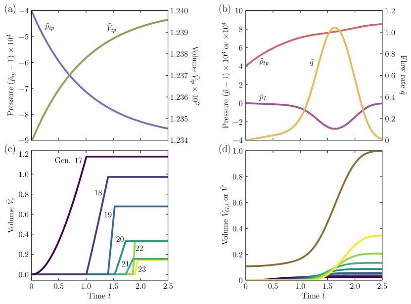
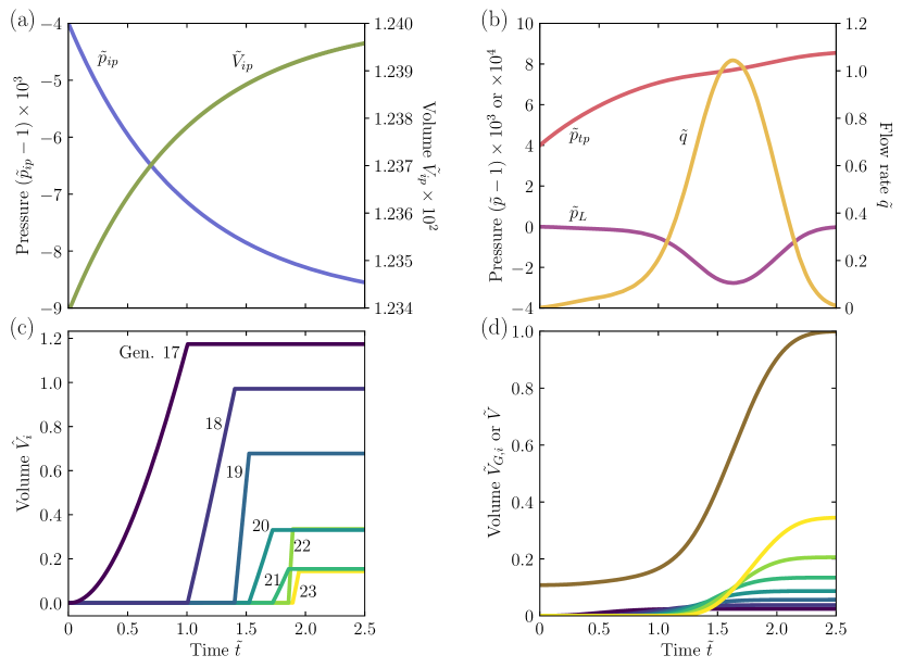
We begin by describing the full inhalation dynamics of a representative healthy lung, using published measurements for the input parameters [56, 72, 73, 60, 61, 63, 64, 65, 66, 67, 68, 69, 70, 71]. The simulation captures the expected dynamics of inhalation. First, the contraction of the respiratory muscles generates a stress on the intrapleural cavity, expanding it (Fig. 2a, green) and reducing its internal pressure (Fig. 2a, blue). The transpulmonary pressure difference between the lung interior and the intrapleural space subsequently builds up (Fig. 2b, red), causing respiratory branches to successively open (Fig. 2c-d, dark blue to green to yellow), reducing the pressure in the respiratory zone (Fig. 2b, purple) and driving air flow into the lungs (Fig. 2b, yellow). This process continues as the intrapleural cavity expands over time. Eventually, however, the applied stress is able to expand the chest by less and less (Fig. 2a, plateau in green curve), and branches open at a slower rate; the flow rate of air into the lungs eventually reaches zero at a time (Fig. 2b, yellow), and inhalation ceases.
Each generation is made of progressively smaller branches having progressively larger threshold pressures for opening. Thus, we expect lung opening to be hierarchical: the larger proximal branches open first, and then smaller distal branches open later, after air is able to propagate to them and exceed the capillary pressure threshold. Our computational model captures this hierarchy of branch opening, as shown in Figs. 2c-d, complementing previous investigations of collective branch opening [77, 78, 79, 55]. Fig. 2c shows an example of individual, connected branches in different generations as they open. The branch in the first generation of the respiratory zone opens first (, dark blue); once it has fully opened, air can propagate into the branch in the next generation (, lighter blue), causing successive opening of branches through the different generations and eventually reaching the terminal alveoli (, yellow). Notably, however, even though these terminal generations must open later, and though their individual branches are smaller, they collectively contribute the largest volume to the open lung, as shown in Fig. 2d; the dark blue to yellow curves show the volume of all the open branches in a given generation , , while the brown curve shows the total open volume of the lung . Though the first generation of the respiratory zone (dark blue) is the first to open during inhalation, it contributes only volume to the open lung; by contrast, the terminal generation (yellow) is the last to open, but contributes of the overall lung volume. Thus, our model quantifies the expectation that opening later generations is key for healthy lung performance.
Influence of changes in biomechanical parameters on inhalation
Having characterized the typical dynamics of inhalation, we next investigate how these dynamics are controlled by key biomechanical factors. Specifically, motivated by their relevance to respiratory distress stemming from the ongoing COVID-19 crisis, as well as from prevalent conditions such as CF, COPD, asthma, and emphysema, we focus on the role of four key factors: (i) Muscle-induced stress , (ii) Mucus viscosity , (iii) Mucus surface tension , and (iv) Airway wall stiffness .
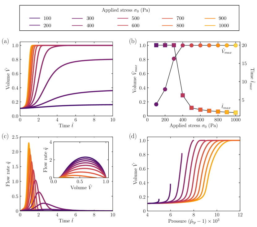
Role of muscle-induced stress
Patients with COVID-19, CF, or COPD frequently exhibit fatigue and muscle weakness [80, 81]; similar symptoms also manifest in patients who have undergone mechanical ventilation as a treatment for prolonged periods of time [82]. The analysis presented in the Theory section suggests that this decrease in reduces the expansion of the intrapleural cavity during inhalation, limiting the amount of air that can be taken into the lungs and giving rise to respiratory distress.
Our simulations with varying confirm this expectation. In particular, we find that the dynamics of lung opening strongly depend on the applied stress (Fig. 3a), indicating that it is a key regulator of breathing; reducing the stress exerted by the thoracic muscles decreases the rate at which air is drawn in and prolongs the overall duration of inhalation (Fig. 3c). Indeed, when is reduced to just half of its typical healthy value Pa [66, 67, 68, 69], the full duration of lung opening takes nearly ten times longer, and only reaches half the fully opened volume, as shown by the squares and circles in Fig. 3b, respectively. The corresponding stress-dependent pressure-volume (Fig. 3d) and flow rate-volume (Fig. 3c, inset) curves obtained in our simulations are strikingly similar to those observed in experimental measurements [83]. Thus, our computational approach provides a way to quantify the impact of specific changes in muscle-induced stress on inhalation, shedding light on its relative influence in causing respiratory distress.
Role of mucus viscosity
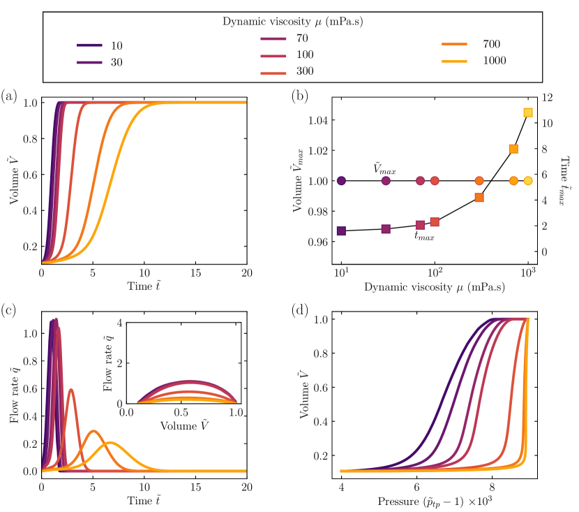
A common symptom of bronchitis, CF, COPD, interstitial lung disease, and possibly COVID-19 is a large increase in the viscosity of lung mucus [64, 84, 85, 86, 87]. The analysis presented in the Theory section suggests that this increase in increases the time scale over which individual lung branches open, possibly slowing the opening dynamics during inhalation and giving rise to respiratory distress.
Our simulations with varying confirm this expectation. In particular, we find that the dynamics of lung opening strongly depend on the mucus viscosity (Fig. 4a), indicating that it is another key regulator of breathing; increasing the mucus viscosity increases the time needed to reach the capillary pressure threshold and open airway branches, decreasing the rate at which air is drawn in and prolonging the overall duration of inhalation (Fig. 4c). Indeed, when is increased by a factor of from its typical healthy value mPa-s, as can be the case in many lung diseases [64, 84, 85, 86, 87], the fully opened lung volume is unchanged, but the full duration of lung opening takes approximately five times longer, as shown by the circles and squares in Fig. 4b, respectively. Thus, alterations in mucus viscosity alter the dynamics, but not full extent, of lung opening during inhalation. The corresponding viscosity-dependent pressure-volume (Fig. 4d) and flow rate-volume (Fig. 4c, inset) curves obtained in our simulations are again strikingly similar to those observed in experimental measurements [83]. Thus, our computational approach provides a way to quantify the impact of specific changes in mucus viscosity on inhalation, shedding light on its relative influence in causing respiratory distress.
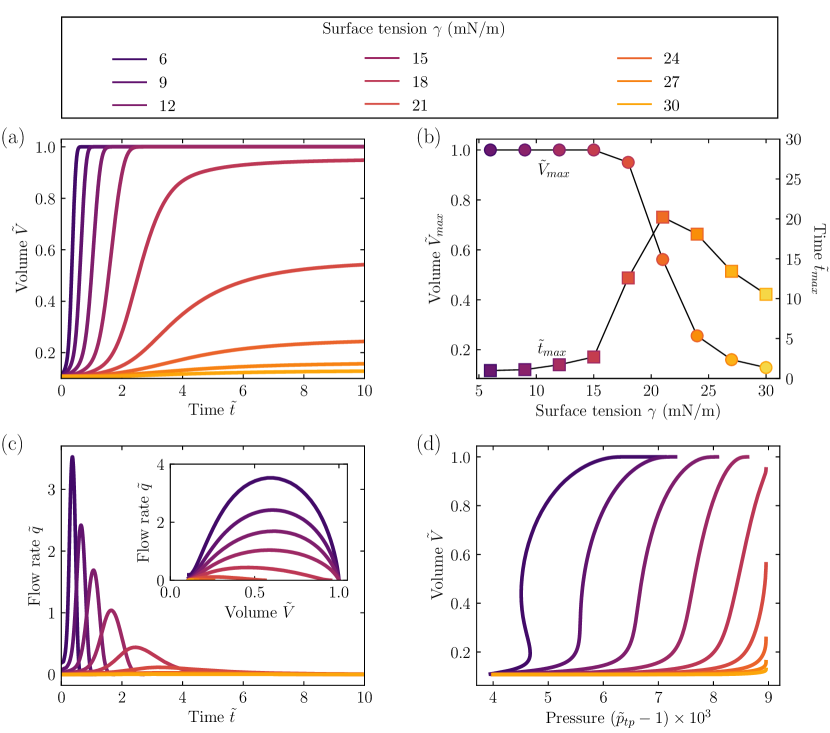
Role of mucus surface tension
One of the most prominent pathological features of COVID-19 is hindered production of lung surfactant due to viral infection, resulting in a large increase in the surface tension of airway mucus [88, 89, 90, 91]. Similar complications arise in COPD and possibly in asthma and emphysema [81, 92, 93]. The analysis presented in the Theory section suggests that this increase in has two key effects, both of which could contribute to respiratory distress. First, it increases the threshold pressure required to open a collapsed branch . Second, it decreases the biomechanical parameter , which quantifies the competition between elastic and capillary stresses in the lung: when is smaller, capillary forces are more likely to overcome the elastic energy penalty of holding lung branches shut. Both effects likely hinder the opening of the lungs during respiration, giving rise to respiratory distress in diseased patients.
Our simulations with varying confirm this expectation. In particular, we find that the dynamics of lung opening strongly depend on the surface tension (Fig. 5a), indicating that it is another key regulator of breathing; increasing the surface tension decreases the rate at which air is drawn in and prolongs the overall duration of inhalation (Fig. 5c). Indeed, when is increased by just a factor of two from its typical healthy value mN/m [72, 73], as can be the case in COVID-19 and COPD [88, 89, 90, 91, 81, 92, 93], the full duration of lung opening takes nearly four times longer, and only reaches a tenth of the fully opened volume, as shown by the squares and circles in Fig. 5b, respectively. Intriguingly, the duration of inhalation varies non-monotically with , as shown by the squares: when is small, the capillary pressure threshold is easily overcome and the lungs open quickly, while as increases, capillarity increasingly resists lung opening and the duration of inhalation increases. However, as increases above mN/m, capillarity holds increasing numbers of branches of the lungs shut, and inhalation is truncated—causing the duration of inhalation to decrease again. This behavior also manifests in the simulated pressure-volume (Fig. 5d) and flow rate-volume (Fig. 5c, inset) curves, which are again strikingly similar to those observed in experimental measurements [83]. Indeed, we even observe the previously-reported [94, 95] non-monotonic variation of with at low , as shown by the dark purple curve: under these conditions, because the capillary pressure threshold is easily overcome, rapid lung opening causes an abrupt decrease in the lung pressure—a phenomenon that has been termed an “elastic shock” [94, 95]. Together, these results indicate that our computational approach provides a way to quantify the impact of specific changes in mucus surface tension on inhalation, isolating its relative influence in causing respiratory distress.
Role of airway wall stiffness
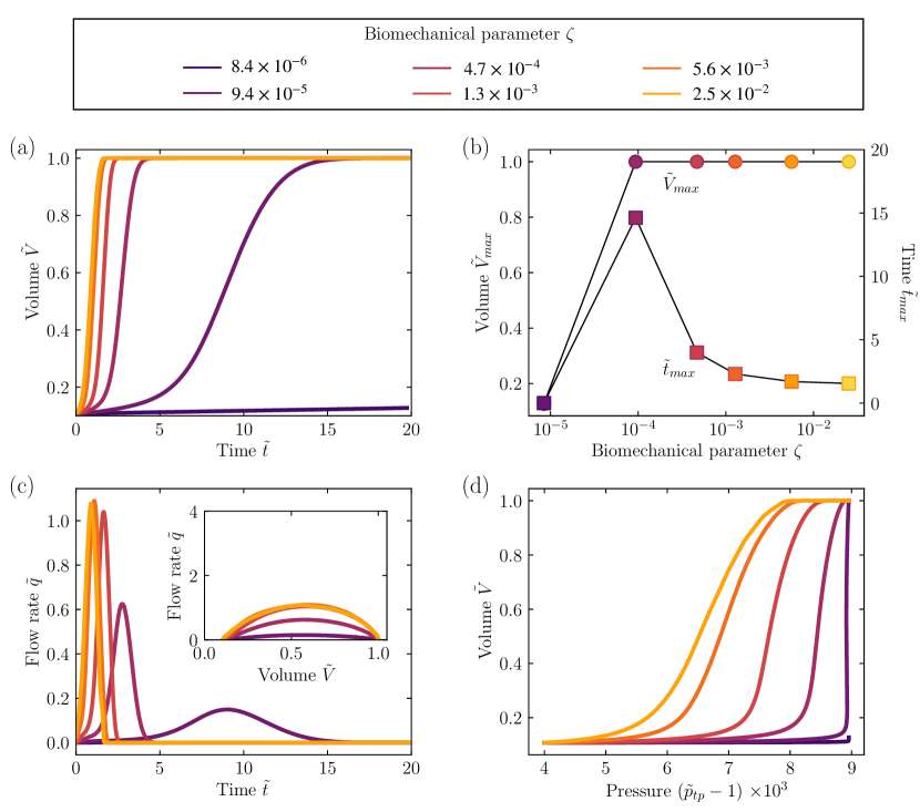
The elasticity of the airway wall changes greatly in disease, often in opposing ways. For example, buildup of excess fibrous connective tissue stiffens the airway walls in CF, COPD, and asthma [96, 97, 98, 99], while weakening of the tissue leads to weaker airway walls in emphysema [11, 100, 101, 102]; whether lung tissue elasticity increases, decreases, or stays unchanged is currently still being studied for COVID-19. The analysis presented in the Theory section suggests that weakening of the airway walls in emphysema hinders lung opening during inhalation, and is likely the main contributor to respiratory distress in this case: capillary forces due to the surface tension of the mucus lining tend to hold the soft walls of closed branches together. Conversely, we expect that stiffening of the airway walls in CF, COPD, and asthma paradoxically promotes lung opening, in opposition to the respiratory distress associated with these conditions: stiffer lungs are more difficult to bend and close shut. Thus, in these cases, we expect that respiratory distress arises instead due to changes in other biomechanical factors, such as mucus viscosity and surface tension, as suggested by clinical studies [103, 104, 105].
This competition between lung elasticity and capillarity is quantified by the biomechanical parameter . When is large, elastic stresses, as quantified by the characteristic bending stiffness , dominate and peel the lung branches open; conversely, while when is small, capillarity, as quantified by the characteristic capillary pressure , dominates and tends to hold the lung branches shut. Our simulations with varying , exploring the full physiological range of using measurements of the variation that arises in and [56, 60, 61, 63, 64, 72, 73], confirm this expectation. Similar to the cases of varying and , the dynamics of lung opening strongly depend on (Fig. 6a), indicating that it is another key regulator of breathing; decreasing decreases the rate at which air is drawn in and prolongs the overall duration of inhalation (Fig. 6c). Intriguingly, similar to the case of , the duration of inhalation varies non-monotically with , as shown by the squares in Fig. 6b: when is large, the lungs open quickly due to elastic stresses, while as decreases, elastic stresses do not pull the lungs open as quickly and the duration of inhalation increases. However, as decreases below , elastic stresses cannot open many branches of the lungs, and inhalation is again truncated. This behavior also manifests in the simulated pressure-volume (Fig. 6d) and flow rate-volume (Fig. 6c, inset) curves, which are again strikingly similar to those observed in experimental measurements [106, 107].
We expect that because decreases in emphysema [11, 100, 101, 102], and possibly increases [81, 92, 93], concurrently decreases and is thus the key biomechanical parameter that controls the onset of respiratory distress in this case. Conversely, because increases in CF, COPD, and asthma [96, 97, 98, 99], may not decrease in these cases—suggesting that the onset of respiratory distress is instead controlled by increases in the mucus viscosity or surface tension, as described above. Thus, our computational approach provides a way to separately quantify the impact of specific changes in airway wall stiffness on breathing, shedding light on its relative influence in causing respiratory distress.
Discussion
The work described here represents a first step toward developing a model of the lungs that accurately describes the multi-scaled spatial and temporal features of respiration, while still managing to be computationally tractable. Our dynamic network approach explicitly resolves the relevant length and time scales of branch opening during inhalation, while also capturing how opening propagates through the interconnected and hierarchical architecture of the lungs. We demonstrate this principle by directly connecting alterations in four key biomechanical factors—the strength of thoracic muscle contraction, the viscosity and surface tension of the airway mucus lining, and the elasticity of the airway wall—to overall alterations in breathing, in qualitative agreement with experimental and clinical findings. Our model thus helps to establish how lung biomechanics impact respiration—both deepening our fundamental understanding of this ubiquitous process, and helping to elucidate how disease-induced changes in tissue-scale factors give rise to respiratory distress. However, despite the similarity between our results and published measurements, direct validation against systematic experimental measurements or more sophisticated models, for which the values of all the input parameters are known, will be a crucial next step.
Given the increasing prevalence of respiratory diseases [108], there is a critical need for computational tools capable of quantitatively assessing the efficacy of different therapeutic interventions, such as mechanical ventilation, exogenous administration of lung surfactant, and exogenous administration of mucus thinners. The work described here addresses this critical need. Specifically, it yields a generally-applicable computational model for which measured treatment-induced changes in biomechanical parameters—e.g. —can be input, and the impact on breathing outcome can be assessed. Because different treatments alter lung biomechanics in different ways, this approach may yield useful insights into how treatments that influence these specific parameters will affect breathing dynamics in general—and may eventually provide a straightforward way to quickly assess the impact of different treatments for a given patient.
The model presented here focused on the case of inhalation starting from a completely closed respiratory zone as a proof of principle; however, the dynamics described in the Theory section can be extended in future work to also describe the closure of individual lung branches due to compression of the thoracic cavity, as well as breathing dynamics in a lung with regional atelectasis, involving a mixture of both open, partially-closed, and fully-closed branches. Accomplishing this extension will require development of a form of Eq. 2 that characterizes branch closure instead of opening. Further, for both inhalation and exhalation, Eq. 2 can be replaced by the results of more sophisticated tube models that incorporate non-axisymmetric deformation modes, possible collapse of the mucus film, mucus-wall liquid-solid interactions arising from the competition between viscous stress, capillary stresses, and wall deformations, and heterogeneities in airway branch geometry [57, 38, 39, 47, 48, 49, 50, 51, 109, 110, 40, 41, 42, 43, 44, 45, 111]. Finally, while our network representation necessarily simplifies many of the rich complexities of the lung in favor of ease of computation, it can be extended by incorporating different lung architectures, by directly inputting specific values of ; heterogeneity in the biomechanical parameter values, by directly inputting specific values of ; and non-Newtonian mucus rheology, by incorporating a rate-dependent viscosity in Eq. 2. Exploring the influence of these different features on breathing will be an important extension of our work.
Methods
To simulate the dynamics of inhalation, we implement the rules given in the Theory section in discretized form, evaluating volumes, pressures, and flow rates at successive time steps separated by using the iterative scheme described below. We use two different notations to differentiate between branch-scale quantities and overall lung-scale quantities. The tilde notation indicates that pressure, volume, flow rate, time, and flow resistance have been normalized by the atmospheric pressure , maximal open lung volume , characteristic flow rate , breathing time , and characteristic flow resistance , respectively. The hat notation indicates that the variables , , , , and have been normalized by the characteristic branch-scale quantities , , , and , respectively, where the subscript 17 refers to the mean value at the first generation of the respiratory zone. For simplicity, we assume that the air is an ideal gas at a fixed temperature.
-
1.
The applied stress forces the volume of the intrapleural cavity to increase:
(3) where is given by Eq. 1.
-
2.
Given a fixed amount of air within the intrapleural space, the expansion of the intrapleural cavity causes the pressure in the intrapleural cavity to concomitantly decrease:
(4) -
3.
This decrease in intrapleural pressure transiently increases the transpulmonary pressure, which we estimate as:
(5) We take to be a constant throughout the respiratory zone due to the low air flow resistance of the respiratory zone. In particular, we compare the flow resistance of air through the conducting zone or through the respiratory zone, or , respectively, where the individual branch flow resistance is given by the Hagen-Poiseuille equation with an air viscosity Pa-s. Using measurements of and throughout the airways [56, 59], we estimate Pa-s/L and Pa-s/L. Since , we assume that the air flow resistance of the lungs is given by that of the conducting zone, and the air pressure is constant throughout the respiratory zone.
-
4.
For each branch that is in contact with the open region of the lungs, if exceeds the threshold , this pressure difference across the branch wall forces it to open, as given by Eq. 2:
(6) -
5.
As branches open, the open volume of the lungs increases, causing the pressure in the respiratory zone to transiently decrease to an intermediate value . In normal respiration, the lungs are an open system, and air in the lungs can be treated as incompressible, so that its density does not vary with the pressure changes that arise during breathing due to inertial and viscous losses in the airways. However, in our discretized representation of the lungs, for sufficiently small , the lungs can be approximated to be a closed system at each intermediate time step: the time scale of volume changes of the lungs is much shorter than the characteristic air inflow time scale. Thus, assuming a constant and a constant amount of air within the respiratory zone during this intermediate step, we estimate the intermediate pressure as:
(7) -
6.
This decrease in pressure draws air into the lungs from the atmosphere with a volumetric flow rate , driven by the pressure difference . To evaluate this flow rate, we first consider the limit of ; in this case, the pressure difference is fully equilibrated at each time step, and conservation of the amount of air exchanged yields , where is an intrinsic resistance that reflects the discrete time formulation of the simulation. For the case of , we then modify this expression to also incorporate :
(8) -
7.
Because , this air flow does not fully equilibrate the pressure difference . Instead, the pressure in the respiratory zone at the end of the time step is given by:
(9)
For each simulation presented in the main text, we iteratively solve Eqs. 3–9 over successive time steps separated by up to . We obtain identical results with even finer discretization, as shown for the case of in Fig. 7, validating the assumptions made in Steps 5–6 above. This iterative solving is done using a C++ framework that explicitly considers a given lung network structure described by the input morphological parameters and the biomechanical parameters . This framework is split into a layer of virtual classes that treat memory management, multithreading, and provide the basic functions to create a branched network; each simulation is then derived from these virtual base classes with data structures defining the specific parameters that are input into the model. This scheme thus enables us to determine the full evolution of the pressures , the volumes and the flow rate over time .
Author Contributions
All authors helped to design simulations; S.S.D. and J-F.L. designed and performed the theoretical analysis; F.K. performed all simulations; J-F.L., F.K., and S.S.D. analyzed the data; J-F.L., F.K., and S.S.D. discussed the results and implications and wrote the manuscript; S.S.D. designed and supervised the overall project.
Acknowledgments
This work was supported by startup funds from Princeton University as well as partial support from the Keller Center REACH program for F.K. It is a pleasure to acknowledge Nathanael Ji and Anvitha Sudhakar for their work on a preliminary version of the network model at the inception of this project.
Data Availability
The code necessary to reproduce the results reported here and to further explore the dynamic network model will be available in a Github repository at https://github.com/FelixKratz/LungFramework.
References
- [1] Zhe Xu, Lei Shi, Yijin Wang, Jiyuan Zhang, Lei Huang, Chao Zhang, Shuhong Liu, Peng Zhao, Hongxia Liu, Li Zhu, et al. Pathological findings of COVID-19 associated with acute respiratory distress syndrome. The Lancet respiratory medicine, 8(4):420–422, 2020.
- [2] Catrin Sohrabi, Zaid Alsafi, Niamh O’Neill, Mehdi Khan, Ahmed Kerwan, Ahmed Al-Jabir, Christos Iosifidis, and Riaz Agha. World health organization declares global emergency: A review of the 2019 novel coronavirus (COVID-19). International Journal of Surgery, 2020.
- [3] Chuan Qin, Luoqi Zhou, Ziwei Hu, Shuoqi Zhang, Sheng Yang, Yu Tao, Cuihong Xie, Ke Ma, Ke Shang, Wei Wang, et al. Dysregulation of immune response in patients with COVID-19 in wuhan, china. Clinical Infectious Diseases, 2020.
- [4] Neo Poyiadji, Gassan Shahin, Daniel Noujaim, Michael Stone, Suresh Patel, and Brent Griffith. COVID-19–associated acute hemorrhagic necrotizing encephalopathy: CT and MRI features. Radiology, page 201187, 2020.
- [5] Wen-Hsiang Chen, Ulrich Strych, Peter J Hotez, and Maria Elena Bottazzi. The SARS-CoV-2 vaccine pipeline: an overview. Current tropical medicine reports, pages 1–4, 2020.
- [6] Xiao-Wei Xu, Xiao-Xin Wu, Xian-Gao Jiang, Kai-Jin Xu, Ling-Jun Ying, Chun-Lian Ma, Shi-Bo Li, Hua-Ying Wang, Sheng Zhang, Hai-Nv Gao, et al. Clinical findings in a group of patients infected with the 2019 novel coronavirus (SARS-Cov-2) outside of wuhan, china: retrospective case series. bmj, 368, 2020.
- [7] Weiren Luo, Hong Yu, Jizhou Gou, Xiaoxing Li, Yan Sun, Jinxiu Li, and Lei Liu. Clinical pathology of critical patient with novel coronavirus pneumonia (COVID-19). Preprints, 2020:2020020407, 2020.
- [8] Richard A Polin, Waldemar A Carlo, et al. Surfactant replacement therapy for preterm and term neonates with respiratory distress. Pediatrics, 133(1):156–163, 2014.
- [9] Rob Mac Sweeney and Daniel F McAuley. Acute respiratory distress syndrome. The Lancet, 388(10058):2416–2430, 2016.
- [10] B Taylor Thompson, Rachel C Chambers, and Kathleen D Liu. Acute respiratory distress syndrome. New England Journal of Medicine, 377(6):562–572, 2017.
- [11] Cláudio LN Oliveira, Ascânio D Araújo, Jason HT Bates, José S Andrade Jr, and Béla Suki. Entropy production and the pressure–volume curve of the lung. Frontiers in Physiology, 7:73, 2016.
- [12] Michael Robinson and Peter TB Bye. Mucociliary clearance in cystic fibrosis. Pediatric pulmonology, 33(4):293–306, 2002.
- [13] Patricia A Newhouse, Fred White, John H Marks, and Douglas N Homnick. The intrapulmonary percussive ventilator and flutter device compared to standard chest physiotherapy in patients with cystic fibrosis. Clinical pediatrics, 37(7):427–432, 1998.
- [14] Pamela B Davis and Paul A di Sant’Agnese. Assisted ventilation for patients with cystic fibrosis. Jama, 239(18):1851–1854, 1978.
- [15] Brigitte Fauroux, Michèle Boulé, Frédéric Lofaso, Françoise Zérah, Annick Clément, Alain Harf, and Daniel Isabey. Chest physiotherapy in cystic fibrosis: improved tolerance with nasal pressure support ventilation. Pediatrics, 103(3):e32–e32, 1999.
- [16] Noel G McElvaney, RC Hubbard, P Birrer, RG Crystal, MS Chernick, MM Frank, and DB Caplan. Aerosol 1-antitrypsin treatment for cystic fibrosis. The Lancet, 337(8738):392–394, 1991.
- [17] Matthias Griese, P Bufler, J Teller, and D Reinhardt. Nebulization of a bovine surfactant in cystic fibrosis: a pilot study. European Respiratory Journal, 10(9):1989–1994, 1997.
- [18] Gehan Devendra and Roger G Spragg. Lung surfactant in subacute pulmonary disease. Respiratory research, 3(1):11, 2002.
- [19] H Clark and K Reid. The potential of recombinant surfactant protein d therapy to reduce inflammation in neonatal chronic lung disease, cystic fibrosis, and emphysema. Archives of disease in childhood, 88(11):981–984, 2003.
- [20] Matthias Griese, P Birrer, and A Demirsoy. Pulmonary surfactant in cystic fibrosis. European Respiratory Journal, 10(9):1983–1988, 1997.
- [21] Douglas N Homnick, Fred White, and Carol de Castro. Comparison of effects of an intrapulmonary percussive ventilator to standard aerosol and chest physiotherapy in treatment of cystic fibrosis. Pediatric pulmonology, 20(1):50–55, 1995.
- [22] Anne E Holland, Linda Denehy, G Ntoumenopoulos, Matthew T Naughton, and John W Wilson. Non-invasive ventilation assists chest physiotherapy in adults with acute exacerbations of cystic fibrosis. Thorax, 58(10):880–884, 2003.
- [23] ME Hodson, BP Madden, MH Steven, VT Tsang, and MH Yacoub. Non-invasive mechanical ventilation for cystic fibrosis patients–a potential bridge to transplantation. European Respiratory Journal, 4(5):524–527, 1991.
- [24] D Keith Walters, Greg W Burgreen, David M Lavallee, David S Thompson, and Robert L Hester. Efficient, physiologically realistic lung airflow simulations. IEEE Transactions on Biomedical Engineering, 58(10):3016–3019, 2011.
- [25] Bela Soni and Shahrouz Aliabadi. Large-scale cfd simulations of airflow and particle deposition in lung airway. Computers & Fluids, 88:804–812, 2013.
- [26] Baoshun Ma and Kenneth R Lutchen. An anatomically based hybrid computational model of the human lung and its application to low frequency oscillatory mechanics. Annals of biomedical engineering, 34(11):1691–1704, 2006.
- [27] Wolfgang A Wall, Lena Wiechert, Andrew Comerford, and Sophie Rausch. Towards a comprehensive computational model for the respiratory system. International Journal for Numerical Methods in Biomedical Engineering, 26(7):807–827, 2010.
- [28] Tina A Lewis, Yang-Sheng Tzeng, Erin L McKinstry, Angela C Tooker, Kwansoo Hong, Yanping Sun, Joey Mansour, Zachary Handler, and Mitchell S Albert. Quantification of airway diameters and 3D airway tree rendering from dynamic hyperpolarized 3He magnetic resonance imaging. Magnetic Resonance in Medicine: An Official Journal of the International Society for Magnetic Resonance in Medicine, 53(2):474–478, 2005.
- [29] Natalya Nowak, Prashant P Kakade, and Ananth V Annapragada. Computational fluid dynamics simulation of airflow and aerosol deposition in human lungs. Annals of biomedical engineering, 31(4):374–390, 2003.
- [30] J Sznitman, S Schmuki, R Sutter, A Tsuda, and Thomas Rösgen. Cfd investigation of respiratory flows in a space-filling pulmonary acinus model. Modelling in Medicine and Biology VII, WIT Transactions on Biomedicine and Health, 12:147–156, 2007.
- [31] D Keith Walters and William H Luke. Computational fluid dynamics simulations of particle deposition in large-scale, multigenerational lung models. Journal of biomechanical engineering, 133(1), 2011.
- [32] D Keith Walters and William H Luke. A method for three-dimensional navier–stokes simulations of large-scale regions of the human lung airway. Journal of Fluids Engineering, 132(5), 2010.
- [33] Robert Francis Kunz, Daniel C Haworth, DP Porzio, and Andres Kriete. Progress towards a medical image through cfd analysis toolkit for respiratory function assessment on a clinical time scale. In 2009 IEEE International Symposium on Biomedical Imaging: From Nano to Macro, pages 382–385. IEEE, 2009.
- [34] Rajnish Kaur Calay, Jutarat Kurujareon, and Arne Erik Holdø. Numerical simulation of respiratory flow patterns within human lung. Respiratory physiology & neurobiology, 130(2):201–221, 2002.
- [35] M Malve, S Chandra, JL Lopez-Villalobos, EA Finol, A Ginel, and M Doblare. Cfd analysis of the human airways under impedance-based boundary conditions: application to healthy, diseased and stented trachea. Computer methods in biomechanics and biomedical engineering, 16(2):198–216, 2013.
- [36] Wolfgang A Wall and Timon Rabczuk. Fluid–structure interaction in lower airways of CT-based lung geometries. International Journal for Numerical Methods in Fluids, 57(5):653–675, 2008.
- [37] T Gemci, Valery Ponyavin, Y Chen, H Chen, and R Collins. Computational model of airflow in upper 17 generations of human respiratory tract. Journal of Biomechanics, 41(9):2047–2054, 2008.
- [38] Andrew L Hazel and Matthias Heil. Three-dimensional airway reopening: the steady propagation of a semi-infinite bubble into a buckled elastic tube. Journal of Fluid Mechanics, 478:47–70, 2003.
- [39] Matthias Heil, Andrew L Hazel, and Jaclyn A Smith. The mechanics of airway closure. Respiratory physiology & neurobiology, 163(1-3):214–221, 2008.
- [40] James B Grotberg. Respiratory fluid mechanics. Physics of Fluids, 23(2):021301, 2011.
- [41] D Halpern and JB Grotberg. Fluid-elastic instabilities of liquid-lined flexible tubes. Journal of Fluid Mechanics, 244:615–632, 1992.
- [42] JB Grotberg. Pulmonary flow and transport phenomena. Annual Review of Fluid Mechanics, 26(1):529–571, 1994.
- [43] James B Grotberg and Oliver E Jensen. Biofluid mechanics in flexible tubes. Annual review of fluid mechanics, 36, 2004.
- [44] Matthias Heil and Andrew L Hazel. Fluid-structure interaction in internal physiological flows. Annual review of fluid mechanics, 43:141–162, 2011.
- [45] Donald P Gaver, David Halpern, Oliver E Jensen, and James B Grotberg. The steady motion of a semi-infinite bubble through a flexible-walled channel. Journal of Fluid Mechanics, 319:25–65, 1996.
- [46] Andrew L Hazel and Matthias Heil. The influence of gravity on the steady propagation of a semi-infinite bubble into a flexible channel. Physics of Fluids, 20(9):092109, 2008.
- [47] Anne Juel and Alexandra Heap. The reopening of a collapsed fluid-filled elastic tube. Journal of Fluid Mechanics, 572:287–310, 2007.
- [48] Matthias Heil. Airway closure: occluding liquid bridges in strongly buckled elastic tubes. Journal of Biomechanical Engineering, 121(5):487–493, 1999.
- [49] Andrew L Hazel and Matthias Heil. Surface-tension-induced buckling of liquid-lined elastic tubes: a model for pulmonary airway closure. Proceedings of the Royal Society A: Mathematical, Physical and Engineering Sciences, 461(2058):1847–1868, 2005.
- [50] Matthias Heil. Stokes flow in collapsible tubes: computation and experiment. Journal of Fluid Mechanics, 353(1):285–312, 1997.
- [51] Matthias Heil and Tim J Pedley. Large post-buckling deformations of cylindrical shells conveying viscous flow. Journal of Fluids and Structures, 10(6):565–599, 1996.
- [52] Chanikarn Wongviriyawong, Tilo Winkler, R Scott Harris, and Jose G Venegas. Dynamics of tidal volume and ventilation heterogeneity under pressure-controlled ventilation during bronchoconstriction: a simulation study. Journal of Applied Physiology, 109(4):1211–1218, 2010.
- [53] Antonio Z Politi, Graham M Donovan, Merryn H Tawhai, Michael J Sanderson, Anne-Marie Lauzon, Jason HT Bates, and James Sneyd. A multiscale, spatially distributed model of asthmatic airway hyper-responsiveness. Journal of theoretical biology, 266(4):614–624, 2010.
- [54] Graham M Donovan. Inter-airway structural heterogeneity interacts with dynamic heterogeneity to determine lung function and flow patterns in both asthmatic and control simulated lungs. Journal of theoretical biology, 435:98–105, 2017.
- [55] Peter S Stewart and Oliver E Jensen. Patterns of recruitment and injury in a heterogeneous airway network model. Journal of The Royal Society Interface, 12(111):20150523, 2015.
- [56] Ewald R. Weibel. Morphometry of the Human Lung. Academic PressInc., Publishers, 1963.
- [57] Keith Horsfield, Gladys Dart, Dan E Olson, Giles F Filley, and Gordon Cumming. Models of the human bronchial tree. Journal of applied physiology, 31(2):207–217, 1971.
- [58] Gianluca Nucci, Simonluca Tessarin, and Claudio Cobelli. A morphometric model of lung mechanics for time-domain analysis of alveolar pressures during mechanical ventilation. Annals of Biomedical Engineering, 30(4):537–545, 2002.
- [59] Robert H Habib, Richard B Chalker, Béla Suki, and Andrew C Jackson. Airway geometry and wall mechanical properties estimated from subglottal input impedance in humans. Journal of Applied Physiology, 77(1):441–451, 1994.
- [60] Jau-Yi Wang, Patrick Mesquida, Prathap Pallai, Chris J Corrigan, and Tak H Lee. Dynamic properties of human bronchial airway tissues. arXiv preprint arXiv:1111.5645, 2011.
- [61] Mark A Lewis and Markus R Owen. The mechanics of lung tissue under high-frequency ventilation. SIAM Journal on Applied Mathematics, 61(5):1731–1761, 2001.
- [62] S. J. Lai-Fook, R. E. Hyatt, and J. R. Rodarte. Elastic constants of trapped lung parenchyma. Journal of Applied Physiology, 44(6):853–858, 1978. PMID: 670008.
- [63] Sonia Baconnais, Rabindra Tirouvanziam, Jean-Marie Zahm, Sophie de Bentzmann, Bruno Péault, Gérard Balossier, and Edith Puchelle. Ion composition and rheology of airway liquid from cystic fibrosis fetal tracheal xenografts. American journal of respiratory cell and molecular biology, 20(4):605–611, 1999.
- [64] E Puchelle, JM Zahm, and C Duvivier. Spinability of bronchial mucus. relationship with viscoelasticity and mucous transport properties. Biorheology, 20(2):239–249, 1983.
- [65] Hirotoshi Matsui, Victoria E Wagner, David B Hill, Ute E Schwab, Troy D Rogers, Brian Button, Russell M Taylor, Richard Superfine, Michael Rubinstein, Barbara H Iglewski, et al. A physical linkage between cystic fibrosis airway surface dehydration and pseudomonas aeruginosa biofilms. Proceedings of the National Academy of Sciences, 103(48):18131–18136, 2006.
- [66] Philip D Hughes, Michael I Polkey, M Lou Harris, Andrew JS Coats, John Moxham, and Malcolm Green. Diaphragm strength in chronic heart failure. American journal of respiratory and critical care medicine, 160(2):529–534, 1999.
- [67] J Martin Bland and Douglas G Altman. Statistical methods for assessing agreement between two methods of clinical measurement. The lancet, 327(8476):307–310, 1986.
- [68] J Ker et al. Respiratory muscle endurance in heart failure–the effect of clinical severity. Cardiovascular Journal of Africa, 9(1):20–23, 1998.
- [69] Michael I Polkey, Dimitris Kyroussis, Carl-Hugo Hamnegard, Gary H Mills, Malcolm Green, and John Moxham. Diaphragm strength in chronic obstructive pulmonary disease. American journal of respiratory and critical care medicine, 154(5):1310–1317, 1996.
- [70] Horacio P D’Agostino and Mary Ann Edens. Physiology, pleural fluid. In StatPearls [Internet]. StatPearls Publishing, 2019.
- [71] Ronald V Christie and CA McIntosh. The measurement of the intrapleural pressure in man and its significance. Journal of Clinical Investigation, 13(2):279, 1934.
- [72] Marianne Geiser, Samuel Schurch, and Peter Gehr. Influence of surface chemistry and topography of particles on their immersion into the lung’s surface-lining layer. Journal of applied physiology, 94(5):1793–1801, 2003.
- [73] Coralie Alonso, Alan Waring, and Joseph A Zasadzinski. Keeping lung surfactant where it belongs: protein regulation of two-dimensional viscosity. Biophysical journal, 89(1):266–273, 2005.
- [74] Laura Ellwein Fix, Joseph Khoury, Russell R Moores Jr, Lauren Linkous, Matthew Brandes, and Henry J Rozycki. Theoretical open-loop model of respiratory mechanics in the extremely preterm infant. PloS one, 13(6):e0198425, 2018.
- [75] Xiangming Zhang and Rong Z Gan. Dynamic properties of human tympanic membrane based on frequency-temperature superposition. Annals of biomedical engineering, 41(1):205–214, 2013.
- [76] JA Blom. Monitoring of respiration and circulation. CRC Press, 2003.
- [77] Béla Suki, Albert-László Barabási, Zoltán Hantos, Ferenc Peták, and H Eugene Stanley. Avalanches and power-law behaviour in lung inflation. Nature, 368(6472):615–618, 1994.
- [78] Béla Suki, Adriano M Alencar, József Tolnai, Tibor Asztalos, Ferenc Peták, Mamatha K Sujeer, Keena Patel, Jignish Patel, H Eugene Stanley, and Zoltán Hantos. Size distribution of recruited alveolar volumes in airway reopening. Journal of Applied Physiology, 89(5):2030–2040, 2000.
- [79] Jason HT Bates and Charles G Irvin. Time dependence of recruitment and derecruitment in the lung: a theoretical model. Journal of Applied Physiology, 93(2):705–713, 2002.
- [80] Weier Wang, Jianming Tang, and Fangqiang Wei. Updated understanding of the outbreak of 2019 novel coronavirus (2019-nCoV) in wuhan, china. Journal of medical virology, 92(4):441–447, 2020.
- [81] Victor Kim and Gerard J Criner. Chronic bronchitis and chronic obstructive pulmonary disease. American journal of respiratory and critical care medicine, 187(3):228–237, 2013.
- [82] Martin J Tobin, Franco Laghi, and Amal Jubran. Narrative review: ventilator-induced respiratory muscle weakness. Annals of internal medicine, 153(4):240–245, 2010.
- [83] Wanda J Kozlowska and Paul Aurora. Spirometry in the pre-school age group. Paediatric respiratory reviews, 6(4):267–272, 2005.
- [84] Samuel K Lai, Ying-Ying Wang, Denis Wirtz, and Justin Hanes. Micro-and macrorheology of mucus. Advanced drug delivery reviews, 61(2):86–100, 2009.
- [85] Bruce K Rubin. Mucus structure and properties in cystic fibrosis. Paediatric respiratory reviews, 8(1):4–7, 2007.
- [86] Jennifer M Sturgess, AJ Palfrey, and Lynne Reid. The viscosity of bronchial secretion. Clinical science, 38(1):145–156, 1970.
- [87] Zheng Ye, Yun Zhang, Yi Wang, Zixiang Huang, and Bin Song. Chest CT manifestations of new coronavirus disease 2019 (COVID-19): a pictorial review. European radiology, pages 1–9, 2020.
- [88] MD Bracco Lorenzo. COVID-19, Type II alveolar cells and surfactant. J Med–Clin Res & Rev, 4(4):1–3, 2020.
- [89] Andreas Günther, Clemens Ruppert, Reinhold Schmidt, Philipp Markart, Friedrich Grimminger, Dieter Walmrath, and Werner Seeger. Surfactant alteration and replacement in acute respiratory distress syndrome. Respiratory research, 2(6):353, 2001.
- [90] Lisa E Gralinski and Ralph S Baric. Molecular pathology of emerging coronavirus infections. The Journal of pathology, 235(2):185–195, 2015.
- [91] Ye Yi, Philip NP Lagniton, Sen Ye, Enqin Li, and Ren-He Xu. COVID-19: what has been learned and to be learned about the novel coronavirus disease. International journal of biological sciences, 16(10):1753, 2020.
- [92] Jens M Hohlfeld. The role of surfactant in asthma. Respiratory research, 3(1):1–8, 2001.
- [93] Edward P Ingenito, Larry W Tsai, Arnab Majumdar, and Bela Suki. On the role of surface tension in the pathophysiology of emphysema. American journal of respiratory and critical care medicine, 171(4):300–304, 2005.
- [94] Adriano M Alencar, Stephen P Arold, Sergey V Buldyrev, Arnab Majumdar, Dimitrije Stamenović, H Eugene Stanley, and Béla Suki. Dynamic instabilities in the inflating lung. Nature, 417(6891):809–811, 2002.
- [95] HD Crane. Switching properties in bubbles, balloons, capillaries and alveoli. Journal of Biomechanics, 6(4):411–422, 1973.
- [96] Nathachit Limjunyawong, Jonathan Fallica, Maureen R Horton, and Wayne Mitzner. Measurement of the pressure-volume curve in mouse lungs. JoVE (Journal of Visualized Experiments), (95):e52376, 2015.
- [97] Harm AWM Tiddens, Scott H Donaldson, Margaret Rosenfeld, and Peter D Paré. Cystic fibrosis lung disease starts in the small airways: can we treat it more effectively? Pediatric pulmonology, 45(2):107–117, 2010.
- [98] George M Coates and Matthew S Ersner. Occurrence of eosinophils in the mucous membrane of the maxillary sinus in asthmatic patients. Archives of Otolaryngology, 11(2):158–168, 1930.
- [99] JL Wright and A Churg. Advances in the pathology of COPD. Histopathology, 49(1):1–9, 2006.
- [100] Béla Suki, Rajiv Jesudason, Susumu Sato, Harikrishnan Parameswaran, Ascanio D Araujo, Arnab Majumdar, Philip G Allen, and Erzsébet Bartolák-Suki. Mechanical failure, stress redistribution, elastase activity and binding site availability on elastin during the progression of emphysema. Pulmonary pharmacology & therapeutics, 25(4):268–275, 2012.
- [101] Keisuke Ohnishi, Michiaki Takagi, Yoshimochi Kurokawa, Susumu Satomi, and Yrjo T Konttinen. Matrix metalloproteinase-mediated extracellular matrix protein degradation in human pulmonary emphysema. Laboratory investigation; a journal of technical methods and pathology, 78(9):1077–1087, 1998.
- [102] Jessica de Ryk, Jacqueline Thiesse, Eman Namati, and Geoffrey McLennan. Stress distribution in a three dimensional, geometric alveolar sac under normal and emphysematous conditions. International journal of chronic obstructive pulmonary disease, 2(1):81, 2007.
- [103] Edith Puchelle, Odile Bajolet, and Michel Abély. Airway mucus in cystic fibrosis. Paediatric respiratory reviews, 3(2):115–119, 2002.
- [104] Duncan F Rogers and Peter J Barnes. Treatment of airway mucus hypersecretion. Annals of medicine, 38(2):116–125, 2006.
- [105] Gioia Piatti, Umberto Ambrosetti, Pierachille Santus, and Luigi Allegra. Effects of salmeterol on cilia and mucus in COPD and pneumonia patients. Pharmacological research, 51(2):165–168, 2005.
- [106] Timothy Barreiro and Irene Perillo. An approach to interpreting spirometry. American family physician, 69(5):1107–1114, 2004.
- [107] Warren M Gold and Laura L Koth. Pulmonary function testing. Murray and Nadel’s Textbook of Respiratory Medicine, page 407, 2016.
- [108] The global impact of respiratory disease – second edition. Forum of International Respiratory Societies, Sheffield, European Respiratory Society, 2017.
- [109] John A Moriarty and James B Grotberg. Flow-induced instabilities of a mucus–serous bilayer. Journal of Fluid Mechanics, 397:1–22, 1999.
- [110] Francesco Romanò, Hideki Fujioka, Metin Muradoglu, and JB Grotberg. Liquid plug formation in an airway closure model. Physical Review Fluids, 4(9):093103, 2019.
- [111] Benjamin Mauroy, Patrice Flaud, Dominique Pelca, Christian Fausser, Jacques Merckx, and Barrett R Mitchell. Toward the modeling of mucus draining from human lung: role of airways deformation on air-mucus interaction. Frontiers in physiology, 6:214, 2015.