Chiral edge currents in confined fibrosarcoma cells
During metastatic dissemination, streams of cells collectively migrate through a network of narrow channels within the extracellular matrix Cheung2016 , before entering into the blood stream. This strategy is believed to outperform other migration modes, based on the observation that individual cancer cells can take advantage of confinement to switch to an adhesion-independent form of locomotion Paluch2016 . Yet, the physical origin of this behaviour has remained elusive and the mechanisms behind the emergence of coherent flows in populations of invading cells under confinement are presently unknown. Here we demonstrate that human fibrosarcoma cells (HT1080) confined in narrow stripe-shaped regions undergo collective migration by virtue of a novel type of topological edge currents, resulting from the interplay between liquid crystalline (nematic) order, microscopic chirality and topological defects. Thanks to a combination of in vitro experiments and theory of active hydrodynamics Marchetti2013 ; Doostmohammadi2018 , we show that, while heterogeneous and chaotic in the bulk of the channel, the spontaneous flow arising in confined populations of HT1080 cells is rectified along the edges, leading to long-ranged collective cell migration, with broken chiral symmetry. These edge currents are fuelled by layers of +1/2 topological defects, orthogonally anchored at the channel walls and acting as local sources of chiral active stress Duclos2018 ; Hoffmann2020 . Our work highlights the profound correlation between confinement and collective migration in multicellular systems and suggests a possible mechanism for the emergence of directed motion in metastatic cancer.
Physiological and pathological conditions, like embryonic morphogenesis, wound healing and cancer invasion, depend upon the ability of cells to migrate collectively Friedl2009 ; Ladoux2017 ; Kawaguchi2017 ; Hakim2017 ; Trepat2018 . While commonly ascribed to the cell signaling machinery, it has recently become obvious that such a process further relies on mechanical cues at length scales several times larger than that of individual cells. Depending on the cell shape and motility, as well as the mechanical and geometrical properties of the environment, eukaryotic cell layers have been observed to display a large variety of dynamical behaviors, ranging from collective jamming to chaotic flows reminescent of turbulence in Newtonian fluids Angelini2011 ; Park2015 ; Garcia2015 ; Atia2018 ; Blanch-Mercader2018m ; Palamidessi2019 . Yet, harnessing collective motion in order to achieve biological functionality, requires a toolbox of reliable and robust control mechanisms, whose exploration and understanding are still in their infancy.
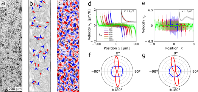
Motivated by studies of sarcoma cancer invasion in vascular networks Weigelin2012 ; Weigelin2016 , here we investigate collective cell migration in cultures of human fibrosarcoma cells (HT1080) confined in adhesive stripes, whose width varies in the range to m, surrounded by repellent coating Duclos2018a (see Movie 1 in Supplementary Note). Although initially sparse upon the plating, cells proliferate and eventually form a confluent monolayer without visible gaps, a process which typically takes on the order of 24 h (Fig. 1a). The spatial organization of the cellular layer is revealed by its orientational anisotropy (Fig. 1b), characterized in terms of the two-dimensional nematic tensor . Where the vector is the nematic director and is the order parameter, describing the average orientation and degree of alignment, respectively. was measured from phase-contrast images with a structure tensor method (see Methods). We find a non-vanishing order parameter (mean standard deviation for stripes), which remains constant over the measurement time, indicating that nematic order is prominent in confined fibrosarcoma cells (see Fig. S1 in Supplementary Note). This value is comparable to human bronchial epithelial cells (HBEC) under similar conditions and larger than Madin-Darby Canine Kidney (MDCK) cell layers Blanch-Mercader2018m . By contrast, the nematic director exhibits significant variations across the channel, resulting into a dense distribution of (marked in red) and (marked in blue) topological defects.
Fig. 1c shows a particle image velocimetry (PIV) reconstruction of the collective cellular flow. Unlike epithelial cell types, such as MDCK or HBEC Blanch-Mercader2018m , which relax toward a jammed state after reaching confluence, in fibrosarcoma layers the root mean square speed remains constant for over h, with m/h (mean standard deviation for , see Fig. S2 in Supplementary Note). The flow is truly chaotic in the bulk, with regions of positive (red) and negative (blue) vorticity tiling the channel in an irregular and yet homogeneous fashion. Remarkably, however, an emergent structure appears at the boundary when the flow fields are averaged over the length of the channel and in time (Fig. 1d). This consists of a net tangential flow localized within a boundary layer of approximate size m, independent on the channel width (Fig. 1d inset), and oppositely directed at the two lateral edges of the channel. Despite the channel being perfectly left-right symmetric, such an edge current is predominantly clockwise (see Fig. S3 in Supplementary Note), thus indicating a break down of chiral symmetry observable at the length scale of the entire system.
To shed light on the emergence of these chiral edge currents, we have complemented our in vitro experiments with a computational model based on the hydrodynamic theory of active nematic liquid crystals. Similar approaches have been successfully used to account for the emergent behavior of a vast class of living systems, such as bacteria suspensions, Dunkel2013 ; Beer2020 ; Copenhagen2020 ; You2018 and epithelial cell monolayers Lee2011 ; Cochet-Escartin2014b ; Banerjee2015 ; Blanch-Mercader2017 ; Tlili2018 ; Recho2020 . A central concept in active nemato-hydrodynamics is the so called active stress. This results from microscopic forces generated by the active sub-units along their longitudinal direction. This force can be written , where is a vector describing the orientation of individual sub-units. At the mesoscopic scale, and in the absence of microscopic chirality, these give rise to an active stress of the form . The phenomenological constant embodies the biomechanical activity of individual sub-units and is given by , with the cell length and the number density Pedley1992 ; Simha2002 . In the presence of chirality at the cellular scale, resulting for instance from a repositioning of the internal organelles, microscopic forces are augmented with a transverse component: i.e. , with . This gives rise to an active stress of the form:
| (1) |
where and is the antisymmetric Levi-Civita tensor Hoffmann2020 . With Eq. (1) in hand, we have numerically integrated the hydrodynamic equations of a chiral active nematic in a two-dimensional rectangular domain, subject to periodic boundary conditions at and stress-free boundary conditions (see Methods for details). Fig. 1e shows the average longitudinal component of the velocity field, , as a function of the distance from the channel center-line. Consistent with our experimental observations, the flow is chaotic in the bulk with vanishing time-averaged velocity, but characterized by chiral edge currents, penetrating inside the bulk by an amount independent on the channel width (Fig. 1e inset).
Analogously, in both our in vitro and in silico cell layers, the nematic director is randomly oriented in the bulk of the channel and parallel to the lateral edges at the boundary, with a slight tilt in the direction of the flow (Figs. 1f,g).
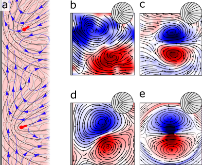
In the following we demonstrate that the edge currents observed in our experiments and simulations have a topological origin, which could be ascribed to the concerted action of disclinations, orthogonally anchored to the channel walls, and the hydrodynamic flow sourcing from the chiral active stresses. In nematic cell monolayers, disclinations are point-like singularities where the orientation of the cells is undefined and around which the nematic director rotates by , with the winding number. Unlike passive nematics, where topological defects annihilate under the effect of their elastic interactions, two-dimensional active nematics can feature a steady density of disclinations resulting from the instability of the nematic director under the flow sourced by the active stress Giomi2015 . Although originating from the active flow, disclinations leave a distinct signature on the flow itself, whose local structure is almost entirely determined by the configuration of the nematic director around the defect core, via the so called backflow mechanism Giomi2013 ; Giomi2014 . In particular, disclinations in non-chiral active nematics drive a Stokeslet-like local flow consisting of two vortices mirror-symmetrically counter-rotating about the defect longitudinal direction vromans2015 (Fig. 2). This characteristic flow pattern causes a local flow at the centre of the defect parallel with its orientation causing defects to self propel.
Chirality affects the flow around a defect by tilting its mirror-symmetry axis with respect to by an angle , with and the active stresses appearing in Eq. (1), Fig. 2b Hoffmann2020 . The latter property can be understood by noticing that the active part of the stress tensor can be expressed as , where represents a counterclockwise rotation by an angle and denotes transposition.
When we analysed the average flows in the vicinity of defects in HT1080 monolayers in boundary-free cell cultures the average flow field revealed a vortex-pair flow pattern, Fig. 2c (see Methods for details). This corresponds to an extensile active stress, , with an average speed of at the defect core (mean standard deviation for defects). This average flow at the core of the defect is tilted relative to the orientation of the defect by (mean standard deviation for) indicating a chiral stress of the order of . When the average flow field was computed for an equal-sized set of random points in boundary-free cell cultures, the vortex-pair pattern was lost, Fig. S5 in Supplementary Note, and the average speed decreased to (mean standard deviation for defects).
When the director field orients tangentially at the boundaries of a channel, elastic interactions tend to align boundary-adjacent disclinations towards the boundaries, namely with the outer unit normal vector, Fig. 2a. This results in an enrichment of defects along the edges and whose mean orientation is parallel to the outward point normal vector . Since each defect generates a vortex pair, this originates vortex street of alternating clockwise and counterclockwise vortices accumulated along the edges Fig. 2a. Now, although representing a spectacular example of self-organization in active matter, in the absence of chirality this emergent defect structure would not give rise to an edge current due to the mirror-symmetry of the local flow generated by defects. The chiral stresses, embodied in the constant in Eq. (1), explicitly breaks this mirror symmetry by titling individual vortex pairs with respect to the defect orientation . This tilt could push the right (or left for ) handed vortex of each pair closer to the boundary, leading to a boundary layer with net vorticity, thus a shear flow. Moreover, as the nematic director can be rotated by a shear flow, the latter feeds back on the cell orientation by inducing a slight but visible rotation of the defect orientation toward the flow, thus further reinforcing the chiral edge current. This is confirmed by analysing the flow around defects located close to the boundary. In both simulations and experiments, the average flow pattern around a defect close to the boundary exhibited a vortex pair that was tilted with respect to boundary normal, Fig. 2d,e.
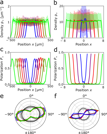
In order to further test the proposed mechanism, we compared space-dependent defect properties for stripes of varying width in both simulations and experiments, Fig. 3. To this end, we quantify the local positive defect density as , where is the number of defects in a thin stripe of width centred at position and running the length of the channel, . This is given by , with the summation extended over all defects in the system at positions . Similarly, the local positive defect polarization can be quantified . The system features a steady and spatially uniform distribution of the disclinations away from the boundaries, Fig. 3a,b. In the interior of the channel, these have no preferential orientation, as marked by the fact that the average amplitude of the polarization vanishes, Fig. 3c,d. By contrast, has a maximum close to the boundaries, indicating the existence of a persistent layer of co-aligned defects, Fig. 3c,d. Figs. 3e,f show the polar histograms of the defect direction in the bulk and near the boundaries of the channel. Whereas there is no alignment in the bulk, at the boundaries we found a predominant orthogonal alignment independent of the channel width with a average chiral tilt of for HT1080 cell monolayers. This confirms the aforementioned orthogonaly anchored boundary layer of defects with a chiral tilt that is independent of the channel geometry.
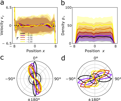
Using numerical simulations we can explore the effect of the chiral active stress on the performance of the edge current. As shown in Fig. 4, both the speed of the edge current (Fig. 4a) and the density of defects (Fig. 4b) increase with . The latter, in particular, implies a reduction of the inter-defect spacing, which, in turn, causes the chiral boundary layer to become narrower and sharper. This results in a higher local shear rate, which further accentuates the rotation of the nematic director (Fig. 4c), hence the tilt of the defect layer (Fig. 4d).
In conclusion, when confined on adhesive stripes, HT1080 monolayers organise into a novel collective state, characterised by the existence of chiral edge currents. Far from the boundaries, the cells exhibit disordered chaotic flows, with no net motion. The cellular flow is, however, rectified along the edges, leading to long-ranged collective cell migration, with broken chiral symmetry. Modeling the system as a chiral active nematic liquid crystal, we demonstrated that, consistently with experimental observations on stripes of various width, the edge currents are fuelled by layers of topological defects, orthogonally anchored at the channel walls and acting as local sources of chiral active stress. Overall, our findings strengthens the idea that biological organization could take advantage of topological mechanisms Ladoux2017 ; Kawaguchi2017 , by demonstrating that topological defects in cell monolayers could act as organising centers for collective migration in the presence of chiral active stresses.
Acknowledgements.
It is a pleasure to thank the members of the Biology-Inspired Physics at Mesoscales (BiPMS) group. This work is partially supported by the Netherlands Organisation for Scientific Research (NWO/OCW), as part of the Vidi scheme and the Frontiers of Nanoscience program and by the ERC-CoG grant HexaTissue (L.G.).I Methods
Cell culture. HT1080 cells (gift from Dr Philippe Chavrier, Institut curie) were cultured in Dulbecco’s modified Eagle’s medium (High glucose + GlutaMAX, Gibco) supplemented with FBS (Sigma) and antibiotics solution [penicillin ( units/mL) + streptomycin ( mg/mL); Gibco] at , , and humidity.
Time-lapse microscopy. Time-lapse multi-field experiments were performed in phase contrast on an automated inverted microscope (Olympus IX71) equipped with thermal and regulations. Typical field of view (FOV) was mm mm. The position of the measured sample and the acquisitions with a CCD camera (Retiga 4000R, QImaging) were controlled by Metamorph (Universal Imaging) software. The typical delay between two successive images of the same field was set to 15 minutes.
Image Processing. Stripe were cropped from the raw images using the ImageJ public domain software (imageJ, ). The orientation field was obtained by computing the local structure tensor with ImageJ plugin OrientationJ (OrientationJ, ) within widows of m m. S corresponds to the anisotropy level and the nematic director was computed from the output angle. Visualisation of the orientation field was performed with Line Integral Convolution (LIC) under Matlab (MATLAB:2018, ). The velocity field in the monolayer was mapped by particle image velocimetry (PIV) analysis. Stacks of images were analysed with a custom made PIV algorithm based on the MatPIV MatPIV ; Petitjean2010 software package. The window size was set to pixels = m with a overlap for m and pixels = m with a overlap for m and m.
Micro-patterning technique. Clean glass substrates were first uniformly coated with a cell-repellent layer (interpenetrated gel of acrylamide and polyethylene glycol). A photoresist mask was then structured directly on top of the layer by classical photolithography methods and air plasma was used to locally etch the protein-repellent coating through this mask. The photoresist was then removed with acetone yielding a cell repellant substrate where bare glass domains on which cells can adhere have been defined (deforet2014, ; Duclos2018a, ).
Statistical analysis. Statistical analysis was performed with Matlab. Experiments were performed in at least 4 replicas, each using 6 well plates with stripes of three distinct widths range (for m and m) or distinct widths (for m, m, m) and plain glass slides. The number of analyzed FOVs for each width is reported in the list below. Error bars represent the SDs over all the FOVs analysed (pooling all experiments for specific stripe width ). Monolayers reach confluency at least 12h after cells were seeded. The orientation and velocity were averaged over a hour period starting after confluence. List of the stripes binned by widths analysed in the present study and the corresponding number N of FOVs used for the orientation field and PIV analysis cells: (L(m), N) = (300, 95); (400, 40); (500, 68); (600,47); (700, 38); (800, 43); (1000, 29); (boundary-free, 24).
Average fields around defects. In the following, we explain the calculation of the averaged velocity and averaged director fields over defect populations. and defect positions were detected by searching for the local minima of an order parameter in the window of m m in the orientation map obtained by OrientationJ plugin. defects and their direction was determined using the procedure explained in Ref. vromans2015 . For each detected defect, the velocity and director maps were aligned with respect to the corresponding defect direction and then cropped over a window of size m m centered at the defect core. Finally, we computed the ensemble averages over different sets of defects to obtain the averaged fields. In the case of the averaged fields for random positions that are shown in Fig. S5 in Supplementary Note, the above-mentioned procedure was the same except that the defect positions were replaced by random positions within the cell monolayer.
Numerical simulations. We numerically solve the hydrodynamic equations for the nematic tensor and the flow velocity . These are given by:
| (2a) | |||
| (2b) | |||
where and are, respectively, the strain rate and vorticity tensor, is the flow alignment parameter, dictating how the nematic director rotates in a shear flow, and is a rotational viscosity. is the hydrostatic pressure and is the identity matrix. The quantity , with
| (3) |
the Landau-de Gennes free energy of the system, is the molecular tensor driving the relaxation of the nematic tensor toward the lowest free energy configuration. is a constant with dimensions of energy, whereas is a length scale setting the typical core radius of the topological defects. In Eq. (2b), is the elastic stress resulting from a flow-driven distortion of the nematic director, whereas is the chiral active stress given in Eq. (1). Eqns. (2) are numerically integrated using a finite difference scheme on a rectangular grid of size , subject to periodic boundary conditions at and Navier boundary conditions, i.e. , with the boundary normal and a drag coefficient, at . The aspect ratio of the channels was simulated at and the resolution of the grid was . To render Eqs. (2) dimensionless we rescale length by core radius , time by the nematic relaxational time scale and stress by elastic stress . All quantities plotted in Figs. 1, 3 and 4 are rescaled accordingly. In this units, we set , , , , , , and .
References
- (1) Cheung KJ, Ewald AJ (2016) A collective route to metastasis: Seeding by tumor cell clusters. Science 352: 167–169.
- (2) Paluch EK, Aspalter IM, Sixt M (2016) Focal adhesion–independent cell migration. Annual Review of Cell and Developmental Biology 32: 469-490.
- (3) Marchetti MC, Joanny JF, Ramaswamy S, Liverpool TB, Prost J, et al. (2013) Hydrodynamics of soft active matter. Reviews of Modern Physics 85: 1143–1189.
- (4) Doostmohammadi A, Ignés-Mullol J, Yeomans JM, Sagués F (2018) Nat. commun. 9: 3246.
- (5) Duclos G, Blanch-Mercader C, Yashunsky V, Salbreux G, Joanny JF, et al. (2018) Spontaneous shear flow in confined cellular nematics. Nature Physics 14: 728–732.
- (6) Hoffmann LA, Schakenraad K, Merks RM, Giomi L (2020) Chiral stresses in nematic cell monolayers. Soft Matter 16: 764–774.
- (7) Friedl P, Gilmour D (2009). Collective cell migration in morphogenesis, regeneration and cancer. doi:10.1038/nrm2720.
- (8) Ladoux B, Mège RM (2017) Mechanobiology of collective cell behaviours. Nature Reviews Molecular Cell Biology 18: 743–757.
- (9) Kawaguchi K, Kageyama R, Sano M (2017) Topological defects control collective dynamics in neural progenitor cell cultures. Nature 545: 327–331.
- (10) Hakim V, Silberzan P (2017). Collective cell migration: A physics perspective. doi:10.1088/1361-6633/aa65ef.
- (11) Trepat X, Sahai E (2018). Mesoscale physical principles of collective cell organization. doi:10.1038/s41567-018-0194-9.
- (12) Angelini TE, Hannezo E, Trepatc X, Marquez M, Fredberg JJ, et al. (2011) Glass-like dynamics of collective cell migration. Proceedings of the National Academy of Sciences of the United States of America 108: 4714–4719.
- (13) Park JA, Kim JH, Bi D, Mitchel JA, Qazvini NT, et al. (2015) Unjamming and cell shape in the asthmatic airway epithelium. Nature Materials 14: 1040–1048.
- (14) Garcia S, Hannezo E, Elgeti J, Joanny JF, Silberzan P, et al. (2015) Physics of active jamming during collective cellular motion in a monolayer. Proceedings of the National Academy of Sciences of the United States of America 112: 15314–15319.
- (15) Atia L, Bi D, Sharma Y, Mitchel JA, Gweon B, et al. (2018) Geometric constraints during epithelial jamming. Nature Physics 14: 613–620.
- (16) Blanch-Mercader C, Yashunsky V, Garcia S, Duclos G, Giomi L, et al. (2018) Turbulent Dynamics of Epithelial Cell Cultures. Physical Review Letters 120: 208101.
- (17) Palamidessi A, Malinverno C, Frittoli E, Corallino S, Barbieri E, et al. (2019) Unjamming overcomes kinetic and proliferation arrest in terminally differentiated cells and promotes collective motility of carcinoma. Nature Materials 18: 1252–1263.
- (18) Weigelin B, Bakker GJ, Friedl P (2012) Intravital third harmonic generation microscopy of collective melanoma cell invasion. IntraVital 1: 32–43.
- (19) Weigelin B, Bakker GJ, Friedl P (2016) Third harmonic generation microscopy of cells and tissue organization. Journal of Cell Science 129: 245–255.
- (20) Duclos G, Deforet M, Yevick HG, Cochet-Escartin O, Ascione F, et al. (2018) Controlling confinement and topology to study collective cell behaviors. In: Methods in Molecular Biology, Humana Press Inc., volume 1749. pp. 387–399. doi:10.1007/978-1-4939-7701-7˙28.
- (21) Dunkel J, Heidenreich S, Drescher K, Wensink HH, Bär M, et al. (2013) Fluid dynamics of bacterial turbulence. Physical Review Letters 110: 228102.
- (22) Be’er A, Ilkanaiv B, Gross R, Kearns DB, Heidenreich S, et al. (2020) A phase diagram for bacterial swarming. Communications Physics 3: 1–8.
- (23) Copenhagen K, Alert R, Wingreen NS, Shaevitz JW (2020) Topological defects induce layer formation in Myxococcus xanthus colonies. arXiv preprint arXiv:200103804 .
- (24) You Z, Pearce DJ, Sengupta A, Giomi L (2018) Geometry and mechanics of microdomains in growing bacterial colonies. Physical Review X 8: 031065.
- (25) Lee P, Wolgemuth CW (2011) Crawling cells can close wounds without purse strings or signaling. PLoS Computational Biology 7: e1002007.
- (26) Cochet-Escartin O, Ranft J, Silberzan P, Marcq P (2014) Border forces and friction control epithelial closure dynamics. Biophysical Journal 106: 65–73.
- (27) Banerjee S, Utuje KJ, Marchetti MC (2015) Propagating Stress Waves during Epithelial Expansion. Physical Review Letters 114: 228101.
- (28) Blanch-Mercader C, Vincent R, Bazellières E, Serra-Picamal X, Trepat X, et al. (2017) Effective viscosity and dynamics of spreading epithelia: a solvable model. Soft Matter 13: 1235–1243.
- (29) Tlili S, Gauquelin E, Li B, Cardoso O, Ladoux B, et al. (2018) Collective cell migration without proliferation: Density determines cell velocity and wave velocity. Royal Society Open Science 5: 172421.
- (30) Recho P, Fouchard J, Wyatt T, Charras G, Kabla A (2020) A tug-of-war between stretching and bending in living cell sheets. arXiv preprint arXiv:200203966 .
- (31) Pedley TJ, Kessler JO (1992) Hydrodynamic phenomena in suspensions of swimming microorganisms. Annual Review of Fluid Mechanics 24: 313-358.
- (32) Aditi Simha R, Ramaswamy S (2002) Hydrodynamic fluctuations and instabilities in ordered suspensions of self-propelled particles. Phys Rev Lett 89: 058101.
- (33) Giomi L (2015) Geometry and topology of Turbulence in active nematics. Physical Review X 5: 031003.
- (34) Giomi L, Bowick MJ, Ma X, Marchetti MC (2013) Defect annihilation and proliferation in active Nematics. Physical Review Letters 110.
- (35) Giomi L, Bowick MJ, Mishra P, Sknepnek R, Marchetti MC (2014) Defect dynamics in active nematics. Philosophical Transactions of the Royal Society A: Mathematical, Physical and Engineering Sciences 372.
- (36) Vromans AJ, Giomi L (2016) Orientational properties of nematic disclinations. Soft matter 12: 6490–6495.
- (37) Ferreira T, Rasband W (2012) Imagej user guide—ij 1.46. Available at: imagej nih gov/ij/docs/guide .
- (38) Rezakhaniha R, Agianniotis A, Schrauwen JTC, Griffa A, Sage D, et al. (2012) Experimental investigation of collagen waviness and orientation in the arterial adventitia using confocal laser scanning microscopy. Biomechanics and modeling in mechanobiology 11: 461–473.
- (39) MATLAB (2018) (R2019b). Natick, Massachusetts: The MathWorks Inc.
- (40) Sveen JK (2004) An introduction to matpiv v. 1.6.1. University of Oslo, Department of Mathematics .
- (41) Petitjean L, Reffay M, Grasland-Mongrain E, Poujade M, Ladoux B, et al. (2010) Velocity fields in a collectively migrating epithelium. Biophysical journal 98: 1790–800.
- (42) Deforet M, Hakim V, Yevick H, Duclos G, Silberzan P (2014) Emergence of collective modes and tri-dimensional structures from epithelial confinement. Nature communications 5: 1–9.