Enhanced polarization transfer to the characteristic x-ray lines near the nonlinear Cooper minimum of two-photon ionization
Abstract
It has been shown that for nonlinear atomic ionization, transfer of the degree of circular polarization from incident to fluorescence light is maximum at the Cooper minimum (see Phys. Rev. A 100, 011401(R) (2019)). Until the present, however, it is still a challenge to produce and detect circularly-polarized light at xuv and x-ray photon energies. We here show, that transfer of linear polarization is strongly enhanced at the Cooper minimum in two-photon ionization and can be readily detected using current free-electron laser facilities by measurements of the degree of linear polarization of the characteristic and lines. Two-photon ionization of electron of tungsten atom is proposed to demonstrate the presented phenomena and realization of such experiment is discussed in detail.
I Introduction
First experimental observations of two-photon absorption mechanism involving core-level atomic states have become possible only rather recently by virtue of the development of x-ray Free Electron Lasers (XFELs) Emma et al. (2010); Amann et al. (2012a). Since the cross-sections of two-photon absorption and ionization process are generally low, it is often necessary to employ high brilliance incident x-ray beams with properties such as coherence, duration of the pulse and intensity, which are otherwise known only from optical lasers. With the advent of XFEL facilities, x-ray beams with femtosecond pulse duration and photon flux of photons/(s cm become achievable. The application of such high-power x-ray beams enabled one to approve nonlinear processes with quite low excitation probabilities, including multiple ionization Young et al. (2010); Vinko et al. (2012), double-core hole creation Tamasaku et al. (2013) and nonsequential two-photon ionization Richardson et al. (2010); Doumy et al. (2011). More recently, experimental observations of two-photon ionization of -shell electrons at hard x-ray energies were reported Tamasaku et al. (2014); Ghimire et al. (2016); Szlachetko et al. (2016); Tamasaku et al. (2018); Kayser et al. (2019).
Core-hole excitation and ionization processes can be effectively investigated by observing the subsequent fluorescence photons. In particular, the photon polarization carries information about the alignment and orientation of an atom, which, in turn, strongly depends on details of the core electron ionization or excitation Fano and Macek (1973); Klar (1980); Starace (1982); Huang (1982). The polarization properties of fluorescence light following single photon ionization is a well developed part of atomic spectroscopy Schmidt (1992); Kabachnik et al. (2007); Sharma et al. (2010); Sukhorukov et al. (2019) in contrast to the nonlinear ionization, which is much less explored. In recent experiments, the total two-photon inner-shell ionization cross sections have been extracted from the subsequent fluorescence yields Tamasaku et al. (2014); Ghimire et al. (2016); Szlachetko et al. (2016); Tamasaku et al. (2018); Kayser et al. (2019). However, these measurements provide only the total two-photon ionization cross section, which generally suffers from low precision. Performing similar experiments together with detection of the polarization of the fluorescence photons would allow to investigate the magnetic population dynamics in the produced ion and reach higher precision of the extracted ionization pathways (channels) contributions.
In one-photon ionization of atoms, the energy at which the dominant ionization channel vanishes is called the Cooper minimum Cooper (1964). It has been shown both theoretically and experimentally, that performing measurements at the Cooper minimum can reveal otherwise hidden contributions to the ionization process, such as the contributions of higher multipole orders of the electromagnetic field Dehmer and Dill (1976); Johnson and Cheng (1978); White et al. (1979); Grum-Grzhimailo (2001); Grum-Grzhimailo and Gryzlova (2014); Ilchen et al. (2018). Cooper minimum in multi-photon ionization can be also noticed in earlier calculations, both in total cross sections Lambropoulos and Tang (1987); Saenz and Lambropoulos (1999); Nikolopoulos (2006) as well as in photoelectron angular distributions Lagutin et al. (2017); Petrov et al. (2019), however, no attention was paid to investigate it in detail. We recently demonstrated, that such Cooper minimum in multi-photon ionization processes can strongly influence the photoelectron angular distributions Hofbrucker et al. (2018), or the magnetic sublevel populations of photoions Hofbrucker et al. (2019, 2020). In Hofbrucker et al. (2019), it has also been shown, that in the case of two-photon ionization of atomic electrons by circularly polarized light, the polarization transfer from the ionizing light to the atom is maximized at this nonlinear Cooper minimum. However, although it is possible to generate intense circularly polarized beams at some free-electron laser facilities Allaria et al. (2013); Lutman et al. (2016), the production of such laser beams are usually more limited in photon energy than linearly polarized light. Moreover, detection of polarization of XUV or x-ray is experimentally very challenging. On the other hand, linearly polarized light can be produced at practically all free-electron facilities, and moreover, detection of the degree of linear polarization in x-ray domain can be carried out with high accuracy Marx et al. (2013).
In this paper, we study the transfer of linear polarization in the two-photon ionization of atomic electrons. In the next section, we describe our theoretical approach based on the second-order perturbation and the density matrix theories as well as independent-particle approximation. In Sec. III, we present our calculations which were chosen to fulfill current experimental possibilities. On the example, we demonstrate the dynamic behavior of the polarization transfer in two-photon ionization of electron of tungsten as a function of incident photon energy. Moreover, we show that significant variation of the polarization transfer occurs at the nonlinear Cooper minimum, whilst the corresponding cross sections are comparable to the already performed experiments Tamasaku et al. (2014); Ghimire et al. (2016); Szlachetko et al. (2016); Tamasaku et al. (2018). A discussion of the possible experimental possibilities to perform the suggested experiment are provided in Sec. IV. In the last section, we conclude the main findings of this paper.
II Theory
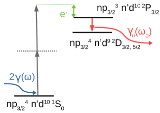
We consider a two-step process. In the first step, an inner-shell ionization of an atom in an initial state by two identical, linearly polarized photons with energy results in emission of a photoelectron with well-defined momentum and spin projection , and production of an ion in a state , which has typically a hole in one of the core-shells. Here, the many-electron wave functions of the atom are classified by the total angular momentum , its projection as well as all further quantum numbers that are needed to specify the state uniquely. In the second step, the excited ion decays into a lower energy state with spontaneous emission of a fluorescence photon . This two-step process can be schematically represented as (see also Fig. 1)
In this paper, we will only consider the two-photon ionization of an electron from the (filled) shell of an atom with total angular momentum , which leads to a photoion with the total angular momentum . In the independent-particle model and by using second-order perturbation theory, the (two-photon) ionization can then be described by single-electron amplitudes Hofbrucker et al. (2016, 2017), including the summation over the complete (single-) electron spectrum of the virtual intermediate states, with symmetries determined by the orbital and total angular momenta and . The final photoelectron wave function is expanded into partial waves, each with the angular momenta and . This expansion allows us to characterize the process in terms of angular momentum ionization channels. In the electric dipole approximation, the ionization of the electron proceeds through intermediate states with and symmetries, into final photoelectron and states. There are eight possible relativistic ionization channels, each described by an amplitude . Once, the photoelectron has left the ion, all further (decay) properties of the photoion can be described by its density matrix . In practice, however, it is often more convenient to characterize the ion polarization in terms of (so-called) statistical tensors that transform like spherical tensor of rank . These tensors are given by
| (2) |
For the two-photon ionization of a electron from a closed shell atom, the statistical tensor takes the form (see Hofbrucker et al. (2019, 2020) for details)
| (3) | |||||
where the exact form of the transition amplitudes in terms of is given in the Appendix appendix. The calculations presented in this work have been also carried out with contributions of higher multipole orders. However, the contributions of higher multipole orders were found negligible, and hence, electric dipole approximation is well justified. In contrast, the results presented in Ref. Hofbrucker et al. (2020) concentrate on a special case of two-photon ionization of electrons by circularly polarized light near the nonlinear Cooper minimum. In this particular scenario, all electric dipole channels drop to zero, and hence, reveal the contributions of higher multipole orders. The total two-photon ionization cross section can be easily obtained from the zero rank statistical tensor as . Moreover, statistical tensors are often utilized also to characterize the magnetic state of an ion. For example, an atom is said to be unpolarized if it is described by just a single nonzero (statistical) scalar , while the atom or ion is called polarized if at least one nontrivial () statistical tensor occurs. Moreover, if only tensors or even rank occurs in the representation of the ion, it is called aligned. Finally, if at least one odd rank statistical tensor is nonzero, the system is called oriented. It is well-known, that for ionization of atom by linearly polarized light, the excited atom can be aligned along the direction of the photon momentum, but cannot be oriented. As seen from the expressions (II), the two-photon ionization by linearly polarized light leads to photoions that are described by even rank tensors only. On the contrary, two-photon ionization by circularly polarized photons produces aligned as well as oriented ion states, which can even reach pure orientation Hofbrucker et al. (2019). To quantify the ion polarization, it is convenient to introduce the normalized statistical tensor defined as
| (4) |
The component is called the alignment parameter (or simply alignment) and expresses the difference in population between electrons with projections and . This alignment parameter depends of course on the prior excitation or ionization process, and is fully described by the transition amplitudes for the two-photon ionization of an initially closed-shell atom.
The alignment of an excited ion propagates also to the characteristics of subsequently emitted photons. We here consider the emission of the characteristic and lines, which arise from the and transitions, respectively. From the statistical tensors, we can easily obtain the general expression for the degree of linear polarization
where specifies the fluorescence photon emission direction with respect to the incident beam propagation direction and where we used the fact that . We can further use the constant factor relation between and from Eqs. (II), which remains valid only for the case of two-photon ionization of electrons by linearly polarized light. The relation of the two statistical tensors together with setting allows us to obtain corresponding simplified expression for the degree of linear polarization of the lines
| (6) |
This simple expression reveals that is generally larger than , and hence, experimental detection of the characteristic will yield a stronger signal. The above expressions also confirm the physical intuition that the degree of linear polarization of the emitted photon increases with the degree of ion alignment. For the process under consideration, . The maximum alignment is reached when the contributions of the amplitude strongly dominates over others. In the case of maximum value of the alignment, the polarization of fluorescence reaches at .
More importantly, Eq. (II) shows that the polarization of the lines is determined by a single variable . Since the alignment expresses the ratio of probabilities of creation of and holes, any variation in the degree of polarization of the lines directly expresses the ratio of populations of the corresponding substates.
III Results and Discussion
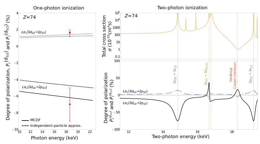
Recently, Kämpfer et al. Kämpfer et al. (2016) used unpolarized photons produced in an x-ray tube to ionize the electron of neutral tungsten atom. The inner-shell hole was subsequently filled by an electron from shell, and the characteristic and fluorescence photons were emitted. In their work, degree of linear polarization of these photons emitted perpendicular to the incident photon propagation was measured and reported together with theoretical calculation employing the multi-configuration Dirac-Fock method Jönsson et al. (2013); Fritzsche (2019). Left side of Fig. 2 presents the values from Kämpfer et al. (2016) together with our calculations performed within independent-particle approximation. The good agreement of our prediction with experimental results as well as many-electron calculations presented in Kämpfer et al. (2016) hints that our methodology may be further used to study x-ray fluorescence polarization phenomena in the regime of nonlinear interactions.
A similar experiment as in Ref. Kämpfer et al. (2016) is suggested here but for a two-photon ionization of a electron of tungsten. For such a nonlinear inner-shell ionization, x-ray beams of sufficient high intensity are needed as produced, for instance, by XFEL. Since most free-electron laser beams are linearly polarized, we will consider two-photon ionization of electron of neutral tungsten atom by linearly polarized photons. Generally, polarization transfer is maximized along the incident beam propagation, for this reason, we consider the degree of linear polarization of the fluorescence photons to be measured at . In an experiment, it is impractical to perform the detection on the beam axis. For such cases, the degree of the photon polarization at an appropriate detection angle can be calculated using Eq. (II).
The right side of Fig. 2 shows the degree of linear polarization (in percentage) of the fluorescence photons following two-photon ionization of a electron of tungsten by linearly polarized light (bottom), together with the corresponding two-photon ionization cross section (top) to guide the eye. The horizontal axis shows the combined energy of the two photons, i.e. the energy transferred to the atomic system. Already from the first glance, it is apparent that ionizing the neutral tungsten atom in nonlinear instead of linear regime results in high sensitivity of the fluorescence photon (or ion) polarization degree on the incident photon energy. Even more intriguing are the physical origins of each of the local maxima and minima in the signal. At low photon energies, in the nonresonant region, the degree of linear polarization is comparable to the case of one-photon ionization, however at higher energies, this value changes dramatically due to intermediate level resonances and the nonlinear Cooper minimum. The first and last local maxima (and minima) arise from the incident photon energy matching an intermediate resonance (7.43 keV and 9.6 keV, respectively), they have been marked by solid gray lines. The two maxima marked with green dashed vertical lines originate from resonant transitions to the two fine-structure electron levels (8.3 keV, 8.4 keV). In tungsten atom, the and orbitals are of course occupied and hence, the electron cannot be promoted into them. However, the resonance behavior arises from two-photon absorption, in which first photon ionizes the or electron and the second is excited the electron into the corresponding hole, as experimentally verified in singly charged neon Kanter et al. (2011). As the lifetime of the core-hole states are in the order of tens or hundreds of attoseconds, the ionization in this case proceeds non-sequentially only. For this reason, the lifetime of the atom in a state with and holes has been neglected. The last maximum, marked with red dashed line, originates from the significantly reduced contributions of the ionization channels, which corresponds to the nonlinear Cooper minimum Hofbrucker et al. (2019).
The increased polarization transfer for incident photon energies matching an intermediate resonance and the nonlinear Cooper minimum have opposite signs, although they both arise due to relative increase of the channel contributions. This is the case because the individual fine-structure channels with intermediate states pass through their corresponding nonlinear Cooper minima at slightly different energies. As a consequence, the marked nonlinear Cooper minimum corresponds only to zero contributions of the channels, while the magnitude of the contributions of channels is comparable to . However, since the channels with higher angular momentum generally dominate Fano (1985), the polarization at the nonlinear Cooper minimum is still dominantly determined by the channel, and hence posses the same sign as the polarization signal at the corresponding resonance. One can also notice, that the resonances in the degree of polarization of the fluorescence photons are slightly shifted with respect to the corresponding resonances in the total cross sections. The shift of resonances of the total cross section with respect to other observables appears already for autoionization resonances in one-photon ionization, where the shift properties have been discussed in detail e.g. in Ref. Grum-Grzhimailo et al. (2005).
The comparison of the left and right plots of Fig. 2 demonstrates just how entirely new behavior of the light-matter interaction can be achieved with nonlinear processes. While the left-hand side figure contains only information about the initial and final electron states, two-photon ionization allows us to probe the electron complete structure of atoms, while utilize already available methods previously used in one-photon ionization case to obtain this information. Moreover, measurements of the fluorescence polarization can be carried out in order to determine the position of the nonlinear Cooper minimum, which would allow us to put the theoretical representation of the electron spectra (including positive and negative continua) to a test.
IV Experimental consideration
Two-photon ionization experiments are typically performed by irradiating a sample with tightly focused XFEL (down to sub size) beams and with the incidence x-ray energy being set below single-photon ionization threshold. Signature of the two-photon ionization mechanism is then confirmed by detection of subsequent x-ray emission signal resulting from relaxation of the produced ion Tamasaku et al. (2014); Kayser et al. (2019). Dedicated experimental procedures are implemented to avoid higher harmonic components of the incident beam (monochromators) as well as energy jitter associated with x-ray generation at XFELs through the self-amplified spontaneous emission process Vinko et al. (2012); Szlachetko et al. (2016). We should note, that the nonlinear interactions of x-rays with a sample are not triggered selectively when using ultra-intense and ultra-short x-ray beams, and the linear contributions often surpass the nonlinear processes. As a consequence, the nonlinear x-ray signals are often accompanied with linear interaction contributions, which requires application of dedicated high energy resolution setups Kayser et al. (2019); Szlachetko et al. (2016) to resolve the linear and nonlinear contributions to the measured signal.
To address the influence of linear ionization on the predicted results explicitly, let us shortly discuss it together with other effects such as the pulse length and time structure. As the cross-sections for two-photon ionization mechanisms are very low, the linear interaction of x rays with matter will surpass the nonlinear interactions. At the considered x-ray energies of 6 keV - 9 keV, the dominant interaction will consist of single photoionization of -shell electrons with corresponding binding energies spanning from 1.809 keV for subshell to the energy of 2.820 keV for the subshell and photoionization cross-sections in the order of cm2 Linstrom and Mallard . The core-holes created by single-photon ionization will decay mostly via Auger process and will thus increase charge state by one for every single Auger decay. The Auger cascades continue until the initial hole is transferred to the upmost subshell. It is safe to assume that single core-hole ionization may lead to the increase of atomic charge up to few hole states Rudek et al. (2012, 2018); Kayser et al. (2019). This high charge state will, however, be very quickly depopulated by electron transfer from surrounding atoms Erk et al. (2013); Rudenko et al. (2017); Yoneda et al. (2014). We calculated the two-photon ionization cross-sections assuming depletion of and shells of tungsten atoms and found that the increase in charge state does not influence significantly the two-photon ionization cross-section values, nor the positions of resonances. We should note here, the expected change in binding energies of inner shell electrons as well as the position shift of the Cooper minimum (around eV) will be within energy bandwidth of the XFEL beam, and therefore will not affect the estimated two-photon ionization rates. Since the energy of the inner-shell levels does not shift significantly, the subsequent x-ray emission will also not be strongly influenced by outer-shell vacancies. This effect may be understood on the basis of strong and uniform screening of the and states by the remaining electron charge. The energy stability of inner-shell electrons was experimentally observed for x-ray emission for different oxidation states Wach et al. (2020); Tiraşoğlu et al. (2003). It is reported that even -satellites lines are reported to be within the linewidth of the x-ray emission line Czarnota et al. (2013) and only -shell satellites lines will be outside the emission linewidth. However, appearance of -satellites lines would indicate presence of three-photon absorption process within the core-hole lifetime which, according to the power law of th order process cm Faisal (2011), will be of much lower probability than the two-photon absorption.
Up to now, cross-section values for two-photon ionization at hard x-ray energies have been determined for various gaseous and solid samples in a diverse range of incidence x-ray energies (see Tyrała et al. (2019) and references therein). The cross-section values are reported in the range of cm4s for two-photon -shell ionization of Ne, cm4s for Cu and cm4s for Ge and Zr. The determined -dependence of the total two-photon ionization cross section has been established to be between and Tyrała et al. (2019). The main differences between reported cross-section values and -dependence originate from employed incidence x-ray energy and/or x-ray pulse length as well as its time structure. Indeed, the effects of second-order coherence and time structure to the determined cross section values are still discussed and the eventual influence have not been validated with experimental verification Sytcheva et al. (2012); Gorobtsov et al. (2018). We should also stress that the time-independent theory employed for cross-section calculations implies the process of simultaneous absorption of both x-rays. The simultaneous absorption mechanism is also maintained in the case of and resonant two-photon ionization at around two-photon energy of 15 keV and 17 keV. It is assumed that first photo-absorption event leads to depletion of or core-level, which opens a strong dipole excitation channel for core-electron into the partially empty levels, and hence, unveils the hidden resonance as explored experimentally by Kanter et al. (2011). The core-hole lifetime for the hole level is 0.04 fs and the lifetime for the hole level amounts to 0.38 fs Campbell and Papp (1995). The typical XFEL beam pulse lengths are in the order of few tens of femtoseconds and are much longer than the intermediate states core-hole lifetimes, therefore, the assumption of simultaneous two-photon ionization mechanism with intermediate atomic and states remains valid. Moreover, the expected polarization effect resulting from the produced tungsten ion via two-photon ionization is mainly determined by the angular momentum properties of the corresponding subshells.
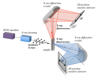
Tungsten atoms ionized through the two-photon absorption process will decay to the ground state by electronic transition from higher electronic orbital. In the present case, we focus explicitly on the () and (3) decay channels. As the and final states are separated by 63 eV, a high energy resolution detection schemes are necessary to precisely measure the two x-ray emission lines. Polarization sensitivity of the experiment will be ensured by horizontal polarization of the XFEL beam in combination with dedicated spatial arrangement of the x-ray emission spectrometers. The x-ray spectrometer will be operated with Si(333) diffraction crystal giving 44.93∘ Bragg angle for and 45.36∘ for x-ray emission. Operation of the spectrometer at Bragg angles close to 45∘ diffraction geometry will provide strong rejection of fluorescence polarized out of the plane of the crystal surface. Typically, the rejection factor of 1000 is expected close to 45∘/45∘ diffraction geometry Lindle et al. (1988); Kämpfer et al. (2016). Moreover, the diffraction spectrometer will ensure energy resolution of around 1-2 eV, which is smaller than the natural line widths of transitions of 6.5 eV Campbell and Papp (1995), and large enough to precisely measure the shape of the x-ray emission. In order to achieve specificity on polarization of x-ray fluorescence, a two-spectrometer setup should be employed, providing measurements of x-ray emission spectra in parallel and perpendicular directions with respect to propagation of the XFEL beam. Schematic layout of the proposed experimental setup is drawn in Fig. 3.
The experimental feasibility may be evaluated from the calculated cross sections for two-photon ionization, parameters of x-ray laser beams and characteristics of the x-ray spectrometer setups. x-ray fluorescence rate at detector of the x-ray spectrometer is given by , where is the spectrometer efficiency, is the two-photon ionization cross-sections, is the incidence x-ray beam flux, is the x-ray fluorescence yield for shell transitions and is the electron decay branching ratio. The efficiency of dispersive-type spectrometer employing cylindrically bent crystal and 2D position sensitive detector is in the range of 10-4/eV Szlachetko et al. (2012). For the XFEL pulse, we assumed intensity of 1011 photons/pulse, 30 fs pulse duration and focus of the x-ray beam down to 55 , which provides the photon flux of around photons /(s cm2) Tamasaku et al. (2013); Doumy et al. (2011); Vinko et al. (2012); Tamasaku et al. (2014). Using the two-photon ionization cross-section value of cm4s at nonlinear Cooper minimum and fluorescence yield of 28% the x-ray rates at detector are estimated to be 280 photons/(s eV). This number confirms the experimental feasibility of detection polarization components of x-ray fluorescence induced by two-photon ionization process within a sub % precision and few hundreds of seconds of acquisition time.
With the present capabilities of XFELs, the maximum x-ray flux is achieved when the machine is operating with Self Amplified Spontaneous Emission mode Andruszkow et al. (2000); Decking et al. (2020). For optimal experiments, short attosecond highly monochromatized x-ray pulses would be desirable in order to maximize nonlinear interaction signals with respect to linear ionization. This, however, is not presently possible, as hard x-ray FEL beam monochromatization via double crystal monochromators or by means of self-seeding modes or short-pulse operation will in turn deliver lower x-ray fluence Amann et al. (2012b). In addition, such beam manipulation will broaden the temporal structure of the x-ray pulse due to the penetration of x-rays into monochromator crystals Shastri et al. (2001). This imposes an experimental limit on exploring either short x-ray pulses or pulses with high monochromaticity. On the other hand, new methods are also developed that may allow us to overcome this physical limit Kayser et al. (2019). However, these methods have not yet been proven experimentally at sub-femtosecond time scales. Thus, while experimental studies of two-photon absorption processes with attosecond x-ray pulses would be of great interest, generation of isolated attosecond pulses in the soft x-ray range have been demonstrated only recently Duris et al. (2020) and are not yet available for hard x-ray energies.
Finally, we would like to address sample preparation issues and eventual possibilities that may be explored during the proposed experiment. The XFEL beams are highly damaging due to their high power which leads to irreversible damage of the sample for every x-ray shot due to the heat dissipation occurring at picosecond time scales David et al. (2011). For this reason, the sample material should be refreshed on a shot-to-shot basis to avoid multiple x-ray hits in the same sample spot. In the case of tungsten, preparation of the sample in the gas phase may be very challenging at high enough densities and around the occupied sample environment at XFEL beamlines. Range of experiments Ghimire et al. (2016); Szlachetko et al. (2016); Tamasaku et al. (2018); Kayser et al. (2019) explored solid state samples, where the sample is moving continuously to obtain fresh spot for each x-ray hit, however this is possible only at low (100 Hz) repetition rate machines and may be unlikely at XFELs such as the European XFEL Decking et al. (2020) operating at MHz repetition rate. Another option is the use of tungsten-based compounds or molecules, because the slight change in the charge state of tungsten atoms does not influence two-photon ionization cross-sections and shifts the positions of resonances and the Cooper minimum only by a few eVs. Such complexes may be prepared in a liquid form and used in liquid-jet systems to ensure sample refreshment even at high repetition rate machines. We should note that comparison of the experimental results obtained for solid and molecular systems could allow us to address for example the effects of plasma creation in solid state samples on the measured x-ray signals.
V Conclusion
In conclusion, we have studied the transfer of linear polarization from incident to fluorescence photons in the case of two-photon ionization of inner-shell electrons. We have shown, that strong variation of the polarization transfer can be achieved not only at intermediate level resonances, but more importantly at the nonlinear Cooper minimum. We proposed an experimentally feasible scenario based on an already realized experiment which could be performed at number of current free-electron laser facilities. The results presented in this work, are however, general and hence could be demonstrated in two-photon ionization of other atomic systems. Measurements of the degree of linear polarization of fluorescence light could serve for the first detection of nonlinear Cooper minimum.
Acknowledgements.
We acknowledge the support from the Bundesministerium für Bildung und Forschung (Grant No. 05K16SJA). This work was partially supported by the National Science Centre (Poland) under grant no. 2017/27/B/ST2/01890.appendix
The electric dipole amplitudes employed in Eq. (II) are
| (8) | |||||
| (9) | |||||
| (10) | |||||
where the short notation for the transition amplitudes was used. In general, these amplitudes are by
| (12) |
The relativistic quantum number is related to the total and orbital angular momentum quantum numbers as . In the transverse (velocity) gauge, these integrals are explicitly given for the magnetic (, or ) transitions
| (13) | |||||
where the radial wave functions and are obtained from single-electron Dirac equation, with a screening potential in the Hamiltonian, which partially accounts for inter-electronic interaction. We compared number of different potential models. The core-Hartree potential, which reproduces binding energies in a good agreement with experimental values, was used to produce the results presented in this work. For the electric transitions (, or )
In the length gauge, this integral is given by
In the above expressions, are the spherical Bessel functions.
References
- Emma et al. (2010) P. Emma, R. Akre, J. Arthur, R. Bionta, C. Bostedt, J. Bozek, et al., Nat. Photonics 4, 641 (2010).
- Amann et al. (2012a) J. Amann, W. Berg, V. Blank, V. F.-J. Decker, Y. Ding, P. Emma, et al., Nat. Photonics 6, 693 (2012a).
- Young et al. (2010) L. Young, E. P. Kanter, B. Krässig, Y. Li, A. M. March, S. T. Pratt, et al., Nature 466, 56 (2010).
- Vinko et al. (2012) S. M. Vinko, O. Ciricosta, B. I. Cho, K. Engelhorn, H.-K. Chung, C. R. D. Brown, et al., Nature 482, 59 (2012).
- Tamasaku et al. (2013) K. Tamasaku, M. Nagasono, H. Iwayama, E. Shigemasa, Y. Inubushi, T. Tanaka, K. Tono, T. Togashi, T. Sato, T. Katayama, T. Kameshima, T. Hatsui, M. Yabashi, and T. Ishikawa, Phys. Rev. Lett. 111, 043001 (2013).
- Richardson et al. (2010) V. Richardson, J. T. Costello, D. Cubaynes, S. Düsterer, J. Feldhaus, H. W. van der Hart, P. Juranić, W. B. Li, M. Meyer, M. Richter, A. A. Sorokin, and K. Tiedke, Phys. Rev. Lett. 105, 013001 (2010).
- Doumy et al. (2011) G. Doumy, C. Roedig, S.-K. Son, C. I. Blaga, A. D. DiChiara, R. Santra, N. Berrah, C. Bostedt, J. D. Bozek, P. H. Bucksbaum, J. P. Cryan, L. Fang, S. Ghimire, J. M. Glownia, M. Hoener, E. P. Kanter, B. Krässig, M. Kuebel, M. Messerschmidt, G. G. Paulus, D. A. Reis, N. Rohringer, L. Young, P. Agostini, and L. F. DiMauro, Phys. Rev. Lett. 106, 083002 (2011).
- Tamasaku et al. (2014) K. Tamasaku, E. Shigemasa, Y. Inubushi, T. Katayama, K. Sawada, H. Yumoto, et al., Nat. Photonics 8, 313 (2014).
- Ghimire et al. (2016) S. Ghimire, M. Fuchs, J. Hastings, S. C. Herrmann, Y. Inubushi, J. Pines, S. Shwartz, M. Yabashi, and D. A. Reis, Phys. Rev. A 94, 043418 (2016).
- Szlachetko et al. (2016) J. Szlachetko, J. Hoszowska, J.-C. Dousse, M. Nachtegaal, W. Błachucki, Y. Kayser, et al., Sci. Rep. 6, 33292 (2016).
- Tamasaku et al. (2018) K. Tamasaku, E. Shigemasa, Y. Inubushi, I. Inoue, T. Osaka, T. Katayama, M. Yabashi, A. Koide, T. Yokoyama, and T. Ishikawa, Phys. Rev. Lett. 121, 083901 (2018).
- Kayser et al. (2019) Y. Kayser, C. Milne, P. Juranić, L. Sala, J. Czapla-Masztafiak, R. Follathand, et al., Nat. Commun. 10, 409 (2019).
- Fano and Macek (1973) U. Fano and J. H. Macek, Rev. Mod. Phys. 45, 553 (1973).
- Klar (1980) H. Klar, J. Phys. B 13, 2037 (1980).
- Starace (1982) A. F. Starace, Phys. Rev. A 25, 842 (1982).
- Huang (1982) K.-N. Huang, Phys. Rev. A 25, 3438 (1982).
- Schmidt (1992) V. Schmidt, Reports on Progress in Physics 55, 1483 (1992).
- Kabachnik et al. (2007) N. Kabachnik, S. Fritzsche, A. Grum-Grzhimailo, M. Meyer, and K. Ueda, Phys. Rep. 451, 155 (2007).
- Sharma et al. (2010) L. Sharma, A. Surzhykov, M. K. Inal, and S. Fritzsche, Phys. Rev. A 81, 023419 (2010).
- Sukhorukov et al. (2019) V. Sukhorukov, I. Petrov, B. Lagutin, A. Ehresmann, K.-H. Schartner, and H. Schmoranzer, Phys. Rep. 786, 1 (2019).
- Cooper (1964) J. W. Cooper, Phys. Rev. Lett. 13, 762 (1964).
- Dehmer and Dill (1976) J. L. Dehmer and D. Dill, Phys. Rev. Lett. 37, 1049 (1976).
- Johnson and Cheng (1978) W. R. Johnson and K. T. Cheng, Phys. Rev. Lett. 40, 1167 (1978).
- White et al. (1979) M. G. White, S. H. Southworth, P. Kobrin, E. D. Poliakoff, R. A. Rosenberg, and D. A. Shirley, Phys. Rev. Lett. 43, 1661 (1979).
- Grum-Grzhimailo (2001) A. N. Grum-Grzhimailo, Journal of Physics B: Atomic, Molecular and Optical Physics 34, L359 (2001).
- Grum-Grzhimailo and Gryzlova (2014) A. N. Grum-Grzhimailo and E. V. Gryzlova, Phys. Rev. A 89, 043424 (2014).
- Ilchen et al. (2018) M. Ilchen, G. Hartmann, E. V. Gryzlova, A. Achner, E. Allaria, A. Beckmann, et al., Nat. Commun. 9, 4659 (2018).
- Lambropoulos and Tang (1987) P. Lambropoulos and X. Tang, J. Opt. Soc. Am. B 4, 821 (1987).
- Saenz and Lambropoulos (1999) A. Saenz and P. Lambropoulos, Journal of Physics B: Atomic, Molecular and Optical Physics 32, 5629 (1999).
- Nikolopoulos (2006) L. A. A. Nikolopoulos, Phys. Rev. A 73, 043408 (2006).
- Lagutin et al. (2017) B. M. Lagutin, I. D. Petrov, V. L. Sukhorukov, P. V. Demekhin, A. Knie, and A. Ehresmann, Phys. Rev. A 95, 063414 (2017).
- Petrov et al. (2019) I. D. Petrov, B. M. Lagutin, V. L. Sukhorukov, N. M. Novikovskiy, P. V. Demekhin, A. Knie, and A. Ehresmann, Phys. Rev. A 99, 013408 (2019).
- Hofbrucker et al. (2018) J. Hofbrucker, A. V. Volotka, and S. Fritzsche, Phys. Rev. Lett. 121, 053401 (2018).
- Hofbrucker et al. (2019) J. Hofbrucker, A. V. Volotka, and S. Fritzsche, Phys. Rev. A 100, 011401(R) (2019).
- Hofbrucker et al. (2020) J. Hofbrucker, A. V. Volotka, and S. Fritzsche, Sci. Rep. 10, 3617 (2020).
- Allaria et al. (2013) E. Allaria et al., Nat. Photonics 7, 913 (2013).
- Lutman et al. (2016) A. A. Lutman et al., Nat. Photonics 10, 468 (2016).
- Marx et al. (2013) B. Marx et al., Phys. Rev. Lett. 110, 254801 (2013).
- Hofbrucker et al. (2016) J. Hofbrucker, A. V. Volotka, and S. Fritzsche, Phys. Rev. A 94, 063412 (2016).
- Hofbrucker et al. (2017) J. Hofbrucker, A. V. Volotka, and S. Fritzsche, Phys. Rev. A 96, 013409 (2017).
- Kämpfer et al. (2016) T. Kämpfer, I. Uschmann, Z. W. Wu, A. Surzhykov, S. Fritzsche, E. Förster, and G. G. Paulus, Phys. Rev. A 93, 033409 (2016).
- Jönsson et al. (2013) P. Jönsson, G. Gaigalas, J. Bieroń, C. F. Fischer, and I. Grant, Comput. Phys. Commun. 184, 2197 (2013).
- Fritzsche (2019) S. Fritzsche, Comput. Phys. Commun. 240, 1 (2019).
- Kanter et al. (2011) E. P. Kanter, B. Krässig, Y. Li, A. M. March, P. Ho, N. Rohringer, R. Santra, S. H. Southworth, L. F. DiMauro, G. Doumy, C. A. Roedig, N. Berrah, L. Fang, M. Hoener, P. H. Bucksbaum, S. Ghimire, D. A. Reis, J. D. Bozek, C. Bostedt, M. Messerschmidt, and L. Young, Phys. Rev. Lett. 107, 233001 (2011).
- Fano (1985) U. Fano, Phys. Rev. A 32, 617 (1985).
- Grum-Grzhimailo et al. (2005) A. N. Grum-Grzhimailo, S. Fritzsche, P. O’Keeffe, and M. Meyer, Journal of Physics B: Atomic, Molecular and Optical Physics 38, 2545 (2005).
- (47) P. Linstrom and W. Mallard, Eds., NIST Chemistry WebBook, NIST Standard Reference Database Number 69, National Institute of Standards and Technology, Gaithersburg MD, 20899 (retrieved 03/05/2019).
- Rudek et al. (2012) B. Rudek, S.-K. Son, L. Foucar, S. W. Epp, B. Erk, R. Hartmann, M. Adolph, R. Andritschke, A. Aquila, N. Berrah, C. Bostedt, J. Bozek, N. Coppola, F. Filsinger, H. Gorke, T. Gorkhover, H. Graafsma, L. Gumprecht, A. Hartmann, G. Hauser, S. Herrmann, H. Hirsemann, P. Holl, A. Hömke, L. Journel, C. Kaiser, N. Kimmel, F. Krasniqi, K.-U. Kühnel, M. Matysek, M. Messerschmidt, D. Miesner, T. Möller, R. Moshammer, K. Nagaya, B. Nilsson, G. Potdevin, D. Pietschner, C. Reich, D. Rupp, G. Schaller, I. Schlichting, C. Schmidt, F. Schopper, S. Schorb, C.-D. Schröter, J. Schulz, M. Simon, H. Soltau, L. Strüder, K. Ueda, G. Weidenspointner, R. Santra, J. Ullrich, A. Rudenko, and D. Rolles, Nat. Photonics 6, 858 (2012).
- Rudek et al. (2018) B. Rudek, K. Toyota, L. Foucar, B. Erk, R. Boll, C. Bomme, J. Correa, S. Carron, S. Boutet, G. J. Williams, K. R. Ferguson, R. Alonso-Mori, J. E. Koglin, T. Gorkhover, M. Bucher, C. S. Lehmann, B. Krässig, S. H. Southworth, L. Young, C. Bostedt, K. Ueda, T. Marchenko, M. Simon, Z. Jurek, R. Santra, A. Rudenko, S.-K. Son, and D. Rolles, Nat. Communications 9, 4200 (2018).
- Erk et al. (2013) B. Erk, D. Rolles, L. Foucar, B. Rudek, S. W. Epp, M. Cryle, C. Bostedt, S. Schorb, J. Bozek, A. Rouzee, A. Hundertmark, T. Marchenko, M. Simon, F. Filsinger, L. Christensen, S. De, S. Trippel, J. Küpper, H. Stapelfeldt, S. Wada, K. Ueda, M. Swiggers, M. Messerschmidt, C. D. Schröter, R. Moshammer, I. Schlichting, J. Ullrich, and A. Rudenko, Phys. Rev. Lett. 110, 053003 (2013).
- Rudenko et al. (2017) A. Rudenko, L. Inhester, K. Hanasaki, X. Li, S. J. Robatjazi, B. Erk, R. Boll, K. Toyota, Y. Hao, O. Vendrell, C. Bomme, E. Savelyev, B. Rudek, L. Foucar, S. H. Southworth, C. S. Lehmann, B. Kraessig, T. Marchenko, M. Simon, K. Ueda, K. R. Ferguson, M. Bucher, T. Gorkhover, S. Carron, R. Alonso-Mori, J. E. Koglin, J. Correa, G. J. Williams, S. Boutet, L. Young, C. Bostedt, S.-K. Son, R. Santra, and D. Rolles, Nature 546, 129 (2017).
- Yoneda et al. (2014) H. Yoneda, Y. Inubushi, M. Yabashi, T. Katayama, T. Ishikawa, H. Ohashi, H. Yumoto, K. Yamauchi, H. Mimura, and H. Kitamura, Nat. Communications 5, 5080 (2014).
- Wach et al. (2020) A. Wach, J. Sá, and J. Szlachetko, Journal of Synchrotron Radiation 27, 689 (2020).
- Tiraşoğlu et al. (2003) E. Tiraşoğlu, U. Çevik, B. Ertuğral, G. Apaydin, M. Ertuğrul, and A. I. Kobya, Eur. Phys. J. D 26, 231 (2003).
- Czarnota et al. (2013) M. Czarnota, D. Banaś, M. Berset, D. Chmielewska, J.-C. Dousse, J. Hoszowska, Y.-P. Maillard, O. Mauron, M. Pajek, M. Polasik, P. A. Raboud, J. Rzadkiewicz, K. Słabkowska, and Z. Sujkowski, Phys. Rev. A 88, 052505 (2013).
- Faisal (2011) F. H. M. Faisal, Lectures on Ultrafast Intense Laser Science (Springer, Berlin, Heidelberg, New York, 2011).
- Tyrała et al. (2019) K. Tyrała, C. Milne, K. Wojtaszek, A. Wach, J. Czapla-Masztafiak, W. M. Kwiatek, Y. Kayser, and J. Szlachetko, Phys. Rev. A 99, 052509 (2019).
- Sytcheva et al. (2012) A. Sytcheva, S. Pabst, S.-K. Son, and R. Santra, Phys. Rev. A 85, 023414 (2012).
- Gorobtsov et al. (2018) O. Y. Gorobtsov, G. Mercurio, F. Capotondi, P. Skopintsev, S. Lazarev, I. A. Zaluzhnyy, et al., Nat. Commun. 9, 4498 (2018).
- Campbell and Papp (1995) J. L. Campbell and T. Papp, X-Ray Spectrom. 24, 307 (1995).
- Lindle et al. (1988) D. W. Lindle, P. L. Cowan, R. E. LaVilla, T. Jach, R. D. Deslattes, B. Karlin, J. A. Sheehy, T. J. Gil, and P. W. Langhoff, Phys. Rev. Lett. 60, 1010 (1988).
- Szlachetko et al. (2012) J. Szlachetko, M. Nachtegaal, E. de Boni, M. Willimann, O. Safonova, J. Sa, G. Smolentsev, M. Szlachetko, J. A. van Bokhoven, J.-C. Dousse, J. Hoszowska, Y. Kayser, P. Jagodzinski, A. Bergamaschi, B. Schmitt, C. David, and A. Lücke, Rev. Sci. Instrum. 83, 103105 (2012).
- Andruszkow et al. (2000) J. Andruszkow, B. Aune, V. Ayvazyan, N. Baboi, R. Bakker, et al., Phys. Rev. Lett. 85, 3825 (2000).
- Decking et al. (2020) W. Decking, S. Abeghyan, P. Abramian, A. Abramsky, A. Aguirre, et al., Nat. Photonics 14, 391 (2020).
- Amann et al. (2012b) J. Amann, W. Berg, V. Blank, F.-J. Decker, Y. Ding, et al., Nature Photonics 6, 693 (2012b).
- Shastri et al. (2001) S. D. Shastri, P. Zambianchi, and D. M. Mills, J. Synchrotron Radiat 8, 1131 (2001).
- Duris et al. (2020) J. Duris, S. Li, T. Driver, E. G. Champenois, J. P. MacArthur, et al., Nat. Photonics 14, 30 (2020).
- David et al. (2011) C. David, S. Gorelick, S. Rutishauser, J. Krzywinski, J. Vila-Comamala, V. A. Guzenko, O. Bunk, E. Färm, M. Ritala, M. Cammarata, D. M. Fritz, R. Barrett, L. Samoylova, J. Grünert, and H. Sinn, Scientific Reports 1, 57 (2011).