Visualizing half-metallic bulk band structure
with multiple Weyl cones of the Heusler ferromagnet
Abstract
Using a well-focused soft X-ray synchrotron radiation beam, angle-resolved photoelectron spectroscopy was applied to a full-Heusler-type Co2MnGe alloy to elucidate its bulk band structure. A large parabolic band at the Brillouin zone center and several bands that cross the Fermi level near the Brillouin zone boundary were identified in line with the results from first-principles calculations. These Fermi level crossings are ascribed to majority spin bands that are responsible for electron transport with extremely high spin polarization especially along the direction being perpendicular to the interface of magneto-resistive devices. The spectroscopy confirms there is no contribution of the minority spin bands to the Fermi surface, signifying half-metallicity for the alloy. Furthermore, two topological Weyl cones with band crossing points were identified around the point, yielding the conclusion that Co2MnGe could exhibit topologically meaningful behavior such as large anomalous Hall and Nernst effects driven by the Berry flux in its half-metallic band structure.
In general, based on their electronic band structures, solids may be classified as either metal or insulator/semiconductor. These two different classes may combine in single magnetic crystals, the so-called half-metallic magnets, in which one spin part of the band structure is metallic and the other semiconducting. Half-metallic magnets have promising uses as spintronics devices because a 100% spin polarization is expected at the Fermi level. From first-principle calculations, some of the Heusler alloys have been predicted to possess half-metallic band structures de Groot et al. (1983); Kübler et al. (1983); Ishida et al. (1995); Picozzi et al. (2002). Among them, Co2MnSi and Co2MnGe are prototypes that are predicted to exhibit a relatively large minority spin gap, which is preserved as long as they are in the ordered phase. Later, it was theoretically pointed out that in-gap minority spin states appear when Co antisite defects are created at Mn sites, resulting in much degraded spin polarizations Picozzi et al. (2004). Indeed, Co2MnSi with excess Mn, which prevented Co atoms from occupying Mn sites, had a substantially improved tunneling magnetoresistance ratio Ishikawa et al. (2009); Liu et al. (2012). Incorporating Fe atoms at the original Mn sites placed the Fermi level in the center of the minority spin gap and further enhanced the magnetoresistance that relies on the spin polarization by up to 2610% at 4.2 K Moges et al. (2016). From recent experimental studies, another striking event happens when the Ge site is substituted with Ga. A huge anomalous Nernst effect takes place Sakai et al. (2018); Guin et al. (2019); Hu et al. (2020) caused by a high Berry flux that originates from the bulk band crossings located near the Fermi level () Xiao et al. (2006, 2010).
We thus claim that the locus of is quite important when engineering the bulk and interface band structures by controlling defects and tuning. The process of computational material design and experimental confirmation has to be iterated to realize the best materials with extremely high functionality. To confirm their highly spin-polarized conducting electrons, numerous experiments employing for example point-contact Andreev reflection spectroscopy Ritchie et al. (2003) and spin-resolved photoelectron spectroscopy Fetzer et al. (2013); Jourdan et al. (2014); Andrieu et al. (2016); Guillemard et al. (2019a, b) were performed. However, the three-dimensional nature of this crystal family has prevented us from approaching their bulk band structures mainly because of the surface and interface sensitivities of these techniques. We note that truly bulk-sensitive hard X-ray photoelectron spectroscopy actually helps in studying the valence band density of states (DOS) of bulk and buried interface, although no momentum-resolved information that is a key to understanding the physical properties has ever been obtained Brown et al. (1998); Miyamoto et al. (2009); Ouardi et al. (2011); Kozina et al. (2014).
A recent soft X-ray angle-resolved photoelectron spectroscopy (ARPES) study of Co2MnSi films, each with Al-Ox capping layer, probed mainly the surface and/or interface states, blocking the band structure underneath Lidig et al. (2019). Therefore, to suppress the influence of the results from the surface and/or interface states, it is mandatory to prepare a clean surface by cleaving the crystal under an ultrahigh vacuum. However, because of the robust 3D nature of its crystal structure [Fig. 1(a)], it is generally difficult to produce a flat surface and consequently multiple steps remain [Fig. 1(b)].
In this Letter, we report the direct evidence of the half-metallic band structure of Co2MnGe and the presence of multiple Weyl cones using ARPES with a beam spot size of m. It enables a single domain with a flat surface to be proved for ARPES.
A high quality single crystalline sample was grown by the Bridgeman method (see Sec. S1 of Supplementary Material for the detailed sample growth conditions). The composition of the sample was Co: 49.3, Mn: 24.9, and Ge: 25.8 (at. %) as determined by energy dispersive X-ray microanalysis (EDX), which is stoichiometric enough to prevent Co anti-site defect and preserve its half-metallicity. The saturation magnetic moment evaluated from the magnetization curve measured with a SQUID at 5 K was 115.3 emu/g and converted to 4.96 , being close to an integer number and consistent with the expected value obtained from the Slater-Pauling rule Galanakis et al. (2002). From differential scanning calorimetry, the Curie temperature of the present specimen was 913 K, being comparable with our previous experimental result Okubo et al. (2010).
ARPES measurements were performed at BL25SU at SPring-8. Circularly polarized synchrotron radiation beam was used to maximize the number of bands that appear in the ARPES results. The beam spot size was adjusted to m. The energy and angular resolutions for ARPES were set to meV and , respectively. A clean surface of Co2MnGe was obtained by cleaving in an ultra-high vacuum (pressure Pa). All measurements were conducted at a temperature of K. The analyzer slit was set along the -axis [parallel to ; see Figs. 1(a, b)].
All first-principles density-functional calculations were performed using the WIEN2k program code Blaha et al. (2001). We used the spin-polarized generalized gradient approximation Perdew et al. (1996) (see Sec. S2 for further calculation details). The Coulomb interaction was not considered because previous studies indicated that plays a minor role in Co2MnGe Tsirogiannis and Galanakis (2015); Sharma and Pandey (2016).
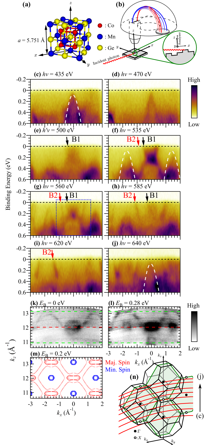
From the photon energy () dependence of the ARPES spectra [Figs. 1(c-j)], we see that the ARPES image evolves with photon energy (also see the Supplemental Movie). At eV, a band that disperses downwards from its peak at eV appears at [overlaid with white dashed line in Fig. 1(c)]. The photoelectron intensity from this band weakens at eV [Fig. 1(d)], and another feature emerges at eV [B1 in Fig. 1(e)] and its intensity is maximized at eV [Fig. 1(f)]. Interestingly, the bands cross (B1, B2) and two bands, each with a downward dispersion, emerge on both sides of the crossing feature (white dashed lines). Increasing the photon energy further, the band indicated by the black arrow moves to . Moreover, a steeply dispersing band crosses [red arrow labeled with B2 in Fig. 1(f)] and moves away from with higher photon energies. A spectral weight of the band, indicated by the red arrow, is maximized at eV [Fig. 1(i)]. The downward-dispersing band re-emerges above eV, and its intensity is maximized at eV [dashed line in Fig. 1(j)].
We realized that the circular features in the - constant energy map at eV [Fig. 1(l)] stem from the downward-dispersing bands near the point. Importantly, they are not seen at [Fig. 1(k)]. This demonstrates that our ARPES measurement tracks the band structure along the out-of-plane momentum () line. We have determined an inner potential of eV from the photon energy dependence of the ARPES spectra with downward dispersions. The computed - constant energy map [Fig. 1(m)] reproduces well the observed features with minority spin character.
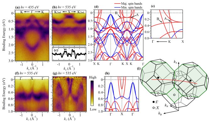
We extracted typical ARPES images acquired at the incident photon energies of 435 eV and 535 eV from Fig. 1 along the -- and -- lines [Figs. 2(a, b), respectively]. A parabolic band with a downward dispersion dominates near at eV. At eV, a spectral feature near around is modified considerably. This feature corresponds to B1 in Fig. 1 and consists of two separated bands that are more clearly identified by the second derivative of the momentum distribution curve at [indicated by inverted triangles in Fig. 2(c)]. By combining this plot with B2 in Fig. 1, we conclude that the three bands cross . Furthermore, we compared these ARPES images with calculated band dispersions along the -- line [Fig. 2(d)]. The calculated results have been shifted in energy by meV (40 meV) toward higher in the majority (minority) spin channel to adjust to the ARPES results. The downward-dispersing ARPES band [Fig. 2(a)] fits very well with the theoretical minority spin band. A band that disperses upwards and crosses [Fig. 2(b)] is ascribed to the three computed majority spin bands.
For the experimental band dispersion along the - line [Fig. 2(f)], the raw ARPES intensity has been normalized to enhance its visibility using the integrated intensity of the momentum distribution curve [Fig. 2(g)]. From a comparison with the calculated band dispersion along the - line [Fig. 2(h)], we claim that the minority and majority spin components contribute respectively to the observed bands that disperse downwards near the points and the band that disperses upwards and crosses .

Figures 3(a-e) shows the experimental constant energy surfaces at , 0.1, 0.2, 0.3 and 0.4 eV. The characteristic features in Figs. 3(a-e) are re-drawn in Figs. 3(f-j). The calculated constant energy surfaces are shown in Figs. 3(k-o). Constant energy surfaces with four-fold symmetry are observed that correlate with the symmetry of the plane of the crystal. Four elliptical pockets [red solid line in Fig. 3(f)] are seen at eV and diminish as increases. At eV [Figs. 3(e, j)], we find four circles that diminish with decreasing and ultimately disappear at . In a comparison with the calculated results [Figs. 3(k-o)], we determined that these circles are ascribable to the minority spin components. All the other features are well explained by the bands of the majority spin channel. This means that only the majority spin bands cross and no minority spin band exists, signifying that a half-metallic band structure is realized in this crystal.
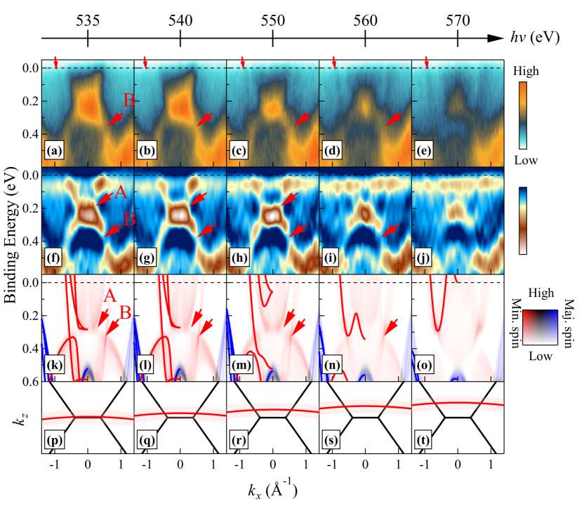
Finally, we discuss the topological aspects of the band structure of Co2MnGe. Weyl fermions appear in materials that break inversion symmetry Xu et al. (2015) or time-reversal symmetry Kuroda et al. (2017). It is expected that the Weyl fermions emerge also in Co2MnGe as a result of time-reversal symmetry breaking as has recently been predicted for some of the Heusler alloys Chang et al. (2017); Belopolski et al. (2019). Figures 4(a-e) show the ARPES images along the -- line acquired at -570 eV. To exclude the effect of a momentum constant background, we differentiated the ARPES intensity twice along the energy axis in Figs. 4(f-j). We see again a band that disperses upwards and crosses , at eV [Figs. 4(a, f)]. The inner band exhibits a strong intensity whereas the outer band [small red arrow in Fig. 4(a)] is weak. Marked by two red arrows A and B, the band dispersing downwards intersects other bands dispersing upwards. These two crossings are persistently seen in the image taken at eV [Fig. 4(h)]. At eV, the crossing of the bands involving the outer band is observed even more clearly at eV. Above eV, all crossing points disappear. To compare the ARPES result with the theoretical band dispersions more rigorously, we considered real momentum “paths” using . It is also necessary to consider broadening effects that stem from the limited probing depth of the photoelectrons (). In Figs. 4(k-o), we show the computed band dispersions integrated in the window together with the band dispersions along the real -lines [Figs. 4(p-t)]. We find that the theoretical ARPES images reproduce the persistent band crossings for -560 eV and its disappearance above eV, although the crossings of the line dispersions are already broken above 550 eV. Our first-principles calculation predicts two band crossings around the point [A and B in Fig. 2(e)]. Further theoretical analysis tells us that all of them form nodal-lines [Figs. S3(a-c)]. We, therefore, conclude that crossing points A and B correspond to the type-II Weyl points produced from the tilted cones. We note that Co2MnGa exhibits similar crossing points [Figs. S3(d-f)] and that these crossings may generate a high Berry flux Chang et al. (2017); Belopolski et al. (2019). The gigantic anomalous electrical and thermal conductivities that give rise to the anomalous Hall effect and the Nernst effect emerge when the gap opens at the crossing point and is tuned inside the gap. Because the crossing points A and B are located above for Co2MnGa, further carrier tuning is required to improve these anomalous conductivities. We propose that the substitution of Ge atoms into the Ga sites may improve the anomalous transport properties. Our study with soft X-ray ARPES confirms that these crossing points are of bulk origin and are maintained even for the end material, Co2MnGe.
In conclusion, we performed ARPES on a full-Heusler-type Co2MnGe bulk crystal utilizing micro-spot-size soft X-ray synchrotron radiation. No contribution of the minority spin band structures to the Fermi surface was observed. All the observed Fermi surfaces were reproduced by the calculated results for the majority spin channel. Moreover, two topological Weyl cones were clearly observed indicating Berry flux sources. Our findings provide strong evidence of half-metallicity coexisting with multiple Weyl cones of the Co2MnGe alloy. They also shed light on the currently elusive spin-gapless Heusler semiconductors, for which one spin part is semimetallic and the other semiconducting. Tuning the highly spin-polarized carriers is possible via an electric field if that becomes feasible. We finally remark that by choosing appropriate elements the Heusler alloys comprising more than three elements give rise to various types of physical behaviors such as the magneto-caloric effect, thermoelectricity and superconductivity, all of which rely on their band structures. Micro-spot ARPES with soft X-ray synchrotron radiation beam affords opportunities to realize highly functional materials for such alloys.
Acknowledgements.
This work was financially supported by KAKENHI (Nos. 17H06152, 17H06138, 18H01690, and 18H03683). The soft X-ray ARPES experiment was performed with the approval of JASRI (Proposal No. 2019A1548). Micro-ARPES instruments was developed by Photon and Quantum Basic Research Coordinated Development Program from MEXT. T.Y. was financially supported by Grants-in-Aid for JSPS Fellows No. 18J22309. We sincerely thank T. Sugawara and I. Narita in Tohoku University for their help to make the single crystals and to perform the EDX experiment.References
- de Groot et al. (1983) R. A. de Groot, F. M. Mueller, P. G. vanEngen, and K. H. J. Buschow, Phys. Rev. Lett. 50, 2024 (1983).
- Kübler et al. (1983) J. Kübler, A. R. Williams, and C. B. Sommers, Phys. Rev. B 28, 1745 (1983).
- Ishida et al. (1995) S. Ishida, S. Fujii, S. Kashiwagi, and S. Asano, J. Phys. Soc. Jpn. 64, 2152 (1995).
- Picozzi et al. (2002) S. Picozzi, A. Continenza, and A. J. Freeman, Phys. Rev. B 66, 094421 (2002).
- Picozzi et al. (2004) S. Picozzi, A. Continenza, and A. J. Freeman, Phys. Rev. B 69, 094423 (2004).
- Ishikawa et al. (2009) T. Ishikawa, H. Liu, T. Taira, K.-i. Matsuda, T. Uemura, and M. Yamamoto, Appl. Phys. Lett. 95, 232512 (2009).
- Liu et al. (2012) H.-x. Liu, Y. Honda, T. Taira, K.-i. Matsuda, M. Arita, T. Uemura, and M. Yamamoto, Appl. Phys. Lett. 101, 132418 (2012).
- Moges et al. (2016) K. Moges, Y. Honda, H.-x. Liu, T. Uemura, M. Yamamoto, Y. Miura, and M. Shirai, Phys. Rev. B 93, 134403 (2016).
- Sakai et al. (2018) A. Sakai, Y. P. Mizuta, A. A. Nugroho, R. Sihombing, T. Koretsune, M.-T. Suzuki, N. Takemori, R. Ishii, D. Nishio-Hamane, R. Arita, P. Goswami, and S. Nakatsuji, Nat. Phys. 14, 1119 (2018).
- Guin et al. (2019) S. N. Guin, K. Manna, J. Noky, S. J. Watzman, C. Fu, N. Kumar, W. Schnelle, C. Shekhar, Y. Sun, J. Gooth, and C. Felser, NPG Asia Materials 11, 16 (2019).
- Hu et al. (2020) J. Hu, Y. Zhang, M. A. Cabero, B. Wei, S. Tu, S. Liu, D. Yu, J.-P. Ansermet, S. Granville, and H. Yu, J. Magn. Magn. Mater. 500, 166397 (2020).
- Xiao et al. (2006) D. Xiao, Y. Yao, Z. Fang, and Q. Niu, Phys. Rev. Lett. 97, 026603 (2006).
- Xiao et al. (2010) D. Xiao, M.-C. Chang, and Q. Niu, Rev. Mod. Phys. 82, 1959 (2010).
- Ritchie et al. (2003) L. Ritchie, G. Xiao, Y. Ji, T. Y. Chen, C. L. Chien, M. Zhang, J. Chen, Z. Liu, G. Wu, and X. X. Zhang, Phys. Rev. B 68, 104430 (2003).
- Fetzer et al. (2013) R. Fetzer, J.-P. Wüstenberg, T. Taira, T. Uemura, M. Yamamoto, M. Aeschlimann, and M. Cinchetti, Phys. Rev. B 87, 184418 (2013).
- Jourdan et al. (2014) M. Jourdan, J. Minár, J. Braun, A. Kronenberg, S. Chadov, B. Balke, A. Gloskovskii, M. Kolbe, H. J. Elmers, G. Schönhense, H. Ebert, C. Felser, and M. Kläui, Nat. Commun. 5, 3974 (2014).
- Andrieu et al. (2016) S. Andrieu, A. Neggache, T. Hauet, T. Devolder, A. Hallal, M. Chshiev, A. M. Bataille, P. Le Fèvre, and F. Bertran, Phys. Rev. B 93, 094417 (2016).
- Guillemard et al. (2019a) C. Guillemard, S. Petit-Watelot, L. Pasquier, D. Pierre, J. Ghanbaja, J.-C. Rojas-Sánchez, A. Bataille, J. Rault, P. Le Fèvre, F. Bertran, and S. Andrieu, Phys. Rev. Applied 11, 064009 (2019a).
- Guillemard et al. (2019b) C. Guillemard, S. Petit-Watelot, J.-C. Rojas-Sánchez, J. Hohlfeld, J. Ghanbaja, A. Bataille, P. Le Fèvre, F. Bertran, and S. Andrieu, Appl. Phys. Lett. 115, 172401 (2019b).
- Brown et al. (1998) D. Brown, M. D. Crapper, K. H. Bedwell, M. T. Butterfield, S. J. Guilfoyle, A. E. R. Malins, and M. Petty, Phys. Rev. B 57, 1563 (1998).
- Miyamoto et al. (2009) K. Miyamoto, A. Kimura, Y. Miura, M. Shirai, M. Ye, Y. Cui, K. Shimada, H. Namatame, M. Taniguchi, Y. Takeda, Y. Saitoh, E. Ikenaga, S. Ueda, K. Kobayashi, and T. Kanomata, Phys. Rev. B 79, 100405(R) (2009).
- Ouardi et al. (2011) S. Ouardi, G. H. Fecher, B. Balke, A. Beleanu, X. Kozina, G. Stryganyuk, C. Felser, W. Klöß, H. Schrader, F. Bernardi, J. Morais, E. Ikenaga, Y. Yamashita, S. Ueda, and K. Kobayashi, Phys. Rev. B 84, 155122 (2011).
- Kozina et al. (2014) X. Kozina, J. Karel, S. Ouardi, S. Chadov, G. H. Fecher, C. Felser, G. Stryganyuk, B. Balke, T. Ishikawa, T. Uemura, M. Yamamoto, E. Ikenaga, S. Ueda, and K. Kobayashi, Phys. Rev. B 89, 125116 (2014).
- Lidig et al. (2019) C. Lidig, J. Minár, J. Braun, H. Ebert, A. Gloskovskii, J. A. Krieger, V. Strocov, M. Kläui, and M. Jourdan, Phys. Rev. B 99, 174432 (2019).
- Galanakis et al. (2002) I. Galanakis, P. H. Dederichs, and N. Papanikolaou, Phys. Rev. B 66, 174429 (2002).
- Okubo et al. (2010) A. Okubo, R. Y. Umetsu, K. Kobayashi, R. Kainuma, and K. Ishida, Appl. Phys. Lett. 96, 222507 (2010).
- Blaha et al. (2001) P. Blaha, K. Schwarz, G. K. H. Madsen, D. Kvasnicka, and J. Luitz, WIEN2K, An Augmented Plane Wave + Local Orbitals Program for Calculating Crystal Properties (Karlheinz Schwarz, Techn. Universität Wien, Austria, 2001).
- Perdew et al. (1996) J. P. Perdew, K. Burke, and M. Ernzerhof, Phys. Rev. Lett. 77, 3865 (1996).
- Tsirogiannis and Galanakis (2015) C. Tsirogiannis and I. Galanakis, J. Magn. Magn. Mater. 393, 297 (2015).
- Sharma and Pandey (2016) S. Sharma and S. K. Pandey, J. Magn. Magn. Mater. 403, 1 (2016).
- Xu et al. (2015) S.-Y. Xu, I. Belopolski, N. Alidoust, M. Neupane, G. Bian, C. Zhang, R. Sankar, G. Chang, Z. Yuan, C.-C. Lee, S.-M. Huang, H. Zheng, J. Ma, D. S. Sanchez, B. Wang, A. Bansil, F. Chou, P. P. Shibayev, H. Lin, S. Jia, and M. Z. Hasan, Science 349, 613 (2015).
- Kuroda et al. (2017) K. Kuroda, T. Tomita, M.-T. Suzuki, C. Bareille, A. A. Nugroho, P. Goswami, M. Ochi, M. Ikhlas, M. Nakayama, S. Akebi, R. Noguchi, R. Ishii, N. Inami, K. Ono, H. Kumigashira, A. Varykhalov, T. Muro, T. Koretsune, R. Arita, S. Shin, T. Kondo, and S. Nakatsuji, Nature Materials 16, 1090 (2017).
- Chang et al. (2017) G. Chang, S.-Y. Xu, X. Zhou, S.-M. Huang, B. Singh, B. Wang, I. Belopolski, J. Yin, S. Zhang, A. Bansil, H. Lin, and M. Z. Hasan, Phys. Rev. Lett. 119, 156401 (2017).
- Belopolski et al. (2019) I. Belopolski, K. Manna, D. S. Sanchez, G. Chang, B. Ernst, J. Yin, S. S. Zhang, T. Cochran, N. Shumiya, H. Zheng, B. Singh, G. Bian, D. Multer, M. Litskevich, X. Zhou, S.-M. Huang, B. Wang, T.-R. Chang, S.-Y. Xu, A. Bansil, C. Felser, H. Lin, and M. Z. Hasan, Science 365, 1278 (2019).