Resolution of Quantum Imaging with Undetected Photons
Abstract
Quantum imaging with undetected photons is a recently introduced technique that goes significantly beyond what was previously possible. In this technique, images are formed without detecting the light that interacted with the object that is imaged. Given this unique advantage over the existing imaging schemes, it is now of utmost importance to understand its resolution limits, in particular what governs the maximal achievable spatial resolution.
We show both theoretically and experimentally that the momentum correlation between the detected and undetected photons governs the spatial resolution — a stronger correlation results in a higher resolution. In our experiment, the momentum correlation plays the dominating role in determining the resolution compared to the effect of diffraction. We find that the resolution is determined by the wavelength of the undetected light rather than the wavelength of the detected light. Our results thus show that it is in principle possible to obtain resolution characterized by a wavelength much shorter than the detected wavelength.
1 Introduction
Quantum imaging is a rapidly developing field with progress fuelled by developments in photon detection technology [1, 2, 3], nonlinear and quantum optics [4, 5, 6, 7, 8], and quantum information theory [9, 10, 11]. Quantum effects applied to conventional imaging techniques have enabled overcoming the classical limits, e.g., shot-noise limit [12, 13, 14] and diffraction limit [15, 16, 17, 18]. Besides this, quantum theory has inspired novel imaging techniques such as ghost imaging [19, 20, 21, 22, 23, 7]. Recently, another quantum imaging technique, “Quantum imaging with undetected photons” (QIUP), has been introduced [24, 25]. QIUP goes significantly beyond the present quantum imaging capabilities. In this technique, the image is formed without detecting the light that interacted with the object. This imaging technique also inspired a number of applications to infrared spectroscopy [26, 27], and optical coherence tomography [28, 29].
The concept of QIUP relies on the creation of a photon pair in quantum superposition at two spatially separated sources [30, 31]. One photon of the pair interacts with the object and is never detected. The image is constructed by detecting the partner photon that does not interact with the object. This distinctive feature of QIUP allows a sample to be probed at a wavelength for which no detector is available, which is an advantage over existing imaging techniques.
Quantum ghost imaging also uses spatially correlated photon pairs to acquire images [19, 20, 21, 22, 23, 7]. While the achievable resolution of ghost imaging has been studied [32, 33, 34], a detailed analysis for QIUP is hitherto lacking. An understanding of the resolution limits is of utmost importance for assessing potential applications of QIUP. Since the principle behind QIUP is fundamentally different from principles of all other quantum imaging techniques, a thorough analysis of the resolution is required to understand its resolution limits.
Here we present a detailed analysis of the resolution for QIUP. We address the problem both theoretically and experimentally. We consider the experimental configuration used in Refs. [24, 25]: the object and the camera are located in the far-field of the nonlinear crystals where the photon pairs are emitted. The transverse dimensions of these crystals are much smaller than the diameter of the lenses employed. We find that in this case, the achievable resolution is limited by the momentum correlation between detected and undetected photons. In addition, we show that in this regime, the resolution of QIUP is regulated by the wavelength of the light that probes the object. Our result highlights that the non-degenerate QIUP scheme can be particularly promising for applications where the wavelength of the light probing the object is shorter than the detected wavelength.
In Sec. 2, we generalize the theory of imaging presented in Ref. [25] without the assumption of perfect momentum correlation. In Sec. 3, we illustrate the experimental setups and discuss the corresponding magnification. Then in Sec. 4, we present a qualitative discussion of the effect of momentum correlation on resolution. In Sec. 5, we present the experimental results and compare them with theoretical predictions. In Sec. 6, we discuss our results, which is followed by proposals of improvements in Sec. 7. Finally, we summarize our results and conclude in Sec. 8.
2 Theory
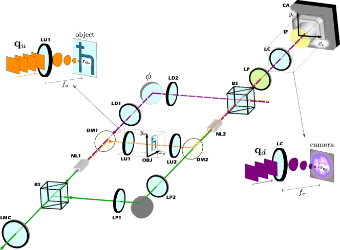
The imaging scheme (Fig. 1) uses two coherently pumped identical nonlinear crystals (NL1 and NL2) as sources of one photon pair ( and ) 111In fact it is not necessary to have two identical sources. This imaging scheme can be realized alternatively with a double pass of the laser in a single crystal [26]. The beams of photon from each source (purple dashed line) are superposed by a beam splitter and subsequently detected on a camera. The beam of the partner photon (; orange dotted line) emitted by NL1 is sent through NL2 and is aligned perfectly with the corresponding beam emitted by NL2 such that the modes of photon emitted by NL1 and NL2 become identical. As a result of this alignment, which-source information is unavailable, and interference is observed in the camera where only photon is detected [30, 31]. Photon is never detected.
In QIUP, the object is placed in the undetected beam between the two crystals and its complex transmission coefficient appears in the interference pattern [24, 25]. The absolute value of the transmission coefficient is proportional to the interference visibility 222Visibility, , is evaluated at each camera pixel.. The phase introduced by the object can be retrieved from the interference pattern.
A detailed theoretical treatment of the imaging scheme was presented in Ref. [25], where it was assumed that the transverse momenta of photons and are perfectly correlated. In another two works, this assumption was dropped and it was suggested that the measured transverse momentum correlation could be associated to the imaging resolution [35, 36]. Here, we generalize the above-mentioned theoretical and experimental work to rigorously show the key role played by partial momentum correlation in the image resolution.
In our experiment, the photons are emitted into paraxial beams. The photon pair () produced by either of the sources can be represented by the normalized quantum state 333Strictly speaking, and are continuous variables and therefore the sum in Eq. (1) is to be understood as an integration.
| (1) |
where and represent the transverse wave vectors associated with photons and , respectively. The transverse momentum correlation of the photon pair is determined by the probability of jointly detecting photon with transverse momentum and photon with transverse momentum . This joint probability is given by .
When the two crystals are weakly pumped, the effect of stimulated emission is negligible [30, 31, 37, 38]. The quantum state of a photon pair, produced jointly by the crystals, is given by a linear superposition of the states generated by the crystals individually. The state is, therefore, given by
| (2) |
where labels the two sources (NL1 and NL2) and we have assumed for simplicity that the sources emit with equal probability. Here, we note that a rigorous derivation of Eq. (2) requires considering the vacuum state generated by SPDC (see Ref. [25]).
Suppose that photon with transverse momentum is detected at a point, , on the camera (Fig. 1). The corresponding partner photon () with transverse momentum passes through a point, , on the object. The object is characterized by the complex amplitude transmission coefficient .
Following the theoretical steps shown in Ref. [25] and using the state given by Eq. (2), one readily finds that the photon counting rate (intensity) at a point on the camera is given by
| (3) |
where is a spatially independent interferometric phase that is varied experimentally. An interference pattern is recorded on the camera. It is clear from Eq. (2) that both the amplitude and the phase of the transmission coefficient of the object are present in the interference pattern. Furthermore, the presence of in the equation suggests that the resolution depends on the momentum correlation 444Note that Eq. (7) of Ref. [36] shows the same result for a phase object..
We note that the object information is only in the interference term of the expression on the right-hand side of Eq. (2). This term can be used to study the spatial resolution quantitatively. For example, when the object is purely absorptive, we have . In this case, it follows from Eq. (2) that the image is represented by the position dependent visibility
| (4) |
where , for , is the conditional probability which depends on the transverse momentum distribution of the pump photon [39].
For a phase object (i.e., ), the resolution may again be studied from the interference term (see Appendix B).
3 Experimental Setups and Magnification
We build two experimental setups characterized by two sets of wavelengths. For setup 1, nm and nm, i.e., the undetected photon’s wavelength is longer than that of the detected photon. For setup 2, nm, nm, i.e., the undetected photon has the shorter wavelength. All other parameters of the imaging system (distances, focal lengths, etc.) are the same in the two setups. This also includes the distance between object and camera. The focal length of positive lenses, LU1 and LU2 (Fig. 1), placed in the path of the undetected photon is mm. The positive lens, LC, placed in front of the camera (Fig. 1) has focal length mm.
The image is constructed by varying the spatially independent interferometer phase, . Subsequently, phase shifts and the visibility of the interference observed on the camera are evaluated using sinusoidal fits to the intensity data for each pixel individually. The experimental study of the resolution considers purely absorptive objects. The same conclusion is expected for a phase object, as we demonstrate in Appendix B.
An important feature of the imaging scheme is that the magnification () depends on the wavelengths of both photons. It has been shown that the magnification is given by [25, 24]
| (5) |
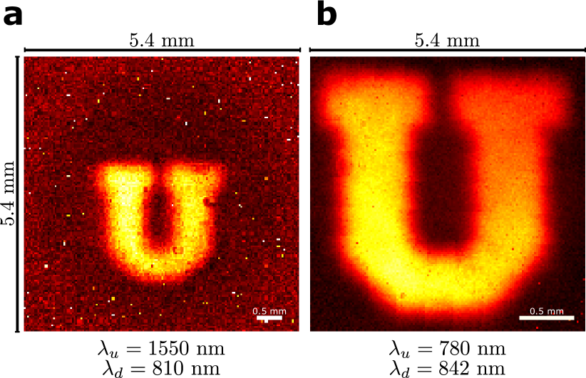
We experimentally verify this formula by imaging the same object using the two setups (Fig. 2). We find that (Fig. 2a) and (Fig. 2b) for setup 1 and setup 2, respectively. The experimentally measured values agree with the theoretical predictions. Using these magnification values, we estimated the far field illumination area. We obtained mm2 for setup 1 and mm2 for setup 2.
The wavelength dependence of the magnification plays an important role in understanding the resolution of the imaging system as we point out later.
4 Resolution and Momentum Correlation
The analysis presented in Sec. 2 (in particular Eqs. (2) and (4)) suggest that a stronger momentum correlation between the two photons leads to a better resolution. We now provide a physical explanation and test the theoretical understanding by analyzing images of the same object taken with different strengths of momentum correlation.

Both the object and the camera are located in the far-field (Fourier plane) of the nonlinear crystals (Fig. 1). If a photon with transverse momentum is detected at a point on the camera, it follows from the lens formula that [25]. In the ideal case of perfect momentum correlation, i.e., when , the detection of a photon () with momentum allows us to infer with certainty that the momentum of its undetected partner () is . Since the object is also located at the Fourier plane of the crystals, the undetected photon () with transverse momentum arrives at a unique point, , on the object plane. Therefore, the interference pattern recorded at point will contain information about only one point on the object when the momentum correlation is perfect. For example, when the transmission coefficient at is zero, the visibility measured at also becomes zero. However, if the momentum correlation is not perfect, one point on the camera does not correspond to only one point on the object. This is because the momentum () of photon now varies over a range when the photon is detected with a particular momentum. This range becomes wider with a weaker momentum correlation. Therefore, the information from a range of points on the object appears at a single point on the camera, i.e., a weaker momentum correlation reduces the spatial resolution.
In the experiment, we use Gaussian pump beams and focus them on the two crystals in such a way that the waists of the two pump beams are equal. The conditional probability governing the momentum correlation is given by [39]
| (6) |
where is the waist of a pump beam, and is the transverse component of the pump wave vector. It follows that a stronger correlation is obtained for a larger beam waist (Fig. 3a). We change the correlation by varying the waist of the two pump beams simultaneously by lens LMC [35, 36].
Figure 3b presents experimentally obtained images (visibility at each point on the camera) of a resolution target obtained with different pump waist sizes for nm and nm. The line of the object has a width of 250 m (see Appendix C for object details). The results clearly show that resolution reduces with a weaker momentum correlation.
5 Quantitative Study and Dependence on Wavelength
We now study the connection between resolution and momentum correlation quantitatively. We also address the question of which wavelength determines the resolution.
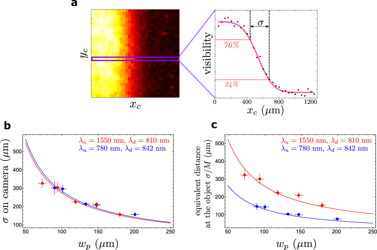
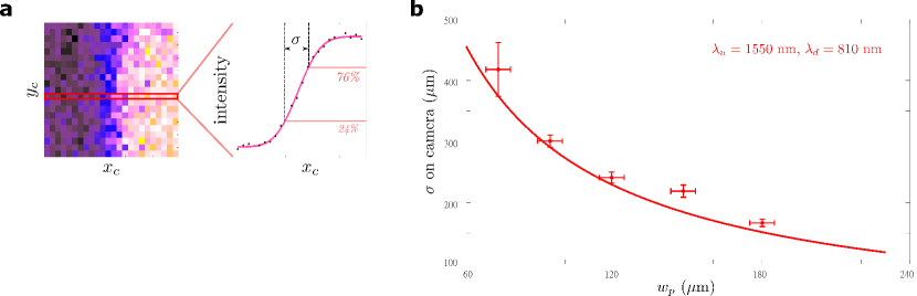
We first consider the edge-spread function (ESF) that allows us to study the unsharpness of the imaging system quantitatively [40]. A knife-edge is used as an object. The ESF is obtained from the image of the knife-edge. Because we use spherical lenses throughout the setup, we assume axial symmetry. We place the knife-edge parallel to the axis and along the line , so that the undetected beam is blocked between the crystals in the region 555The knife-edge is theoretically modeled by the Heaviside step function, which has the form for and for .. It now follows from Eqs. (4) and (4) that 666We replace the sum by an integration and use the relations and . the ESF is given by , where erf is the error function, is the magnification, and is given by
| (7) |
It can be checked from the formula of ESF that is the distance on the camera for which the value of ESF (visibility) rises from approximately to of the maximum attainable value. Experimentally, the quantity is determined from the image (position dependent visibility) of the knife-edge (Fig. 4a).
In Fig. 4b, we plot the experimentally measured values of (data points with error bars) against the pump waist () for both setup 1 and setup 2. The theoretically predicted results (solid lines) are also given for comparison. All measured data differ by less than two standard deviations from the corresponding theoretical values 777The experimental error (standard deviation) was obtained from the sinusoidal fit of the experimental data, and was propagated through the visibility calculations.. It is clear that increases as the pump waist () decreases. We discussed in Sec. 4 that the momentum correlation decreases as the pump waist decreases. Therefore, Fig. 4b demonstrates that the resolution decreases as the momentum correlation reduces.
We note from Eq. (7) that the quantity does not depend on the wavelength () of the light that illuminates the object. It only depends on the wavelength () of the detected light. The experimental results (Fig. 4b) confirm this fact: although the values of are widely different (1550 nm and 780 nm) for the two setups, the values of are relatively close because the values of are also close (810 nm and 842 nm) for the two setups. However, this does not mean that the resolution is characterized by the wavelength of the detected light. The reason is that the quantity represents a distance measured on the camera, whereas resolution refers to minimum resolvable distance at the object. In order to obtain a measure of spatial resolution, one needs to divide by the magnification [41]. An appropriate measure of resolution for our experiment is therefore
| (8) |
where and are given by Eqs. (5) and (7), respectively. We find that depends only on the wavelength of the undetected photon, i.e., the light illuminating the object. The experimentally obtained values of are shown and compared with the theoretical prediction in Fig. 4c. The results show that for the same pump waist at the crystal and consequently for the same strength of the momentum correlation (see Eq. (4)), the resolution is significantly better when the wavelength of the light illuminating the sample is shorter. This holds despite the fact that the wavelength (842 nm) of the detected light is longer than that in the other case (810 nm). Figure 4c strongly suggests that the wavelength of the undetected photon characterizes the resolution.

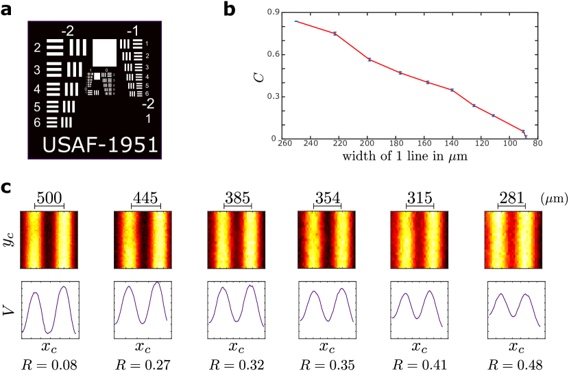
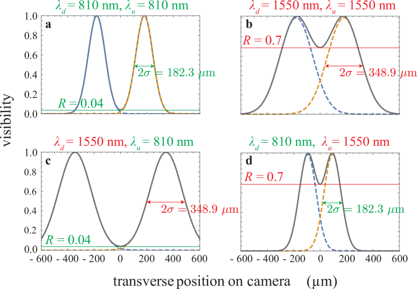
We now perform a direct test of the wavelength dependence of the resolution using the 1951 USAF resolution test chart (Fig. 5). We experimentally obtain images (position dependent visibilities) of different parts of the test target. Images (position dependent visibility) of two consecutive slits of the test chart overlap and form a double-humped distribution (see Appendix C). In analogy to the Rayleigh criterion ([42], Sec. 7.6.3), we assume that two consecutive slits are just resolved when the ratio () of the visibility at the dip (minimum) to that at a peak (maximum) is equal to . Consequently, we choose the minimum resolvable distance as the “width of line in m” when two consecutive slits are just resolved.
In Fig. 5a, we plot the experimentally measured minimum resolvable distances against the pump waist (for both setups) and compare them with the theoretical predictions. Data points (filled circles with error bars) and the numerically simulated curves labeled by (1) and (2) correspond to setup 1 ( nm, nm) and setup 2 ( nm, nm), respectively. Since the width of line in m cannot be varied continuously in the 1951 USAF resolution test chart, the experimentally measured values of are never exactly . We choose the slits for which is less than and nearest to 0.81. The theoretical predictions (solid lines with shaded areas) are made considering the mean values and standard deviations of the experimentally measured ( for setup 1 and for setup 2). In Fig. 5b, we show images of the same set of slits obtained by the two imaging setups for a fixed pump waist ( m). Our results clearly demonstrate that the image resolution is higher if the object is probed using a shorter wavelength, although the detected wavelength is longer in this case.
6 Discussion
Our results show that the width () of the edge-spread function on the camera is determined by the wavelength of the detected light, while the resolution is determined by the wavelength of the undetected light that illuminates the object. The roles of the two wavelengths can be summarized by analyzing the simulated image of a pair of points in different wavelength configurations. Figure 6 depicts the computed visibility cross section (image) on the camera for an object that consists of two points with a separation m. Focal lengths and distances are assumed to be the same as in our experiment and the pump waist is m.
The image of an individual point turns out to be a Gaussian distribution of visibility. This distribution effectively represents the point-spread function for our imaging system. Since we have assumed axial symmetry, the width of this distribution on the camera () is directly related to the width of the edge-spread function (). For a pair of points, a double humped distribution is formed (Fig. 6). The ratio of the visibility at the minimum to the visibility at a peak is denoted by . The lower the value of , the better we can resolve the two points.
In Fig. 6a, the wavelengths of detected and undetected photons are nm, whereas in Fig. 6b both wavelengths are nm. Since the magnification is linearly proportional to the ratio (see Eq. (5)), it has the same value for these two cases. The width of the point-spread function () on the image is given by the respective detected wavelength, which results in a better contrast between the two points in the case of the shorter wavelength (Fig. 6a; ) compared to the longer wavelength (6b; ). Figure 6c shows a situation in which the same object is imaged with nm and nm, i.e., the shorter wavelength is used to probe the object. While the width of the point spread function on the image is given by the detected wavelength, the wavelength dependent magnification results in a contrast between the two points of the same value () as in Fig. 6a. Thus, the resolution can in principle exceed the limit of the detected wavelength and is instead given by the undetected wavelength. The opposite case is depicted in Fig. 6d: although the width of the point spread function on the camera is determined by the shorter detected wavelength, the contrast between the two points attains the same value () as if only the longer wavelength were used. This is due to the fact that the wavelength ratio leads to a demagnification in this case.
In our theoretical analysis, we have not explicitly considered the effect of diffraction at the lenses. The theoretical predictions still match with the experimental observations. The reason for this can be understood as follows: the transverse dimensions of the nonlinear crystals (1 mm 2 mm) constrain the maximum pump spot size and thus the maximally achievable momentum correlation (cf. Eq. (4)), i.e. how precisely is determined if the partner photon as an exact value of . In our setup, 1 inch optics are used in the lens systems that map a transverse wave-vector to a point on the object (camera). Due to the diffraction limit imposed by the lenses [43], the position of a point on the object (or camera) corresponding to a particular transverse wave vector has an uncertainty of m. However, this uncertainty is much smaller than the uncertainty due to the imperfect correlation between and ; the latter corresponds to approximately m for a pump waist of m (roughly equivalent to the effect of a pump-spot sized aperture in the Fourier plane of the object). Thus, the diffraction limit imposed by the lenses is negligible in our experiment when compared to the resolution limit given by the imperfect momentum correlation.
7 How to increase the resolution of future experiments
In our analysis, we have considered experiments that use the scheme of Ref. [24], in which nonlinear crystals are located in the far field of the object and the momentum correlation between detected and undetected photons limits the resolution. In this case, the best resolution can be achieved if in Eq. (8) is minimized. Equation (8) implies that in order to increase the resolution in this type of setup (at the cost of a decreased field-of-view (FOV)), it is beneficial to use lenses of short focal length in the undetected beam. Short focal lengths allow to decrease the distance between two points on an object, that correspond to two “adjacent” wave-vectors, i.e., to a fixed distance in the momentum space. A second alternative is decreasing the FOV by an external optical system.
The resolution can also be enhanced by decreasing the wavelength of the light illuminating the object. In order to increase the momentum correlation between the two photons, a large pump beam and hence nonlinear crystals with large transverse dimensions are beneficial.
A different approach could be to change the geometry of the setup in a way that the momentum correlation does not limit the resolution. This could be achieved, for example, by imaging the nonlinear crystals onto the object and the camera. In this case, the imaging system would rely on photon pair correlations in the transverse position rather than in momentum. Therefore, the small size of the nonlinear crystals is expected to limit the FOV instead of the resolution.
8 Summary and Conclusions
We have studied the resolution in Quantum Imaging with Undetected photons both theoretically and experimentally. Our experiment uses the scheme developed in Ref. [24], in which object and camera are located in the far-field of the photon pair sources. In this case, we have identified the momentum correlation between detected and undetected photons as the dominant factor that limits the resolution. The physical reason for this limit is that the spatial dimensions of the nonlinear crystals are much smaller than that of the lenses, which is a relevant case for practical applications.
Our results show that in this regime, the width of the edge-spread function at the camera is determined by the wavelength of the detected light, whereas the resolution is dependent upon the wavelength of the undetected light illuminating the object. This shows that QIUP can be particularly promising in cases where the wavelength to which available cameras are sensitive is larger than the wavelength required for object illumination. In a recent experiment, photon pairs — with one photon having wavelength in the range of x-ray and the other at optical wavelengths — have been generated [44]. Therefore, it is certainly possible to design an experiment, where an object is illuminated by x-ray and the detected photon has optical wavelength. In such an experiment, it would in principle be possible to obtain x-ray resolution, using readily available cameras to detect only at optical wavelengths.
Despite the fact that the results turned out to be analogous to that of quantum ghost imaging (QGI) enabled by momentum correlation, the theory and experiment of these two imaging schemes are fundamentally different. In QGI, the photon probing the object is detected and the image is acquired through coincidence measurement (post selection), whereas in QIUP the photon probing the object is never detected and the image is acquired through intensity (single-photon counting rate) measurement. Furthermore, QIUP is an interferometric scheme and QGI is not. Therefore, our finding that the resolution of QIUP based on momentum correlation is limited in the same way as QGI is quite remarkable, because it suggests that QIUP accesses the information encoded in two-photon correlations as efficiently as QGI.
We expect that our results will enable future works leading to further improvement of this unique method of imaging and will thereby lead to a deeper understanding of its resolution limit in more general scenarios. It will be interesting to explore to what extent established super-resolution techniques can be adapted and to what extent the resulting implementations using other geometries or image acquisition methods will further increase the achievable resolution888While this manuscript was in submission, new articles were published with interesting discussion on QIUP resolution [45, 46, 47, 48, 49]..
Acknowledgements
The authors thank Markus Gräfe and Marta Gilaberte for encouraging us to finish this project. This work was supported by the Austrian Academy of Sciences (ÖAW) and the FWF project W 1210-N25 (CoQuS). JF and EAO also acknowledge ANID for the financial support (Becas de doctorado en el extranjero “Becas Chile”/ 2015 – No. 72160487 and 2016 – No. 72170402). RL acknowledges the funding by the National Science Centre (Poland) Project No. 2015/17/D/ST2/03471 and the Foundation for Polish Science within the FIRST TEAM project ‘Spatiotemporal photon correlation measurements for quantum metrology and super-resolution microscopy’ co-financed by the European Union under the European Regional Development Fund (POIR.04.04.00-00-3004/17-00).
References
- [1] C. Bruschini et al. “Single-photon avalanche diode imagers in biophotonics: review and outlook” In Light Sci. Appl. 8.1 Nature Publishing Group, 2019, pp. 87 URL: https://doi.org/10.1038/s41377-019-0191-5
- [2] G. Lubin et al. “Quantum correlation measurement with single photon avalanche diode arrays” In Opt. Express 27.23 Optical Society of America, 2019, pp. 32863–32882 URL: https://doi.org/10.1364/OE.27.032863
- [3] A. Nomerotski “Imaging and time stamping of photons with nanosecond resolution in Timepix based optical cameras” In Nucl. Instrum. Methods Phys. Res., Sect. A 937 Elsevier, 2019, pp. 26–30 URL: https://doi.org/10.1016/j.nima.2019.05.034
- [4] A.. Burlakov et al. “Interference effects in spontaneous two-photon parametric scattering from two macroscopic regions” In Phys. Rev. A 56 American Physical Society, 1997, pp. 3214–3225 URL: https://doi.org/10.1103/PhysRevA.56.3214
- [5] A. Mosset, F. Devaux and E. Lantz “Spatially Noiseless Optical Amplification of Images” In Phys. Rev. Lett. 94 American Physical Society, 2005, pp. 223603 URL: https://doi.org/10.1103/PhysRevLett.94.223603
- [6] S.. Walborn, C.. Monken, S. Pádua and P… Ribeiro “Spatial correlations in parametric down-conversion” In Phys. Rep. 495.4-5 Elsevier, 2010, pp. 87–139 URL: https://doi.org/10.1016/j.physrep.2010.06.003
- [7] P.. Moreau, E. Toninelli, T. Gregory and M.. Padgett “Imaging with quantum states of light” In Nat. Rev. Phys. 1.6 Nature Publishing Group, 2019, pp. 367–380 URL: https://doi.org/10.1038/s42254-019-0056-0
- [8] M. Kutas et al. “Terahertz quantum sensing” In Sci. Adv. 6.11 American Association for the Advancement of Science, 2020, pp. eaaz8065 URL: https://doi.org/10.1126/sciadv.aaz8065
- [9] R. Nair and M. Tsang “Far-Field Superresolution of Thermal Electromagnetic Sources at the Quantum Limit” In Phys. Rev. Lett. 117 American Physical Society, 2016, pp. 190801 URL: https://doi.org/10.1103/PhysRevLett.117.190801
- [10] M. Parniak et al. “Beating the Rayleigh Limit Using Two-Photon Interference” In Phys. Rev. Lett. 121 American Physical Society, 2018, pp. 250503 URL: https://doi.org/10.1103/PhysRevLett.121.250503
- [11] O.. Magaña-Loaiza and R.. Boyd “Quantum imaging and information” In Rep. Prog. Phys. 82.12 IOP Publishing, 2019, pp. 124401 URL: https://doi.org/10.1088/1361-6633/ab5005
- [12] G. Brida, M. Genovese and I.. Berchera “Experimental realization of sub-shot-noise quantum imaging” In Nat. Photon. 4.4 Nature Publishing Group, 2010, pp. 227–230 URL: https://doi.org/10.1038/nphoton.2010.29
- [13] J. Sabines-Chesterking et al. “Twin-beam sub-shot-noise raster-scanning microscope” In Opt. Express 27.21 Optical Society of America, 2019, pp. 30810–30818 URL: https://doi.org/10.1364/OE.27.030810
- [14] G.. Garces et al. “Quantum-enhanced stimulated emission detection for label-free microscopy” In Appl. Phys. Lett. 117.2 AIP Publishing LLC, 2020, pp. 024002 URL: https://doi.org/10.1063/5.0009681
- [15] O. Schwartz et al. “Superresolution microscopy with quantum emitters” In Nano Lett. 13.12 ACS Publications, 2013, pp. 5832–5836 URL: https://doi.org/10.1021/nl402552m
- [16] A. Classen, J. Zanthier, M.. Scully and G.. Agarwal “Superresolution via structured illumination quantum correlation microscopy” In Optica 4.6 Optical Society of America, 2017, pp. 580–587 URL: https://doi.org/10.1364/OPTICA.4.000580
- [17] M. Unternährer et al. “Super-resolution quantum imaging at the Heisenberg limit” In Optica 5.9 Optical Society of America, 2018, pp. 1150–1154 URL: https://doi.org/10.1364/OPTICA.5.001150
- [18] R. Tenne et al. “Super-resolution enhancement by quantum image scanning microscopy” In Nat. Photon. 13.2 Nature Publishing Group, 2019, pp. 116–122 URL: https://doi.org/10.1038/s41566-018-0324-z
- [19] D.. Strekalov, A.. Sergienko, D.. Klyshko and Y.. Shih “Observation of two-photon “ghost” interference and diffraction” In Phys. Rev. Lett. 74.18 APS, 1995, pp. 3600 URL: https://doi.org/10.1103/PhysRevLett.74.3600
- [20] T.. Pittman, Y.. Shih, D.. Strekalov and A.. Sergienko “Optical imaging by means of two-photon quantum entanglement” In Phys. Rev. A 52.5 APS, 1995, pp. R3429 URL: https://doi.org/10.1103/PhysRevA.52.R3429
- [21] R.. Bennink, S.. Bentley and R.. Boyd ““Two-photon” coincidence imaging with a classical source” In Phys. Rev. Lett. 89.11 APS, 2002, pp. 113601 URL: https://doi.org/10.1103/PhysRevLett.89.113601
- [22] A. Gatti, E. Brambilla and L. Lugiato “Quantum imaging” In Progress in Optics 51, 2008, pp. 251–348
- [23] R.. Aspden, D.. Tasca, R.. Boyd and M.. Padgett “EPR-based ghost imaging using a single-photon-sensitive camera” In New J. Phys. 15.7, 2013, pp. 073032 URL: https://doi.org/10.1088/1367-2630/15/7/073032
- [24] G.. Lemos et al. “Quantum imaging with undetected photons” In Nature 512.7515, 2014, pp. 409–412 URL: https://doi.org/10.1038/nature13586
- [25] M. Lahiri, R. Lapkiewicz, G.. Lemos and A. Zeilinger “Theory of quantum imaging with undetected photons” In Phys. Rev. A 92.1 APS, 2015, pp. 013832 URL: https://doi.org/10.1103/PhysRevA.92.013832
- [26] G.. Lemos et al. “Quantum Imaging with Undetected Photons” In US Patent 9,557.262 B2; EP Patent 2 887 137 B1
- [27] D.. Kalashnikov, A.. Paterova, S.. Kulik and L.. Krivitsky “Infrared spectroscopy with visible light” In Nat. Photon. 10.2 Nature Publishing Group, 2016, pp. 98–101 URL: https://doi.org/10.1038/nphoton.2015.252
- [28] A. Vallés, G. Jiménez, L.. Salazar-Serrano and J.. Torres “Optical sectioning in induced coherence tomography with frequency-entangled photons” In Phys. Rev. A 97.2 APS, 2018, pp. 023824 URL: https://doi.org/10.1103/PhysRevA.97.023824
- [29] A.. Paterova et al. “Tunable optical coherence tomography in the infrared range using visible photons” In Quantum Sci. Technol. 3.2 IOP Publishing, 2018, pp. 025008 URL: https://doi.org/10.1088/2058-9565/aab567
- [30] X.. Zou, L.. Wang and L. Mandel “Induced coherence and indistinguishability in optical interference” In Phys. Rev. Lett. 67.3 APS, 1991, pp. 318 URL: https://doi.org/10.1103/PhysRevLett.67.318
- [31] L.. Wang, X.. Zou and L. Mandel “Induced coherence without induced emission” In Phys. Rev. A 44.7 APS, 1991, pp. 4614 URL: https://doi.org/10.1103/PhysRevA.44.4614
- [32] R.. Bennink, S.. Bentley, R.. Boyd and J.. Howell “Quantum and Classical Coincidence Imaging” In Phys. Rev. Lett. 92 American Physical Society, 2004, pp. 033601 URL: https://doi.org/10.1103/PhysRevLett.92.033601
- [33] K… Chan, M.. O’Sullivan and R.. Boyd “Two-color ghost imaging” In Phys. Rev. A 79 American Physical Society, 2009, pp. 033808 URL: https://doi.org/10.1103/PhysRevA.79.033808
- [34] P.. Moreau et al. “Resolution limits of quantum ghost imaging” In Opt. Express 26.6 Optical Society of America, 2018, pp. 7528–7536 URL: https://doi.org/10.1364/OE.26.007528
- [35] A. Hochrainer et al. “Quantifying the momentum correlation between two light beams by detecting one” In Proc. Natl. Acad. Sci. USA 114.7 National Acad Sciences, 2017, pp. 1508–1511 URL: https://doi.org/10.1073/pnas.1620979114
- [36] M. Lahiri et al. “Twin-photon correlations in single-photon interference” In Phys. Rev. A 96.1 APS, 2017, pp. 013822 URL: https://doi.org/10.1103/PhysRevA.96.013822
- [37] H.. Wiseman and K. Mølmer “Induced coherence with and without induced emission” In Phys. Lett. A 270.5 Elsevier, 2000, pp. 245–248 URL: https://doi.org/10.1016/S0375-9601(00)00314-5
- [38] M. Lahiri et al. “Nonclassicality of induced coherence without induced emission” In Phys. Rev. A 100 American Physical Society, 2019, pp. 053839 URL: https://doi.org/10.1103/PhysRevA.100.053839
- [39] C.. Monken, P… Ribeiro and S. Pádua “Transfer of angular spectrum and image formation in spontaneous parametric down-conversion” In Phys. Rev. A 57.4 APS, 1998, pp. 3123 URL: https://doi.org/10.1103/PhysRevA.57.3123
- [40] A.. Harms and A. Zeilinger “A new formulation of total unsharpness in radiography” In Phys. Med. Biol. 22.1 IOP Publishing, 1977, pp. 70 URL: https://doi.org/10.1088/0031-9155/22/1/009
- [41] G.. Boreman “Modulation transfer function in optical and electro-optical systems” SPIE press Bellingham, WA, 2001
- [42] M. Born and E. Wolf “Principles of optics” Cambridge University Press, 1999
- [43] J.. Goodman “Introduction to Fourier optics” RobertsCompany Publishers, 2005
- [44] A. Schori et al. “Parametric down-conversion of x rays into the optical regime” In Phys. Rev. Lett. 119.25 American Physical Society, 2017, pp. 253902 URL: https://doi.org/10.1103/PhysRevLett.119.253902
- [45] A.. Paterova et al. “Hyperspectral infrared microscopy with visible light” In Sci. Adv. 6.44 American Association for the Advancement of Science, 2020, pp. eabd0460 URL: https://doi.org/10.1126/sciadv.abd0460
- [46] I. Kviatkovsky et al. “Microscopy with undetected photons in the mid-infrared” In Sci. Adv. 6.42 American Association for the Advancement of Science, 2020, pp. eabd0264 URL: https://doi.org/10.1126/sciadv.abd0264
- [47] A.. Paterova, H. Yang, Z… Toa and L.. Krivitsky “Quantum imaging for the semiconductor industry” In Appl. Phys. Lett. 117.5 AIP Publishing LLC, 2020, pp. 054004 URL: https://doi.org/10.1063/5.0015614
- [48] A.. Paterova et al. “Non-linear interferometry with infrared metasurfaces” In Nanophotonics 10.6 De Gruyter, 2021, pp. 1775–1784 URL: https://doi.org/10.1515/nanoph-2021-0011
- [49] M. Gilaberte Basset et al. “Video-Rate Imaging with Undetected Photons” In Laser Photonics Rev. 15.6 Wiley Online Library, 2021, pp. 2000327 URL: https://doi.org/10.1002/lpor.202000327
Appendix A Details of the Setups
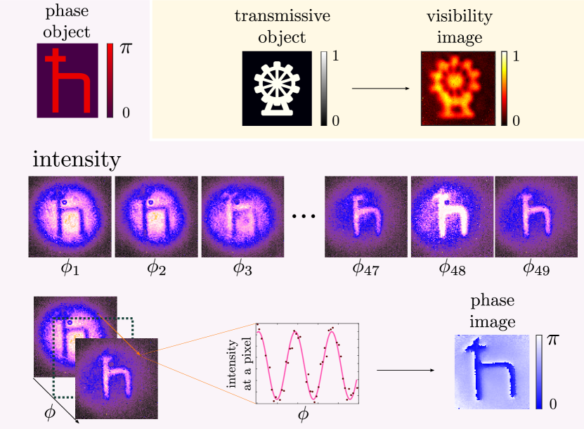
In the first setup were used the set of wavelengths nm, nm, and in front of the camera a long-pass filter 635 nm and an interference filter 810 nm. In the second setup were used the set of wavelengths nm, nm, and in front of the camera a long-pass filter 635 nm and an interference filter 842 nm. The optical distances in both setups were identical. The lenses to focus at the crystals were and (all in mm). The imaging systems were built with the lenses mm, and mm. In both setups the non-linear crystals were PPKTP type-0 of dimensions 2x2x1 mm (lxwxh).
The acquisition process is shown in Fig. 7. The object produces a phase-shift at the wavelength =1550 nm. The acquisition time of each intensity image was 10 seconds.
Appendix B Resolution for Phase Imaging
In this appendix, we justify that our main results are also true for a phase object. We have shown that the intensity at a point on the camera is given by Eq. (2). For a phase object, we have , i.e. . The resolution of a phase object can be studied by the use of the interference term, which now reduces to the form
| (9) |
We find that the edge-spread function is characterized by the parameter
| (10) |
Comparing this equation with Eq. (7), we find that the quantity (and consequently ) has the same value for an absorptive and a phase edge.
The experiment is done using the setup for which nm and nm. To create a phase edge, we use a phase plate, TEM01, which produces a phase difference for nm between two parts of a beam cross-section. In a cross section of an intensity image showing constructive (or destructive) interference, is evaluated an error function to obtain the parameter in different scenarios of momentum correlation. We evaluate the parameter that characterizes the edge-spread function. In Fig. 8, we illustrate the method and the experimental results. We conclude that the spatial resolution of both transmission and phase objects are limited by the imperfect momentum correlation of the photon pairs.
Appendix C Details on the Test Target Evaluation
The minimum resolvable distance between two slit is a method for obtaining the resolvable power of an optical system. We used the object ‘1951 USAF resolution test chart’, shown in Fig. 9(a). The object is formed by groups of six elements with three slits each (horizontal and vertical oriented). The slits have different width and separation through the object. In the method, we evaluated an image of two consecutive slits, with a waist of m, and the wavelengths , and . In Fig. 9(c), we present the minimum resolvable distance between two slits adapted from Rayleigh’s criterion [42]. The threshold for two slit to be resolvable is . An image of two consecutive slit that gives a coefficient , it is considered resolved. Additionally, we adapted the contrast transfer function. We define this function as . In fig 9(b), we present the experimental results of .