2D Surface Phase Diagram of a Multicomponent Perovskite Oxide: La0.8Sr0.2MnO3(110)
Abstract
The many surface reconstructions of (110)-oriented lanthanum–strontium manganite (La0.8Sr0.2MnO3, LSMO) were followed as a function of the oxygen chemical potential (O) and the surface cation composition. Decreasing O causes Mn to migrate across the surface, enforcing phase separation into A-site-rich areas and a variety of composition-related, structurally diverse B-site-rich reconstructions. The composition of these phase-separated structures was quantified with scanning tunneling microscopy (STM), and these results were used to build a 2D phase diagram of the LSMO(110) equilibrium surface structures.
The surfaces of oxide materials, both in their bulk form and as (ultra)thin films, are rarely bulk-terminated. Instead they form a multitude of diverse and complex structural configurations as a function of their oxygen content Surnev et al. (2003); Li et al. (2009); Sedona et al. (2005); Shaikhutdinov and Freund (2012); Netzer (2010); Shimizu et al. (2012), their cation composition (for multielement materials) Wang et al. (2016); Franceschi et al. (2020), and, in case of metal-supported ultrathin films, the support itself Sedona et al. (2005); Netzer (2010). This manifold of phases is determined by a delicate interplay between polarity compensation Noguera (2000), variability of the oxidation state of the metal atoms as a function of the oxygen chemical potential (1/2O2 Reuter and Scheffler (2001); Franceschi et al. (2019), henceforth O for simplicity), strain and defect-formation energies Wang et al. (2014), and, depending on the preparation conditions, kinetic effects Netzer (2010).
The surface structure of metal oxides dictates their surface properties through the coordination, arrangement, and electronic structure of the topmost atoms DeBenedetti et al. (2018); McBriarty et al. (2018); Pan et al. (2020); Jakub et al. (2019a, b); Riva et al. (2018); Zhu et al. (2016); Qin et al. (2020). Hence, it is important to map out the parameter space of the equilibrium surface phases of these materials, including their evolution with O. Such a characterization has come a long way for binary oxides, but is still in its infancy for perovskite oxides (ABO3), despite the undisputed role of their surfaces in many emerging and established technologies Zubko et al. (2011); Bhalla et al. (2000); Peña and Fierro (2001); Kumah et al. (2020).
One prominent example is lanthanum–strontium manganite (LSMO). Owing to its many and diverse physical properties (among others, half metallicity, colossal magnetoresistance, metal–insulator transition, anti/ferromagnetic to paramagnetic transitions, high electronic conductivity at elevated temperatures), LSMO is used in a wide range of applications, e.g., spintronics Majumdar and van Dijken (2013); Park et al. (1998); Haghiri-Gosnet et al. (2004), catalysis Hwang et al. (2017); Ponce et al. (2000), energy production Jiang (2008); Ruiz-Morales et al. (2011), and various thin-film technologies Liao and Zhang (2019); Huang et al. (2018a, b). Because many relevant processes and interactions take place at the LSMO surfaces and interfaces, gaining a comprehensive understanding of the equilibrium surface phases of LSMO at the local (i.e., atomic) level is of paramount importance. Such investigations must be performed as a function of both, the cation, and the oxygen concentrations (the latter depends on the value of O at which the surface is treated): Both these parameters define a rich phase diagram including the physical properties named above Hemberger et al. (2002), the performance of LSMO-based devices Kim et al. (2012); Adler (2004); Bristowe et al. (2011); Chen et al. (2015), and emergent phenomena in LSMO thin films Fan et al. (2019). To date, however, there exists no comprehensive investigation that simultaneously maps out the equilibrium surface phases of LSMO (nor of any other multielement oxide) as a function of the cation composition and O. The few studies available have unveiled that perovskite oxide surfaces typically consist of B-site-rich structures made of one or two atomic layers of differently linked polyhedra Andersen et al. (2018); Enterkin et al. (2010), and have focused on their relation to the A:B cation ratio at the surface. In some cases, quantitative compositional relations between the different structures have been established Wang et al. (2016); Franceschi et al. (2020); Gerhold et al. (2014); Feng et al. (2013).
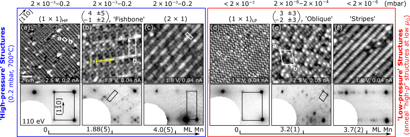
This work pushes the experimental characterization of perovskite oxide surfaces, by investigating the effect of the cation composition and O on the local surface properties of (110)-oriented LSMO. Single-crystalline films of La0.8Sr0.2MnO3 were grown on Nb-doped SrTiO3(110) substrates by pulsed laser deposition (PLD) ( 100 nm, 700∘C, 1 Hz, 2.2 J/cm2, 4 10-2 mbar O2) Franceschi et al. (2020). Their surfaces were investigated in an ultra-high vacuum (UHV) surface science setup attached to the PLD chamber, equipped with scanning tunneling microscopy (STM), low-energy electron diffraction (LEED), x-ray photoelectron spectroscopy (XPS), and low-energy He+-ions scattering (LEIS) (for details about the experimental methods, see Refs. Franceschi et al., 2020; Gerhold et al., 2016, and Section S1). Previous studies have shown that these films exhibit composition-related surface reconstructions at 700∘C and 0.2 mbar O2, and have established the differences in cation coverages between them Franceschi et al. (2020). These structures, here referred to as “high-pressure” (HP), are shown for reference at the left-hand side of Fig. 1, and are described in detail in Section S2. The current work is based on the behavior of the HP reconstructions over a wide range of O values (between eV and eV, corresponding to annealing between UHV and 0.2 mbar O2 at 700∘C): When decreasing O (i.e., at more reducing conditions), new surface structures are formed, and a rich phase diagram emerges. These new structures, referred to as “low-pressure” (LP), are collected at the right-hand side of Fig. 1, and are described in detail in Section S3. Their relative compositions, as derived in this work, are reported in the bottom axis in terms of monolayers (ML) of Mn.
After exploring in detail the behavior of the HP structures with decreasing O and the process of phase separation, this work will propose a novel, STM-based approach to organize the equilibrium surface phases of LSMO(110) (both HP and LP) in a quantitative diagram as a function of the cation composition and O.
The first HP structure whose behavior with O is considered is the A-site-rich HP of Fig. 1(a): This structure stays unaltered upon annealing at 700∘C and 2 10-3 mbar O2 0.2 mbar, but transforms into the LP of Fig. 1(d) at lower pressures (down to UHV). The LP is characterized by the same periodicity as its HP counterpart, and the same cation composition [see LEIS data in Fig. S1(b)]. However, it displays a glide plane that is not present in the HP , and possesses a smaller oxygen content [Fig. S1(b)].
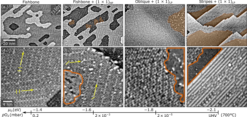
The other HP phases of LSMO(110) behave differently than the HP with decreasing O, as shown below. These structures are significantly Mn-richer and belong to a different family with respect to the HP , and are henceforth referred to as HP, ‘Mn-rich’ phases. Notably, they continuously evolve into one other as a function of the Mn content, such that various surface structures exist between the fishbone and the of Fig. 1(b, c) Franceschi et al. (2020). The qualitative behavior of these HP, Mn-rich phases with decreasing O is exemplified by Fig. 2, which shows the evolution of the fishbone phase of Fig. 1(b), initially prepared at 0.2 mbar and 700∘C. When decreasing the O2 pressure to 2 10-3 mbar at 700∘C, small HP areas appear (orange in Fig. 2). These patches become larger as O decreases, and change their atomic structure to LP . Meanwhile, the remaining fishbone-reconstructed surface undergoes a minor structural change: The small features highlighted by the short yellow lines in Fig. 2(a2, b2) orient closer to the [10] direction, an indication of a slight Mn enrichment Franceschi et al. (2020). Between and mbar [Fig. 2(c)], larger LP patches form, while the remaining surface exposes the oblique structure of Fig. 1(e). Below mbar and down to UHV [Fig. 2(d)], even larger LP patches are observed, while the remaining areas exhibit the stripes of Fig. 1(f). Importantly, the process is reversible: STM confirms that the initial surface is regained when annealing back at high O2. A semi-ordered phase assigned to a transition state between the fishbone and the oblique structures appears around mbar (not shown).
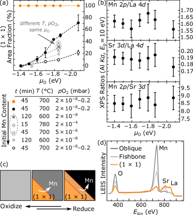
Quantifying the HP/LP areal coverages at each step of the phase separation with STM [Fig. 3(a)] allows to determine the composition of the LP Mn-rich structures, as shown below. Note that because the following discussion focuses on relative cation compositions, which is the same on the two HP and LP phases, these will be henceforth referred to simply as , disregarding the change in atomic structure and oxygen content occurring below mbar O2 at 700∘C. In Fig. 3(a), full, black symbols represent the experiment of Fig. 2: As discussed, decreasing O produces increasingly larger coverages. The same trend is observed for starting Mn-richer surface compositions (gray), albeit with a smaller slope: Mn-richer surfaces form less of the Mn-poorer areas. Importantly, the same phases with the same quantitative coverages are observed under the same value of O, obtained from different combinations of temperature and O2 [dashed oval in Fig. 3(a)]: This indicates that the observed surface structures are equilibrium phases, and that the phase separation is not kinetically limited. Recall that no phase separation occurs when starting from a surface (orange): The remains always monophase, hence the plot with O shows a constant area fraction of 100%.
What drives the phase separation? One can rule out evaporation of cations (due to the reversibility), as well as cation diffusion to or from the bulk (at the employed conditions, cations can travel in bulk LSMO at most one atomic layer Kubicek et al. (2014)): The phase separation thus occurs by mass transport across the surface. This is consistent with the fact that the average surface cation composition is conserved, as indicated by the reversibility of the process, and from the XPS data of Fig. 3(b): These show that the intensity ratios of selected core level peaks have no trend with O. Given the sensitivity of XPS to the cation composition of monophase LSMO(110) structures Franceschi et al. (2020), the absence of a trend indicates that the average surface composition is conserved. The proposed mechanism, sketched in Fig. 3(c), is that Mn travels across the surface and exposes the very stable, Mn-poor phase, while enriching the remaining areas. The LEIS data of Fig. 3(d), showing that the monophase oblique phase is Mn-richer than the monophase fishbone, further support this scenario. Moreover, the stabilization mechanism of the Mn-rich phases at reducing conditions via the increase in their Mn content is consistent with the lower Mn oxidation state displayed by Mn-richer HP structures Franceschi et al. (2020).
Because of the current lack of knowledge on the surfaces of perovskite oxides, one cannot determine whether the O-dependent phase separation witnessed for LSMO(110) can be generalized to this material class. The only other perovskite oxide whose surfaces have been investigated in significant depth, SrTiO3, shows a phase separation among stable surface structures with different A:B ratio Riva et al. (2019a), but not as a function of O. This is presumably because SrTiO3 does not possess the same flexibility in the oxidation state of the B cation as LSMO Kozakov et al. (2015); Abbate et al. (1992); Saitoh et al. (1995), and because of its much smaller tendency to form oxygen vacancies. Materials with easily reducible cations are expected to behave similarly to LSMO(110): After all, structurally complex B-site-rich reconstructions that depend on the cation- and anion-composition seem to be a general trait of perovskite oxides Andersen et al. (2018); Enterkin et al. (2010); Kolpak et al. (2008). Moreover, an AO termination was preferentially exposed at reducing conditions also in another manganite Tselev et al. (2015), and a similar effect was predicted for LSMO(001) Hess and Yildiz (2020).
The remarkable diversity of the surface phases of perovskite oxides calls for methods capable of accessing and controlling their local surface properties as a function of different cation and anion compositions. Here, such a method is showcased for LSMO(110). As detailed below, it yields the quantitative cation compositions of Mn-rich, LP phases relative to LP , based on the measured coverages of the phase-separated structures [Fig. 3(a)], and on the known differences in Mn content of the HP phases Franceschi et al. (2020). The outcome is shown in Figs. 4(a) and 4(b), displaying the experimental 2D surface phase diagram of LSMO(110) as a function of O and the cation composition, and the corresponding sketch, respectively. In Fig. 4(a), each curve describes the evolution of a surface with given composition (horizontal axis) prepared at high O2, and then annealed at decreasing O (vertical axis). For instance, the left-most curve represents the evolution of the initially monophase surface: Its cation composition never changes, hence its evolution with O is a vertical line. The solid black circles represent the evolution of the initial fishbone surface, which phase-separates into areas and regions characterized by LP, Mn-richer structures (oblique or stripes, see Fig. 2). The other curves represent surfaces that have been prepared initially to exhibit a slightly Mn-richer structure [between the fishbone and the of Fig. 1(b, c)].
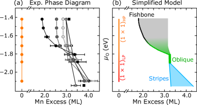
To derive the compositions of the LP phases formed during the phase separation, the number of cations on the whole surface was taken as constant at each annealing step, as supported by the reasonings above. This condition can be expressed as , where is the density of Mn cations for a surface with multiple structures exposed, whose density of Mn cations and areal fractions are and , respectively Riva et al. (2019b). Initially (at high pressure) the surface exposes only a Mn-rich, HP surface, and . Referring this quantity to the density of Mn cations of the , , one has , which is known from Fig. 1: for the fishbone, ML. By annealing at low pressure, the surface phase-separates into areas and new LP Mn-rich areas. Now, , where LP is the (unknown) density of Mn cations on the new LP phase [referred to ], and LP is the corresponding areal coverage (measured by STM). Since by assumption, one obtains that . The only unknown, , can be derived and plotted in the phase diagram of Fig. 4(a).
Note that most of the investigated structures belong to a common family in which a minor change of the cation composition induces a slight modification of the surface structure [consider the reconstructions between the fishbone and the Franceschi et al. (2020), or the stripes discussed in Section S3]. As a result, they appear as ‘coexistence’ regions in the phase diagram, not as lines [grey and blue in Fig. 4(b), respectively].
In conclusion, this study demonstrates that the surfaces of perovskite oxides like LSMO(110) are very sensitive to the cation composition and O. Depending on the treatment conditions, the local structural properties and compositions may be affected drastically: Even if a monophase surface is obtained at some oxygen chemical potential, different annealing conditions might cause a phase separation and the emergence of new structures. This rich behavior agrees with Gibbs’ phase rule, showcased here for the first time for the surface of a multielement oxide. The surfaces of other multielement oxides with easily reducible cations are expected to behave similarly, and to thus affect the performance of oxide-based-devices at different operating conditions. This work advances the needed knowledge and control of the surfaces of multielement oxides: It demonstrates how one can establish an experimental surface phase diagram over a wide range of cation and anion compositions, and how to quantify surface cation concentrations using phase separations.
Acknowledgements.
This work was supported by the Vienna Science and Technology Fund (WWTF), the City of Vienna and Berndorf Privatstiftung through project MA 16-005. GF, UD, and MR were supported by FWF project F45-07 “Functional Oxide Surfaces and Interfaces” (FOXSI). MS was supported by FWF project F45-05 “Functional Oxide Surfaces and Interfaces” (FOXSI). GF acknowledges support by the Doctoral School TU-D of the TU Wien.References
- Surnev et al. (2003) S. Surnev, M. G. Ramsey, and F. P. Netzer, Vanadium oxide surface studies, Prog. Surf. Sci. 73, 117 (2003).
- Li et al. (2009) F. Li, G. Parteder, F. Allegretti, C. Franchini, R. Podloucky, S. Surnev, and F. P. Netzer, Two-dimensional manganese oxide nanolayers on Pd(100): the surface phase diagram, J. Phys. Condens. Matter 21, 134008 (2009).
- Sedona et al. (2005) F. Sedona, G. A. Rizzi, S. Agnoli, F. X. Llabrés i Xamena, A. Papageorgiou, D. Ostermann, M. Sambi, P. Finetti, K. Schierbaum, and G. Granozzi, Ultrathin TiOx films on Pt(111): a LEED, XPS, and STM investigation, J. Phys. Chem. B 109, 24411 (2005).
- Shaikhutdinov and Freund (2012) S. Shaikhutdinov and H.-J. Freund, Ultrathin oxide films on metal supports: structure-reactivity relations, Annu. Rev. Phys. Chem. 63, 619 (2012).
- Netzer (2010) F. P. Netzer, “Small and beautiful”–The novel structures and phases of nano-oxides, Surf. Sci. 604, 485 (2010).
- Shimizu et al. (2012) R. Shimizu, K. Iwaya, T. Ohsawa, S. Shiraki, T. Hasegawa, T. Hashizume, and T. Hitosugi, Effect of oxygen deficiency on SrTiO3(001) surface reconstructions, Appl. Phys. Lett. 100, 263106 (2012).
- Wang et al. (2016) Z. Wang, A. Loon, A. Subramanian, S. Gerhold, E. McDermott, J. A. Enterkin, M. Hieckel, B. C. Russell, R. J. Green, A. Moewes, J. Guo, P. Blaha, M. R. Castell, U. Diebold, and L. D. Marks, Transition from reconstruction toward thin film on the (110) surface of strontium titanate, Nano Lett. 16, 2407 (2016).
- Franceschi et al. (2020) G. Franceschi, M. Schmid, U. Diebold, and M. Riva, Atomically resolved surface phases of La0.8Sr0.2MnO3(110) thin films, J. Mater. Chem. A (2020), 10.1039/D0TA07032G, in press.
- Noguera (2000) C. Noguera, Polar oxide surfaces, J. Phys.: Condens. Matter 12, R367 (2000).
- Reuter and Scheffler (2001) K. Reuter and M. Scheffler, Composition, structure, and stability of RuO2(110) as a function of oxygen pressure, Phys. Rev. B 65, 035406 (2001), Entropy and Enthalpy were taken from Ref. Chase et al., 1998.
- Franceschi et al. (2019) G. Franceschi, M. Wagner, J. Hofinger, T. Krajňák, M. Schmid, U. Diebold, and M. Riva, Growth of In2O3(111) thin films with optimized surfaces, Phys. Rev. Mater. 3, 103403 (2019).
- Wang et al. (2014) Z. Wang, X. Hao, S. Gerhold, M. Schmid, C. Franchini, and U. Diebold, Vacancy clusters at domain boundaries and band bending at the SrTiO3(110) surface, Phys. Rev. B 90, 035436 (2014).
- DeBenedetti et al. (2018) W. J. I. DeBenedetti, E. S. Skibinski, D. Jing, A. Song, and M. A. Hines, Atomic-scale understanding of catalyst activation: Carboxylic acid solutions, but not the acid itself, increase the reactivity of anatase (001) faceted nanocatalysts, J. Phys. Chem. C 122, 4307 (2018).
- McBriarty et al. (2018) M. E. McBriarty, J. E. Stubbs, P. J. Eng, and K. M. Rosso, Potential-specific structure at the hematite–electrolyte interface, Adv. Funct. Mater. 28, 1705618 (2018).
- Pan et al. (2020) Y. Pan, C. Zhang, Z. Liu, C. Chen, and Y. Li, Structural regulation with atomic-level precision: From single-atomic site to diatomic and atomic interface catalysis, Matter 2, 78 (2020).
- Jakub et al. (2019a) Z. Jakub, F. Kraushofer, M. Bichler, J. Balajka, J. Hulva, J. Pavelec, I. Sokolović, M. Müllner, M. Setvin, M. Schmid, et al., Partially dissociated water dimers at the water–hematite interface, ACS Energy Lett. 4, 390 (2019a).
- Jakub et al. (2019b) Z. Jakub, J. Hulva, M. Meier, R. Bliem, F. Kraushofer, M. Setvin, M. Schmid, U. Diebold, C. Franchini, and G. S. Parkinson, Local structure and coordination define adsorption in a model Ir1/Fe3O4 single-atom catalyst, Angew. Chem. 131, 14099 (2019b).
- Riva et al. (2018) M. Riva, M. Kubicek, X. Hao, G. Franceschi, S. Gerhold, M. Schmid, H. Hutter, J. Fleig, C. Franchini, B. Yildiz, and U. Diebold, Influence of surface atomic structure demonstrated on oxygen incorporation mechanism at a model perovskite oxide, Nat. Commun. 9, 3710 (2018).
- Zhu et al. (2016) Y. Zhu, P. A. Salvador, and G. S. Rohrer, Controlling the relative areas of photocathodic and photoanodic terraces on the SrTiO3(111) surface, Chem. Mater. 28, 5155 (2016).
- Qin et al. (2020) R. Qin, K. Liu, Q. Wu, and N. Zheng, Surface coordination chemistry of atomically dispersed metal catalysts, Chem. Rev. (2020), 10.1021/acs.chemrev.0c00094.
- Zubko et al. (2011) P. Zubko, S. Gariglio, M. Gabay, P. Ghosez, and J.-M. Triscone, Interface physics in complex oxide heterostructures, Annu. Rev. Condens. Matter Phys. 2, 141 (2011).
- Bhalla et al. (2000) A. S. Bhalla, R. Guo, and R. Roy, The perovskite structure–a review of its role in ceramic science and technology, Mater. Res. Innovations 4, 3 (2000).
- Peña and Fierro (2001) M. A. Peña and J. L. G. Fierro, Chemical structures and performance of perovskite oxides, Chem. Rev. 101, 1981 (2001).
- Kumah et al. (2020) D. P. Kumah, J. H. Ngai, and L. Kornblum, Epitaxial oxides on semiconductors: From fundamentals to new devices, Adv. Funct. Mater. 30, 1901597 (2020).
- Majumdar and van Dijken (2013) S. Majumdar and S. van Dijken, Pulsed laser deposition of La1-xSrxMnO3: thin-film properties and spintronic applications, J. Phys. D: Appl. Phys. 47, 034010 (2013).
- Park et al. (1998) J.-H. Park, E. Vescovo, H.-J. Kim, C. Kwon, R. Ramesh, and T. Venkatesan, Direct evidence for a half-metallic ferromagnet, Nature 392, 794 (1998).
- Haghiri-Gosnet et al. (2004) A. M. Haghiri-Gosnet, T. Arnal, R. Soulimane, M. Koubaa, and J. P. Renard, Spintronics: perspectives for the half-metallic oxides, Phys. Status Solidi A 201, 1392 (2004).
- Hwang et al. (2017) J. Hwang, R. R. Rao, L. Giordano, Y. Katayama, Y. Yu, and Y. Shao-Horn, Perovskites in catalysis and electrocatalysis, Science 358, 751 (2017).
- Ponce et al. (2000) S. Ponce, M. A. Peña, and J. L. G. Fierro, Surface properties and catalytic performance in methane combustion of Sr-substituted lanthanum manganites, Appl. Catal. B 24, 193 (2000).
- Jiang (2008) S. P. Jiang, Development of lanthanum strontium manganite perovskite cathode materials of solid oxide fuel cells: a review, J. Mater. Sci. 43, 6799 (2008).
- Ruiz-Morales et al. (2011) J. C. Ruiz-Morales, D. Marrero-López, J. Canales-Vázquez, and J. T. S. Irvine, Symmetric and reversible solid oxide fuel cells, RSC Adv. 1, 1403 (2011).
- Liao and Zhang (2019) Z. Liao and J. Zhang, Metal-to-insulator transition in ultrathin manganite heterostructures, Appl. Sci. 9, 144 (2019).
- Huang et al. (2018a) J. Huang, H. Wang, X. Sun, X. Zhang, and H. Wang, Multifunctional La0.67Sr0.33MnO3 (LSMO) thin films integrated on mica substrates toward flexible spintronics and electronics, ACS Appl. Mater. Interfaces 10, 42698 (2018a).
- Huang et al. (2018b) B.-C. Huang, P. Yu, Y. H. Chu, C.-S. Chang, R. Ramesh, R. E. Dunin-Borkowski, P. Ebert, and Y.-P. Chiu, Atomically resolved electronic states and correlated magnetic order at termination engineered complex oxide heterointerfaces, ACS Nano 12, 1089 (2018b).
- Hemberger et al. (2002) J. Hemberger, A. Krimmel, T. Kurz, H.-A. Krug von Nidda, V. Y. Ivanov, A. A. Mukhin, A. M. Balbashov, and A. Loidl, Structural, magnetic, and electrical properties of single-crystalline La1-xSrxMnO3 (), Phys. Rev. B 66, 094410 (2002).
- Kim et al. (2012) Y.-M. Kim, J. He, M. D. Biegalski, H. Ambaye, V. Lauter, H. M. Christen, S. T. Pantelides, S. J. Pennycook, S. V. Kalinin, and A. Y. Borisevich, Probing oxygen vacancy concentration and homogeneity in solid-oxide fuel-cell cathode materials on the subunit-cell level, Nat. Mater. 11, 888 (2012).
- Adler (2004) S. B. Adler, Factors governing oxygen reduction in solid oxide fuel cell cathodes, Chem. Rev. 104, 4791 (2004).
- Bristowe et al. (2011) N. C. Bristowe, P. B. Littlewood, and E. Artacho, Surface defects and conduction in polar oxide heterostructures, Phys. Rev. B 83, 205405 (2011).
- Chen et al. (2015) J. Chen, H. Arandiyan, X. Gao, and J. Li, Recent advances in catalysts for methane combustion, Catal. Surv. Asia 19, 140 (2015).
- Fan et al. (2019) J. Fan, Y. Xie, Y. Zhu, F. Qian, Y. Ji, D. Hu, W. Tong, L. Zhang, L. Ling, C. Wang, et al., Emergent phenomena of magnetic skyrmion and large DM interaction in perovskite manganite La0.8Sr0.2MnO3, J. Magn. Magn. Mater. 483, 42 (2019).
- Andersen et al. (2018) T. K. Andersen, D. D. Fong, and L. D. Marks, Pauling’s rules for oxide surfaces, Surf. Sci. Rep. 73, 213–232 (2018).
- Enterkin et al. (2010) J. A. Enterkin, A. K. Subramanian, B. C. Russell, M. R. Castell, K. R. Poeppelmeier, and L. D. Marks, A homologous series of structures on the surface of SrTiO3(110), Nat. Mater. 9, 245 (2010).
- Gerhold et al. (2014) S. Gerhold, Z. Wang, M. Schmid, and U. Diebold, Stoichiometry-driven switching between surface reconstructions on SrTiO3(001), Surf. Sci. 621, L1 (2014).
- Feng et al. (2013) J. Feng, X. Zhu, and J. Guo, Reconstructions on SrTiO3(111) surface tuned by Ti/Sr deposition, Surf. Sci. 614, 38 (2013).
- Gerhold et al. (2016) S. Gerhold, M. Riva, B. Yildiz, M. Schmid, and U. Diebold, Adjusting island density and morphology of the SrTiO3(110)-(41) surface: Pulsed laser deposition combined with scanning tunneling microscopy, Surf. Sci. 651, 76 (2016).
- Kubicek et al. (2014) M. Kubicek, G. M. Rupp, S. Huber, A. Penn, A. K. Opitz, J. Bernardi, M. Stöger-Pollach, H. Hutter, and J. Fleig, Cation diffusion in La0.6Sr0.4CoO3-δ below 800∘C and its relevance for Sr segregation, Phys. Chem. Chem. Phys. 16, 2715 (2014).
- Riva et al. (2019a) M. Riva, G. Franceschi, M. Schmid, and U. Diebold, Epitaxial growth of complex oxide films: Role of surface reconstructions, Phys. Rev. Res. 1, 033059 (2019a).
- Kozakov et al. (2015) A. T. Kozakov, A. G. Kochur, K. A. Googlev, A. V. Nikolskii, V. I. Torgashev, V. G. Trotsenko, and A. A. Bush, Valence state of manganese and iron ions in La1-xAxMnO3 (A = Ca, Sr) and Bi1-xSrxFeO3 systems from Mn 2p, Mn 3s, Fe 2p and Fe 3s x-ray photoelectron spectra. Effect of delocalization on Fe 3s spectra splitting, J. Alloys Compd. 647, 947 (2015).
- Abbate et al. (1992) M. Abbate, F. M. F. de Groot, J. C. Fuggle, A. Fujimori, O. Strebel, F. Lopez, M. Domke, G. Kaindl, G. A. Sawatzky, M. Takano, Y. Takeda, H. Eisaki, and S. Uchida, Controlled-valence properties of La1-xSrxFeO3 and La1-xSrxMnO3 studied by soft-x-ray absorption spectroscopy, Phys. Rev. B 46, 4511 (1992).
- Saitoh et al. (1995) T. Saitoh, A. E. Bocquet, T. Mizokawa, H. Namatame, A. Fujimori, M. Abbate, Y. Takeda, and M. Takano, Electronic structure of La1-xSrxMnO3 studied by photoemission and x-ray-absorption spectroscopy, Phys. Rev. B 51, 13942 (1995).
- Kolpak et al. (2008) A. M. Kolpak, D. Li, R. Shao, A. M. Rappe, and D. A. Bonnell, Evolution of the Structure and Thermodynamic Stability of the BaTiO3(001) Surface, Phys. Rev. Lett. 101, 036102 (2008).
- Tselev et al. (2015) A. Tselev, R. K. Vasudevan, A. G. Gianfrancesco, L. Qiao, P. Ganesh, T. L. Meyer, H. N. Lee, M. D. Biegalski, A. P. Baddorf, and S. V. Kalinin, Surface control of epitaxial manganite films via oxygen pressure, ACS Nano 9, 4316 (2015).
- Hess and Yildiz (2020) F. Hess and B. Yildiz, Polar or not polar? The interplay between reconstruction, Sr enrichment, and reduction at the La0.75Sr0.25MnO3(001) surface, Phys. Rev. Mater. 4, 015801 (2020).
- Riva et al. (2019b) M. Riva, G. Franceschi, Q. Lu, M. Schmid, B. Yildiz, and U. Diebold, Pushing the detection of cation nonstoichiometry to the limit, Phys. Rev. Mater. 3, 043802 (2019b).
- Chase et al. (1998) M. W. Chase, Jr., C. A. Davies, J. R. Downey, Jr., D. J. Frurip, R. A. McDonald, and A. N. Syverud, NIST-JANAF thermochemical tables, National Institute of Standards and Technology, Gaithersburg, MD (1998), doi:10.18434/T42S31.