Extrusion without a motor: a new take on the loop extrusion model of genome organization
Abstract
Chromatin loop extrusion is a popular model for the formation of CTCF loops and topological domains. Recent HiC data have revealed a strong bias in favour of a particular arrangement of the CTCF binding motifs that stabilize loops, and extrusion is the only model to date which can explain this. However, the model requires a motor to generate the loops, and although cohesin is a strong candidate for the extruding factor, a suitable motor protein (or a motor activity in cohesin itself) has yet to be found. Here we explore a new hypothesis: that there is no motor, and thermal motion within the nucleus drives extrusion. Using theoretical modelling and computer simulations we ask whether such diffusive extrusion could feasibly generate loops. Our simulations uncover an interesting ratchet effect (where an osmotic pressure promotes loop growth), and suggest, by comparison to recent in vitro and in vivo measurements, that diffusive extrusion can in principle generate loops of the size observed in the data.
Extra View on : C. A. Brackley, J. Johnson, D. Michieletto, A. N. Morozov, M. Nicodemi, P. R. Cook, and D. Marenduzzo “Non-equilibrium chromosome looping via molecular slip-links”, Physical Review Letters 119 138101 (2017)
The development of a high-throughput version of chromosome conformation capture experiments (HiC) has led to some paradigm-shifting discoveries about the three-dimensional (3D) organisation of chromosomes within the nucleus. First, it was found that the genome can be split into two “compartments” LiebermanAiden2009 , where active chromatin preferentially interacts with other active regions, and inactive chromatin preferentially interacts with other inactive regions. Active and inactive regions are normally called A and B compartments respectively. Next came the identification of topologically-associating domains, or “TADs” Dixon2012 ; Sexton2012 . A TAD is defined in a HiC contact map as a genomic block in which interactions between loci within a block are enriched compared to those between loci in neighbouring blocks. More recently Rao2014 , HiC experiments have led to the discovery of “loop domains”, which are a subset of TADs that are enclosed within a loop (i.e., there is a direct interaction between the two boundaries). Such loops are normally anchored by the CCCTC-binding factor (CTCF) bound at its cognate sites.
The CTCF loops have an intriguing property. As the CTCF binding motif is non-palindromic, it has a direction on the DNA and can be thought of as an arrow pointing along the chromatin fibre. A pair of sites on the same chromosome can therefore be in one of four possible arrangements: the two motifs could point towards each other or away from each other, both could point forward, or both could point backwards. High-resolution HiC data Rao2014 revealed that in over 90% of CTCF loops, the motifs point towards each other (they are convergent). This striking observation is difficult to explain, as it requires that large scale information on the nature of a genomic loop is somehow transmitted to a protein complex containing CTCF. A simple picture of loop formation might entail a thermodynamic model where two CTCF sites come into contact through random 3D diffusion, and then stick together thanks to some biochemical affinity. But then how could such a pair of sites “know” about the large scale arrangement, and determine whether they should bind or not?
One way in which information about genome organisation could be transferred along the chromosome, is through a tracking mechanism where a protein binds at one point, and then tracks along the chromatin to reach another. The loop extrusion model is a popular idea proposed by several groups Nasmyth2001 ; Alipour2012 ; Sanborn2015 ; Fudenberg2016 to explain the CTCF bias: some loop-extruding factor binds to the chromatin at a single point, folds it into a loop, and then tracks along it in opposite directions to grow, or “extrude”, this loop. Thus, information about the direction of the loop is transmitted down the fibre; in the model the factor is halted when it meets a CTCF bound to a site with its motif oriented towards it, but continues extruding if it meets a CTCF pointing the other way. This naturally explains the looping bias.
Loop extrusion is an appealing model: as well as explaining the motif orientation bias, computer simulations have shown that it can also give a very good prediction of the TAD structure observed in HiC data (though this requires a constant flux of extruders and depends on the choice of parameters Fudenberg2016 ). Since the motif bias was first discovered, disruption of CTCF binding (using genome editing to remove, or even reverse the orientation of binding sites) has been shown to alter domain organisation and affect promoter-enhancer interactions DeWit2015 ; Sanborn2015 ; Guo2015 : loop extrusion can successfully predict many of these observations. A strong candidate for the loop-extruding factor is the SMC complex cohesin Uhlmann2016 . This ring-like complex topologically embraces DNA and chromatin Ivanov2005 ; Stigler2016 ; Davidson2016 , and is found with CTCF at loop anchors (showing a bias to be found to one side of the CTCF motif – justifying the assumption that the interaction between CTCF and extruders is directional). Additionally, cohesin has been observed to translocate away from its loading sites to become enriched elsewhere Lengronne2004 ; Busslinger2017 .
While extrusion can seemingly predict many of the interaction patterns observed in HiC data, the idea remains controversial. One crucial requirement of the model is a motor activity, needed to push cohesin and generate the loop. Which protein is the motor? How much biochemical energy is required? How is the direction of extrusion maintained to promote loop growth (and not shrinking)? These are all as yet unanswered questions. Though cohesin does have an ATPase activity, this is thought to be involved in ring opening and closing, and not directional motion. Interestingly, the related condensin complex has recently been shown to be able move unidirectionally along DNA in the presence of ATP Terakawa2017 , but under similar conditions cohesin only shows diffusive motion Davidson2016 ; Kanke2016 . An alternative possibility is that cohesin is pushed by another motor. In any case the motor must generate loops of 100-1000 kbp within the residence time of cohesin on chromatin (about 20-25 min Gerlich2006 ; Ladurner2014 ; Hansen2017 ). This means that the motor must travel, at the very least, at speeds of 2-20 kbp/min. While some bacterial translocases can travel even faster, the required speed far outstrips that of RNA polymerase (1 kbp/min) which is one of the most processive motors active in interphase.
The loop extrusion hypothesis has inspired many recent publications on CTCF and cohesin, so it would seem that the search is on for the mystery motor which does the extrusion. Here, however, we consider an alternative. What if there is no motor at all? What if a cohesin ring encircles the chromatin fibre in a way that allows it to diffuse freely along that fibre? Can the thermal energy in the nucleus provide enough diffusive motion to extrude loops without a motor? We explore this possibility using theoretical modelling, computer simulations, and the latest in vitro and in vivo data.
Diffusive extrusion: a non-equilibrium model.
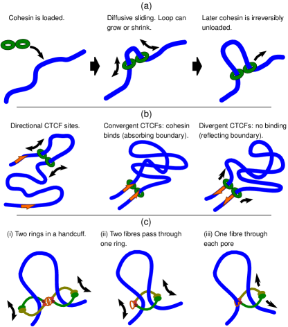
We consider a simple picture where a pair of cohesin complexes are loaded at adjacent positions on a chromatin fibre in a handcuff configuration [Fig. 1(a)]. This is one easy-to-visualize arrangement – everything below also holds for a single ring encircling the fibre at two points [various alternative arrangements are shown in Fig 1(c)]. We then assume that the handcuff can diffuse by sliding along the fibre(s), and a loop will grow and shrink diffusively. Then, sometime later, the cohesin will be unloaded from the chromatin. Importantly, even though the motion is diffusive, this is still a “non-equilibrium”, or active system. In the language of statistical physics, detailed balance is broken since cohesin is always loaded at adjacent points on the chromatin, a loop can grow or shrink, and cohesin can be unloaded (but not loaded) where there is a finite-sized loop (i.e., the system is not time reversible). Biologically, it is thought that chemical energy is required both to load and unload cohesin from the fibre (requiring both ATP hydrolysis and specific loading/unloading factors Murayama2015 ): this provides a mechanistic justification for considering a non-equilibrium model. If, when a diffusing cohesin meets a DNA-bound CTCF protein, it either forms a complex with CTCF or it reflects off it (i.e. just diffuses away again) depending on the CTCF orientation, then this explains the bias for convergent CTCF motifs in loops. Diffusive extrusion is in many ways similar to the active extrusion model discussed above.
Now the question is whether diffusion can generate loops of the required size within the allowed time (the mean residence time of cohesin on DNA). In the active extrusion case the motor would either have to track along the DNA contour (negotiating nucleosomes and other obstacles along the way), or it would have to step along the nucleosomal fibre while maintaining a fixed direction of motion. In the diffusive case, the cohesin ring instead diffuses over whatever fibre structure is present in vivo. The important quantities are therefore the effective diffusion constant for 1D motion along the fibre, and the linear compaction of that fibre [e.g., the number of bp per nanometre (nm)]. A simple theoretical model (full details are given in Ref. PRL ) can put some limits on what these quantities can be in order that diffusive extrusion is viable. For example if we need to generate 100 kbp loops within 25 min, the theory tells us that a 1D diffusion constant of at least 10 kbp2/s is required: if chromatin exists as a 30 nm fibre with about 100 bp/nm, this equates to 0.001m2/s as a minimum diffusion constant. If a more conservative estimate of 20 bp/nm is used (corresponding to a relatively open fibre), then diffusive extrusion is viable if 0.025m2/s or above. Recent in vitro experiments of acetylated cohesin diffusing on chromatin fibres reconstituted in Xenopus egg extract found m2/s; although this was on stretched chromatin in a dilute solution, if the in vivo value is anywhere near this, diffusive extrusion may well be feasible. Other recent in vitro work Stigler2016 studied cohesin on DNA with nucleosome-like obstacles: they found that cohesin did not translocate over obstacles larger than 20 nm, and extrapolating crossing times for smaller obstacles suggested that cohesin would be able to travel 7 kbp in 1 hour (this would correspond to m2/s and a compaction of 3.4 bp/nm, only suitable for naked DNA, hence this extrapolation is in practice a lower bound). If diffusion in vivo is closer to that estimate, then diffusive extrusion would seem less feasible (but see below).
3D simulations of diffusing extruders
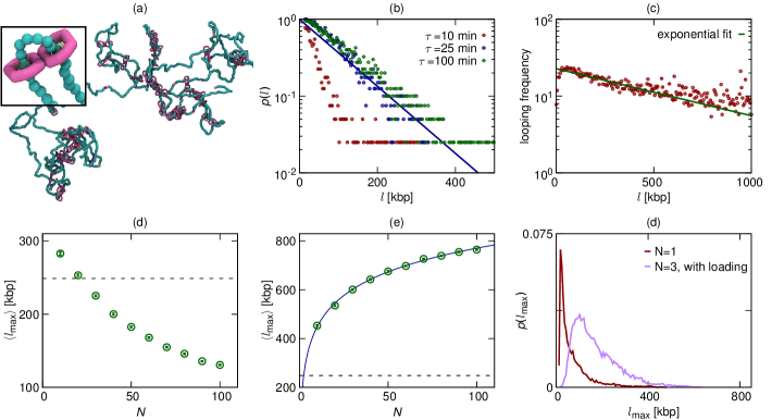
As well as theoretical modelling, we also performed 3D Brownian dynamics simulations (full details are given in Ref. PRL ) to assess whether diffusive extrusion can generate loops, rearranging large stretches of chromatin within the crowded nuclear environment. In these polymer-physics based simulations (which are similar to those in previous studies Brackley2013 ; Barbieri2012 ; Brackley2016 , but with some additions described below) the chromatin fibre is represented as a simple chain of beads connected by springs. Each bead represents 3 kbp of chromatin (though similar results are obtained with different values), and we simulate stochastic diffusive motion of the chain. In previous works on the active extrusion model Sanborn2015 ; Fudenberg2016 , extruding factors were represented by extra springs which move actively along the fibre. Here we explicitly simulate a pair of molecular handcuffs (made up of beads similar to the chromatin) which can slide diffusively on the chromatin. The handcuffs are attached to, and removed from, the fibre at time intervals according to a Poisson process (having a mean residence time ); they are always loaded as a pair onto two adjacent chromatin beads. These dynamics mimic an active, ATP-dependant, loading-unloading process which drives the system away from equilibrium. Figure 2(a) shows a snapshot of part of a simulated fibre.
Figure 2(b) shows a plot of the probability that the simulation generates a loop of a given size, for different values of the unloading rate. There is a significant probability of finding loops of several hundred kbp, implying that the diffusive extrusion mechanism is likely capable of the rearrangement of the chromatin fibre necessary to form a loop. The diffusion constant for cohesin sliding is m2s-1 (which arises naturally from the geometry of our simple bead-based model); this is much smaller than the in vitro value for chromatin in Xenopus extract quoted above, so the simulations provide a conservative estimate of the probability to form loops (still, this value is sufficient to create large loops diffusively).
An interesting feature of the plot in Fig. 2(b) is that the probability of forming a loop is approximately an exponential function of loop length (theoretical modelling predicts an exponential decay with a power-law correction). Standard equilibrium polymer physics would predict that the probability of forming a loop of length is a simple power-law function of deGennes , so the non-equilibrium binding/un-binding kinetics have indeed altered the looping behaviour. HiC data shows that in vivo the probability of two loci interacting decreases with a power-law function of their genomic separation on average LiebermanAiden2009 ; however, ChIA-PET data Tang2015 obtained using an antibody targeting CTCF (which therefore only includes interactions between CTCF bound loci), fit better to an exponential decay [see Fig. 2(c)]. Though there are likely many other factors affecting these data, this suggests that different mechanisms are at play for CTCF loop formation to those behind chromosome interactions in general.
A ratchet effect promotes loop growth over shrinking
In the simulations and theory discussed so far we have considered only a single bound cohesin handcuff, whereas in vivo we might expect many bound cohesins to from a complicated pattern of loops. In the active loop extrusion simulations presented in Refs. Fudenberg2016 ; Sanborn2015 there are many extruding factors which bind at random locations throughout the genome. However, in vivo the cohesin-loading factor (NIPBL in humans, or Scc2 in yeast) binds at preferred genomic locations, and there is some evidence that cohesin is loaded near the promoters of active genes Kagey2010 . Our simulations allow us to investigate both loading at random locations and at preferred sites.
Interestingly the dynamics are very different in the two cases. Figures 2(d) and (e) show results from simple 1D simulations (full details are given in Ref. PRL ) where different numbers of handcuffs are continually being loaded and unloaded from a fibre with a mean residence time of 20 min. If the handcuffs are loaded at randomly chosen locations each time, a series of loops form side by side, competing with each other for space. The average loop size decreases as the number of loops increases. If handcuffs are loaded only at a single location, then the loops tend to be nested inside each other; this leads to an interesting ratchet effect, where the inner loops promote growth over shrinking of the outer loops. This has a simple explanation: when the first handcuff binds it follows a 1D random walk; when the second binds at the same site, it prevents the first from diffusing back towards the loading site, i.e., it exerts an osmotic pressure on the outer handcuff. This osmotic pressure means that the size of the largest loop increases with the number of cohesins. This ratchet effect gives a possible mechanism through which diffusive extrusion might be accelerated, meaning it could be feasible even for smaller 1D diffusion coefficients. Further 3D simulations show that the effect is at work even for a small number of handcuffs [Fig. 2(f)]. Another recent work Yamamoto2017 proposed that the osmotic pressure can be further enhanced by interspersing pairs of cohesin complexes arranged as handcuffs with single cohesins which diffuse along the fibre but are not linked at multiple points so do not form loops.
Domains with diffusive extruders
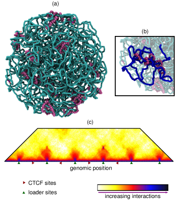
Large-scale Brownian dynamics simulations can also be used to investigate whether diffusing cohesins can generate domains and interaction patterns similar to those seen in HiC data. We performed a simulation of a 15 Mbp region with realistic chromatin density; 32 pairs of handcuffs were continuously added and removed from the fibre with 16 preferred loading sites, and a mean residence time equivalent to 25 min. CTCF sites were placed at 750 or 1500 kbp intervals in either convergent or divergent arrangements. Eight repeat simulations were performed, with CTCF sites populated stochastically such that each simulation could have a different set of sites (to model cell-to-cell variation in CTCF binding); diffusing handcuffs only stick at CTCFs pointing towards them, and when there is a CTCF bound at each side of the handcuff, the unbinding rate is reduced by 10 fold. Figure 3(a) shows a snapshot of the simulation system, and Fig. 3(b) shows a zoom with the region between one pair of convergent CTCF sites (i.e. a TAD) highlighted in blue.
Figure 3(c) shows a contact map generated from these simulations. As in HiC interaction maps, red triangles show domains, and dark spots are seen at the edges of convergent CTCF loop domains. Dark spots are also seen close to the diagonal at loading sites – a feature not normally seen in HiC (though note that there remain few publicly available data sets showing genomic locations where the loader is enriched, and it has yet to be confirmed that cohesin is preferentially loaded at these sites).
While these simulations show that some aspects of the domain structure can indeed be reproduced by our diffusion-based model, we urge caution in expecting such a simple model to be able to replicate interaction maps exactly. For example, the model does not include other DNA-binding proteins that might affect cohesin motion, nor do we attempt to account for active processes such as transcription. Elongating polymerases generate forces and torques (leading to supercoiling Gilbert2014 ; Benedetti2017 ) which may affect cohesin diffusion; indeed recent experiments where the WAPL cohesin unloader protein is knocked down in mouse nuclei show that cohesin collects preferentially between convergent genes, indicating that polymerase can push cohesin along the fibre Haarhuis2017 ; Busslinger2017 . These caveats apply equally to the active loop extrusion model.
Discussion
In this work we have argued that 1D diffusion of cohesin along chromatin can lead to loop extrusion without the need to invoke an explicit motor action. Of course experimental verification of this remains a significant challenge. Nevertheless, we suggest that diffusive extrusion cannot be dismissed in favour of an active extrusion model in the absence of additional experimental evidence.
The in vitro experiments mentioned above Stigler2016 ; Davidson2016 studied the topological loading and diffusion of cohesin rings on stretched DNA templates, and over obstacles. No directed motion was observed, but the diffusivity was found to strongly depend on ATPase activity, salt concentration, and on the way in which the cohesin complex was loaded onto the substrate. Diffusion on reconstituted chromatin in Xenopus egg extracts was also measured Kanke2016 , and it was found that acetylation of the Smc3 sub-unit strongly increased the diffusion coefficient. Together these results suggest that the pore size and diffusivity of cohesin might be regulated by ATP hydrolysis and acetylation in vivo. Recent in vivo studies have shown that knocking-down the loader NIPBL (which leads to loss of chromosome-bound cohesin) leads to loss of looped domains Schwarzer2016 , whereas a knock-down of CTCF affects intra-domain interactions Nora2017 . All these observations are consistent with both the active and diffusive extrusion models. A third possibility is that there is some active translocation, but that the direction is not fixed and the cohesin is “kicked” randomly back and forth along the fibre (the overall effect would look like diffusive motion, but with an increased diffusion constant).
Unlike cohesin, the condensin complex can perform unidirectional active stepping along a stretched DNA template in the presence of ATP Terakawa2017 . This points to the possibility that active extrusion may be at work during mitosis, where condensin plays a central role Nasmyth2001 ; Goloborodko2016 .
Active loop extrusion is often cited as a model for the formation of topological domains, but this is not the only possible mechanism. Another popular model is that chromatin interactions are mediated by transcription factors (or complexes thereof) which can diffuse freely in 3D through the nucleus, and which are multivalent, meaning they can from molecular bridges between different genomic loci Brackley2016 ; Brackley2016b ; Barbieri2017 . This idea has been extensively studied using molecular dynamics and Monte Carlo simulations of simple bead-and-spring polymer physics models (sometimes referred to as the strings-and-binders-switch (SBS) model Barbieri2012 ). Using only limited data about where proteins bind (or using histone modification data to infer protein binding) it is possible to reproduce the TAD patterns observed in HiC data. For example a model using only two factors, one binding to active and one to inactive regions, correctly predicted the locations of 85% of TAD boundaries on chromosome 19 in HUVECs (human umbilical vein endothelial cells) Brackley2016 . This model naturally describes promoter-enhancer interactions mediated by polymerase-transcription factor complexes, or heterochromatin and polycomb repressed regions organised by HP1 and PRC complexes respectively. It can explain the formation of the domains which do not have looping between their boundaries, as well as the larger scale A/B compartment formation, and the fact that compartments are preserved upon loss of chromosome-bound cohesin or CTCF (which is difficult to reconcile with a loop extrusion model). The transcription factor model cannot, however, explain the CTCF motif bias.
It seems likely then, that a complete explanation of genome organisation will require a combination of loop extrusion and multivalent transcription factor models. Even so, as noted above, there are many additional processes which are not yet included in either of these models, so one should not expect to be able to reproduce, for example, all the features of a HiC interaction map. The aim of modelling and simulations therefore should not be to reproduce carbon-copies of experimental results, but should rather be to provide insight, propose new hypothesis, and help direct new experiments.
Acknowledgements This work was supported by ERC (CoG 648050,THREEDCELLPHYSICS), the NIH ID 1U54DK107977-01, CINECA ISCRA Grants No. HP10CYFPS5 and HP10CRTY8P, and by the Einstein BIH Fellowship Award to MN.
References
- (1) E. Lieberman-Aiden et al., “Comprehensive Mapping of Long-Range Interactions Reveals Folding Principles of the Human Genome”, Science 326 289 (2009).
- (2) J. R. Dixon et al., “Topological domains in mammalian genomes identified by analysis of chromatin interactions”, Nature 485 376 (2012).
- (3) T. Sexton et al., “Three-Dimensional Folding and Functional Organization Principles of the Drosophila Genome”, Cell 148 458 (2012).
- (4) S. Rao et al., “A 3D Map of the Human Genome at Kilobase Resolution Reveals Principles of Chromatin Looping”, Cell 159 1665 (2014).
- (5) K. Nasmyth, “Disseminating the Genome: Joining, Resolving, and Separating Sister Chromatids During Mitosis and Meiosis”, Annual Review of Genetics 35 673 (2001).
- (6) E. Alipour and J. F. Marko, “Self-organization of domain structures by DNA-loop-extruding enzymes”, Nucleic Acids Research 40 11202 (2012).
- (7) A. L. Sanborn et al., “Chromatin extrusion explains key features of loop and domain formation in wild-type and engineered genomes”, Proceedings of the National Academy of Sciences USA 112 E6456 (2015).
- (8) G. Fudenberg et al., “Formation of Chromosomal Domains by Loop Extrusion”, Cell Reports 15 2038 (2016).
- (9) E. de Wit et al., “CTCF Binding Polarity Determines Chromatin Looping”, Molecular Cell 60 676 (2015).
- (10) Y. Guo et al., “CRISPR Inversion of CTCF Sites Alters Genome Topology and Enhancer/Promoter Function”, Cell 162 900 (2015).
- (11) F. Uhlmann, “SMC complexes: from DNA to chromosomes.”, Nature Reviews Molecular Cell Biology 17 (2016).
- (12) D. Ivanov and K. Nasmyth, “A Topological Interaction between Cohesin Rings and a Circular Minichromosome”, Cell 122 849 (2005).
- (13) J. Stigler et al., “Single-Molecule Imaging Reveals a Collapsed Conformational State for DNA-Bound Cohesin”, Cell Reports 15 988 (2016).
- (14) I. F. Davidson et al., “Rapid movement and transcriptional re-localization of human cohesin on DNA”, The EMBO Journal 35 2671 (2016).
- (15) A. Lengronne et al., “Cohesin relocation from sites of chromosomal loading to places of convergent transcription”, Nature 430 573 (2004).
- (16) G. A. Busslinger et al., “Cohesin is positioned in mammalian genomes by transcription, CTCF and Wapl”, Nature 544 503 (2017).
- (17) T. Terakawa et al., “The condensin complex is a mechanochemical motor that translocates along DNA”, Science (2017).
- (18) M. Kanke et al., “Cohesin acetylation and Wapl-Pds5 oppositely regulate translocation of cohesin along DNA”, The EMBO Journal 35 2686 (2016).
- (19) D. Gerlich et al., “Live-Cell Imaging Reveals a Stable Cohesin-Chromatin Interaction after but Not before DNA Replication”, Current Biology 16 1571 (2006).
- (20) R. Ladurner et al., “Cohesin’s ATPase Activity Couples Cohesin Loading onto DNA with Smc3 Acetylation”, Current Biology 24 2228 (2014).
- (21) A. S. Hansen et al., “CTCF and cohesin regulate chromatin loop stability with distinct dynamics”, eLife 6 e25776 (2017).
- (22) Y. Murayama and F. Uhlmann, “DNA Entry into and Exit out of the Cohesin Ring by an Interlocking Gate Mechanism”, Cell 163 1628 (2015).
- (23) C. A. Brackley et al., “Non-equilibrium chromosome looping via molecular slip-links”, Physical Review Letters 119 138101 (2017).
- (24) Z. Tang et al., “CTCF-Mediated Human 3D Genome Architecture Reveals Chromatin Topology for Transcription”, Cell 163 1611 (2015).
- (25) C. A. Brackley et al., “Nonspecific bridging-induced attraction drives clustering of DNA-binding proteins and genome organization”, Proceedings of the National Academy of Sciences USA 110 E3605 (2013).
- (26) M. Barbieri et al., “Complexity of chromatin folding is captured by the strings and binders switch model”, Proceedings of the National Academy of Sciences USA 109 16173 (2012).
- (27) C. A. Brackley et al., “Simulated binding of transcription factors to active and inactive regions folds human chromosomes into loops, rosettes and topological domains”, Nucleic Acids Research 44 3503 (2016).
- (28) P.-G. d. Gennes. Scaling concepts in polymer physics, (Cornell University Press, Ithaca [N.Y.] and London 1979).
- (29) M. H. Kagey et al., “Mediator and cohesin connect gene expression and chromatin architecture”, Nature 467 430 (2010).
- (30) T. Yamamoto and H. Schiessel, “Osmotic mechanism of the loop extrusion process”, Physical Review E 96 030402(R) (2017).
- (31) N. Gilbert and J. Allan, “Supercoiling in DNA and chromatin”, Current Opinion in Genetics & Development 25 15 (2014).
- (32) F. Benedetti et al., “Transcription-induced supercoiling explains formation of self-interacting chromatin domains in S. pombe”, Nucleic Acids Research 45 9850 (2017).
- (33) J. H. Haarhuis et al., “The Cohesin Release Factor WAPL Restricts Chromatin Loop Extension”, Cell 169 693 (2017).
- (34) W. Schwarzer et al., “Two independent modes of chromatin organization revealed by cohesin removal”, Nature (2017).
- (35) E. P. Nora et al., “Targeted Degradation of CTCF Decouples Local Insulation of Chromosome Domains from Genomic Compartmentalization”, Cell 169 930 (2017).
- (36) A. Goloborodko et al., “Compaction and segregation of sister chromatids via active loop extrusion”, eLife 5 e14864 (2016).
- (37) C. A. Brackley et al., “Predicting the three-dimensional folding of cis-regulatory regions in mammalian genomes using bioinformatic data and polymer models”, Genome Biology 17 59 (2016).
- (38) M. Barbieri et al., “Active and poised promoter states drive folding of the extended HoxB locus in mouse embryonic stem cells”, Nature Structural & Molecular Biology 24 515 (2017).