:
\theoremsep
\jmlrvolume
\jmlryear
\jmlrsubmitted
\jmlrpublished
\jmlrworkshop
Improving Lesion Detection by exploring bias on
Skin Lesion dataset
Abstract
All datasets contain some biases, often unintentional, due to how they were acquired and annotated. These biases distort machine-learning models’ performance, creating spurious correlations that the models can unfairly exploit, or, contrarily destroying clear correlations that the models could learn. With the popularity of deep learning models, automated skin lesion analysis is starting to play an essential role in the early detection of Melanoma. The ISIC Archive Codella et al. (2019) is one of the most used skin lesion sources to benchmark deep learning based tools. Bissoto et al. Bissoto et al. (2019) experimented with different bounding-box based masks and showed that deep learning models could classify skin lesion images without clinically meaningful information in the input data. Their findings seem confounding since the ablated regions (random rectangular boxes) are not significant. The shape of the lesion is a crucial factor in the clinical characterization of a skin lesion Silveira et al. (2009). In that context, we performed a set of experiments that generate shape-preserving masks instead of rectangular bounding-box based masks. A deep learning model trained on these shape-preserving masked images does not outperform models trained on images without clinically meaningful information. That strongly suggests spurious correlations guiding the models. We propose use of general adversarial network (GAN) to mitigate the underlying bias.
keywords:
bias, health, skin, cancer1 Introduction
Skin cancer is a growing problem globally, and it is the most common cause of cancer in the U.S Guy Jr et al. (2015b). The World Health Organization (WHO) predicts that a 10% decrease in the depleting ozone layer will cause more than 300,000 additional cases of skin cancer WHO (2019). Like any other cancer, early lesion detection is key to improved clinical outcomes. However, lack of access to medical experts, especially in developing nations or areas of the world with inadequate screening facilities, implies higher mortality due to missed diagnosis or diagnosis once cancer has metastasized Sandru et al. (2014). Manually diagnosing is ineffective and expensive Guy Jr et al. (2015a). Automated skin cancer lesion detection has potential benefits such as increasing efficiency, reproducibility, coverage of screening programs, reducing barriers to access, and improving patient outcomes by providing early detection and treatment Tripp et al. (2016). An algorithm to detect referable skin cancer is needed to maximize the clinical utility of automated lesion detection. Researchers have used machine learning for various classification tasks in recent years, including automated lesion detection for skin cancer. Identifying candidate regions in medical images is of the most significant importance since it provides intuitive illustrations for doctors and patients of inferred diagnosis. Recently, advances in Deep Learning have dramatically improved the performance of skin lesion detection. Most of these Deep Learning systems treat Convolutional Neural Network (CNN) as a black box, lacking comprehensive explanation. Diagnoses using deep neural nets with high precision and low recall on a mobile phone are feasible de Carvalho et al. (2019). However, the state-of-the-art models are trained on mostly skin lesion datasets that belong to fair-skinned individuals rather than dark-skinned persons. An AI model trained on predominantly lighter skin background in the image may perform sub-optimally in darker skin backgrounds poorly represented in the training set. Even though the risk of developing skin cancer is relatively high among the light-skinned population, people with dark skin are also at risk. They are frequently diagnosed at later stages Hu et al. (2006). Skin cancer accounts for 4 to 5%, 2 to 4%, 1 to 2% of all cancers in Hispanics, Asians, and Blacks, respectively Gloster Jr and Neal (2006). Hence, deep learning frameworks validated for skin cancer diagnosis in fair-skinned people have a greater risk of misdiagnosing those with darker skin Marcus and Davis (2019). In a recent study, Han et al. Han et al. (2018) trained a deep learning algorithm on a training dataset consisting of skin lesions from Asians. They reported an accuracy of 81% on the Asian testing set, whereas they reported accuracy of only 56% on the Dermofit dataset, which consists of Caucasian people’s skin lesions. Therefore, this drop-in accuracy signifies a lack of transferability of the learned features of deep learning algorithms across datasets that contain persons of a different race, ethnicity, or population. We argue that using the immediate area around the lesion can reduce the pressure on having a well-diversified skin color representation in the training data set. This approach’s inspiration stems from the observation that physicians grading lesions are primarily driven by the morphology of the lesion and less so by the color of the skin surrounding the lesion Silveira et al. (2009) Soceity (2019). Gathering a fair amount of balanced benign and malignant skin lesion images for different skin profiles is a gigantic task. Bissoto et al. Bissoto et al. (2019) highlighted that the challenge is due to the vast visual variability of skin lesion and the subtlety of the cues that differentiate benign and malignant cases. They experimented with different bounding box based masks and showed that deep learning models could classify skin lesion images without clinically meaningful information. The shape of the lesion is a crucial factor in the clinical characterization of a skin lesion Silveira et al. (2009). In that context, we decided to extend the work by Bissoto et al. Bissoto et al. (2019) and experiment with shape-preserving masks. In this paper, we revisit a classical background segmentation approach using Otsu based adaptive thresholding and combine it with binary morphology operations to reduce noise while preserving the lesion shape in the image M et al. (1998); Khan et al. (2016).
2 Dataset
In this study we use an open-source dataset - HAM10000 Tschandl et al. (2019). HAM10000 dataset is commonly used as a benchmark database for academic machine learning purposes. We used the training set part of this dataset consisting of 10015 dermatoscopic images with a size of 450x600. In this paper, we use a binary classification model, and we need to create a binary label - Benign vs. Malignant for the dataset. However, HAM10000 Metadata provides labels for different types of lesions classified by histopathology. For example, the BCC label stands for basal cell carcinoma, and MEL stands for melanoma, representing cancer. The label BKL stands for benign keratosis like lesions. This information is provided as a part of the Kaggle data set Tschandl et al. (2019). We used popular web search engines such as Bing and Google to map the labels provided to malignant or benign categories. This map is used to relabel the original labels into malignant and benign categories.
3 Image Processing Segmentation
Segmentation and classification of skin lesions using deep learning techniques have been of key interest Esteva et al. (2017). The basic segmentation is obtained by applying the following filters to the source image:
-
(1)
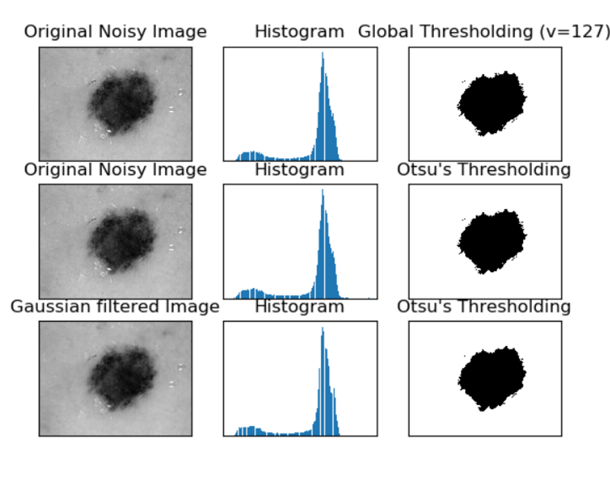
Gray-scale conversion of the color lesion image (Fig. 1 column 1). We experimented with using the original image and also with Gaussian smoothing.
-
(2)
Extract a binary mask using global thresholding and Otsu thresholding Otsu (1979). We found Otsu to be more robust to different image variations. It automatically can find the optimal partitioning point in the grayscale image into the foreground and background, as shown by the bi-modal histogram in the middle column of Figure 1.
- (3)
-
(4)
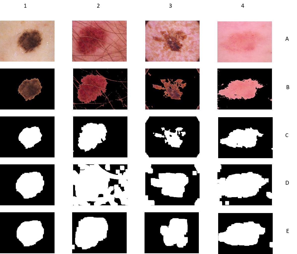
We apply dilation binary morphology operation Haralick et al. (1987) to preserve the shape of the lesion and also capture the nearby region of the lesion similar to what a physician would do (Fig. 2, row D). However, as you can see, Fig. 2(D2-D4) dilation operation also increases the noise. Fig. 2(row E) shows how combining binary erosion and dilation can get accurate capturing of the skin lesion. In Figure 2(column 4), we can see even when the lesion is very faint, the segmentation mask captures the lesion area with reasonably good accuracy.
4 Lesion Classification using Preprocessed Images
A key parameter when using binary morphology is the size and shape of the morphology kernel. Our goal was to generate different masks by varying the opening kernel’s shapes and the dilation kernels. We varied the size of square kernel shapes (0, 5, 8, 10, 12, 15 pixels) and generated masks with different combinations (a total of 15 mask set) for the experiments. We trained a simple CNN model to prove the hypothesis using shape-preserving masks. We use an ImageNet Russakovsky et al. (2015) pre-trained VGG16 Simonyan and Zisserman (2014) model and finetune it with our own data. VGG16 is a 16-layer CNN used by the Visual Geometry Group (VGG) at Oxford University. In 2014, the model won the 2nd place in the ILSVRC (ImageNet) competition, achieving a 7.5% top 5 error rate on the validation set. After defining the fully connected layer architectures, we load the ImageNet pre-trained weight to the model. We then truncate the original softmax layer and replace it with our number of class labels. Then we finetune the model by minimizing the cross-entropy loss function using the stochastic gradient descent (SGD) algorithm. We keep the finetuned CNN very simple to run the evaluation quickly and directionally observe how changing the different mask sizes affect the accuracy of skin lesion characterization.
| Experiment# | #Dilate_#Clean | TestAccuracy(in %) |
|---|---|---|
| RGB Only | ||
| 1 | Baseline | 82.2 |
| Mask Only | ||
| 2 | (10_10) | 80.88 |
| 3 | (15_15) | 80.88 |
| 4 | (10_5) | 80.88 |
| 5 | (50_80) | 80.88 |
| Mask with RGB | ||
| 6 | (10_10) | 80.88 |
| 7 | (15_15) | 81.7 |
| 8 | (10_5) | 80.06 |
| 9 | (50_80) | 80.88 |
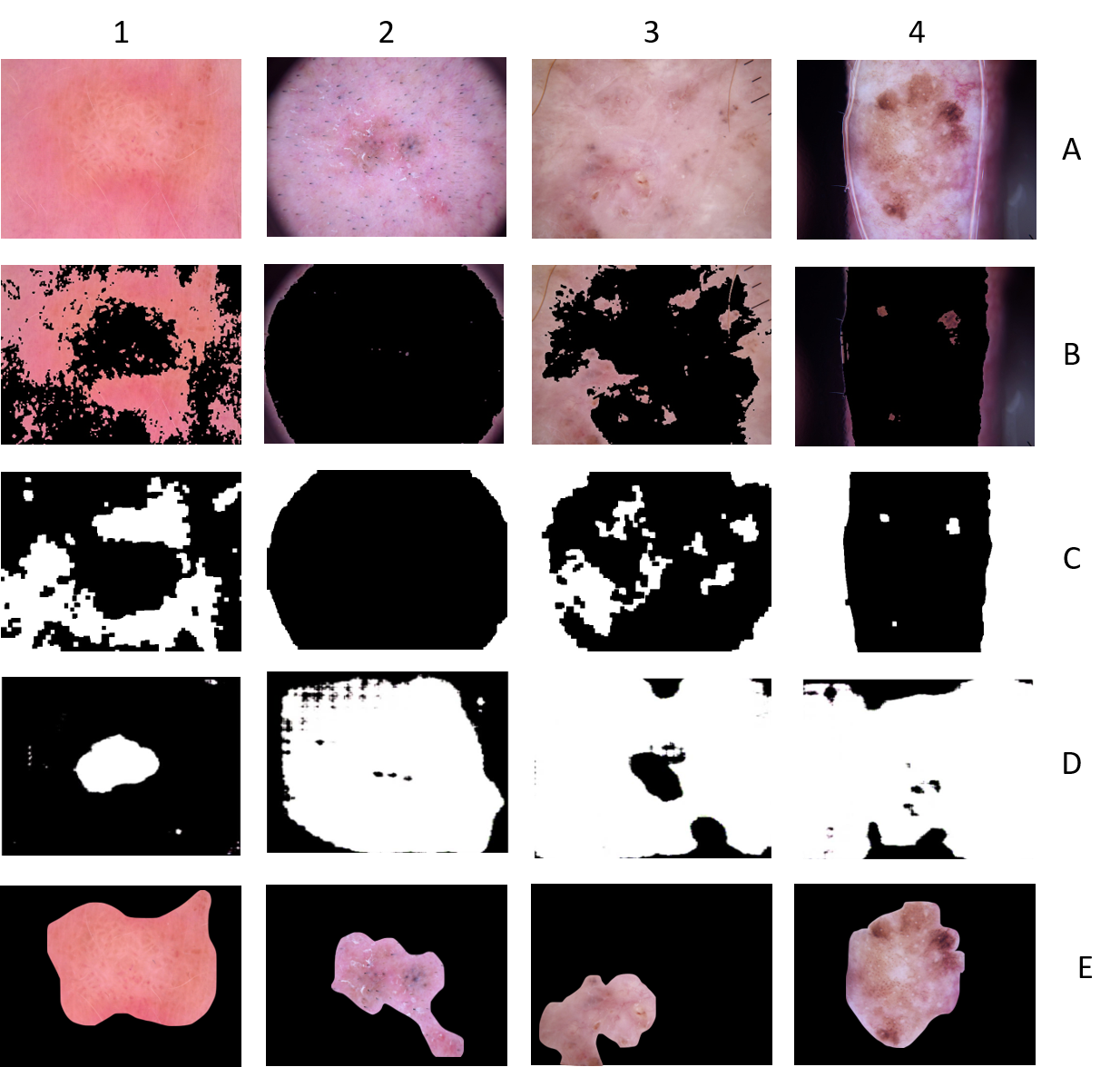
In Table 1, we explore different mask-only and RGB with mask options with CNN. In experiment 1, we ran a baseline test to see how well the RGB does. Although the numbers might seem promising, the model does not take into account different skin tones. In experiments 2, 3, and 4, we started experimenting with varying commands of morphology. After trying different amounts of the mask to maximize the accuracy, we noticed that the accuracy numbers were coming out to be very similar, irrespective of the various masks. We also tried with extreme numbers such as experiment 5, but identical accuracy numbers showed up. This result confirmed the results shown by Bissoto et al. Bissoto et al. (2019). Next, we experimented with shape-preserving masks. In experiments 6,7 and 8, we tried using the mask on top of the RGB images. However, the accuracy numbers came in a similar range of 80/82 percent accuracy as with mask-only experiments in 2, 3, and 4. We also tried extreme masking with RBG, as shown in experiment 9, but similar accuracy numbers showed up. In conclusion, regardless of the amount of dilation, the classifier’s accuracy does not change, remaining between (81%-82.7%). Additionally, in Figure 3, we can see the segmentation algorithm fails in images that have low contrast or very fuzzy boundaries. Fig 3A show very poor image contrast. The mask captures the periphery of the lesion, but the low contrast and low-intensity interior are missed. This is clearly illustrated in Fig 3 row B1-3 and C1-3. Fig 3A2 is so subtle that the mask completely fails. Note row E corresponds to outlines drawn by a certified Radiologist. For these four images, the radiologist remarked that they were challenging because of poor contrast and, in some cases, skin damaged due to scratching or inflammatory effects from localized irritations. The radiologist also consulted another colleague with dermatology experience. They concluded that for these types of samples, the inter-expert disagreement would be very high.
5 Generative Model based Segmentation
As we show with the demonstration mentioned above, skin cancer lesion classification fails with simple segmentation masks, and we want to evaluate more sophisticated deep generative models like Pix2PixGAN Isola et al. (2017). In recent years, deep learning is shown to be successful in visual perception and the semantic segmentation of images. General Adversarial Networks or GANs can be briefly divided into two parts - encoder and decoder. The encoder is a convolution neural network that extracts features from the input image, such as the skin image’s features. The decoder will up-sample the extracted features to the resulting image that we desired as the skin lesion segmentation in our case. Researchers have already used variations of U-NET Ronneberger et al. (2015), Residual U-NET Zhang et al. (2018), and R2U-Net Alom et al. (2018) on ISIC data set as shown in Kaggle (2019). However, regardless of race/background, we want anyone to have immediate and less costly access to find out if they had skin cancer. Bissoto et al. Bissoto et al. (2018) have shown the use of Pix2PixHD GAN for skin images. However, it requires the use of high quality altered images, which is not a guarantee for people in developing nations without access to technology equipped with better quality cameras. This leads us to the idea of improving lesion detection without bias by using Pix2PixGAN. We trained a Pix2PixGAN to make the results more precise and universal.
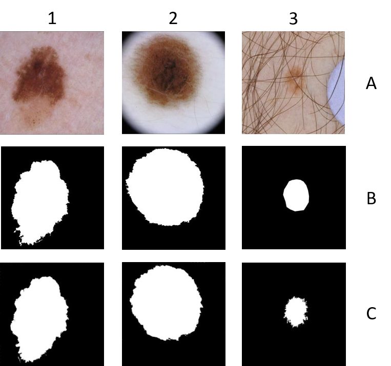
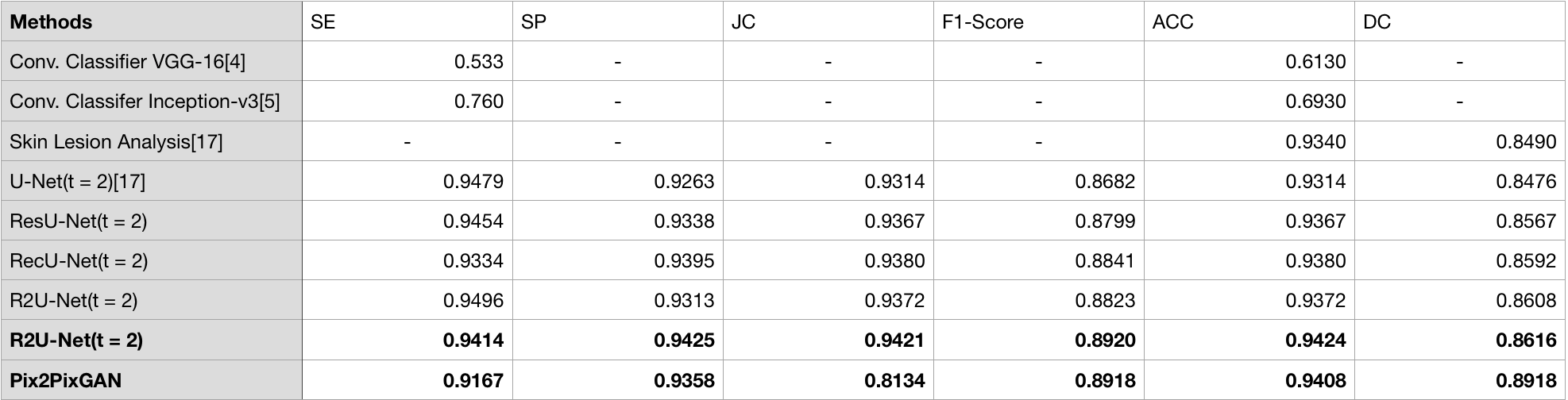
In Fig 4, it shows the results of our trained Pix2PixGAN model on the ISIC dataset. We chose different types of images of skin lesions to point out that Pix2PixGAN was able to segment the correct part of the lesion regardless of the background. In column 1, the model was able to correctly detect the lesion, matching exactly to the ground truth of the corresponding image. In column 2, Fig 4, there is a bit of a background for the skin lesion. However, the GAN was still able to predict the segmented image flawlessly. The skin lesion in column 3 Fig 4 proves how accurate our model is because of the noises in the image, such as hair and random patch in the corner. We cannot always guarantee that users will have images with no distraction, and Pix2PixGAN proves to be still successful under those situations. Figure 4 Fig 4 also supports the strong evaluation metrics provided in table 2, as evidenced by the model’s accuracy. In Fig 3, we compare the mask corresponding to the input images compared to the Pix2PixGAN(using the Kaggle dataset). In all the columns(except column 3), Pix2PixGAN was able to detect the skin lesion at a much higher accuracy than the mask. The mask(Row C) detected the background in the images, whereas the GAN was able to detect parts of the skin lesion. In columns 1 and 2, Fig 4, Pix2PixGAN was able to detect the lesion pretty accurately. However, in column 3 Fig 4, the GAN was unable to detect the skin lesions due to them being scattered on the image, and the model was never trained in such cases. In the last column, Fig 4, again, the GAN was met with an unexpected image and was unable to detect the skin lesion. However, the model produced a better image than the mask(only to some extent). The Pix2PixGAN model could not detect as accurately because it was met with an unexpected input that it was not trained on. We shared these images with two expert doctors with experience in radiology and dermatology. After manually outlining the images, they confirmed that these images were challenging to outline even humans. This indicates our approach is robustly tested against diverse test cases. Fig 3D(2-4) shows the GAN labeled regions were more expansive. This gives us an additional signal in the application to flag such images for manual intervention. For Fig 3D(1), the GAN outlined the lesion area while the physician included additional areas around the lesion. Such subjective variations in interpretation make skin-lesion detection challenging even for well-trained specialists. We plan to incorporate this additional clinical insight into future GAN refinements. As seen in Fig 5, different types of metrics were used to provide quantitative results with the Pix2PixGAN model: Accuracy(ACC), Sensitivity(SE), Specificity(SP), F-1 score, and Dice Coefficient(DC). Compared to the R2-Unet model, Pix2PixGAN had approximately the same accuracy(0.94). Accuracy is calculated using this formula:(TP + TN)/(TP + TN + FP + FN). TP is true positive; TN is true negative; FP is false positive; FN is false negative. Moreover, the high accuracy of the Pix2PixGAN model proves it can perform the segmentation task more effectively than the U-Net and ResUnet models, which had lower accuracies. Pix2PixGAN’s accuracy portrays its accurate results in end-to-end segmentation tasks. The sensitivity of the Pix2PixGAN model was 0.91, and the specificity of the model was roughly 0.93. The formula used to calculate sensitivity is TP/(TP + FN). The formula used to calculate specificity is TN/(TN + FP). Pix2PixGAN’s F1-score proves the model as successful as R2-Unet, as both have similar scores(0.89). The F1-score takes the average of both precision(for Pix2PixGAN, it was 0.89) and recall. The Dice Coefficient(DC) of Pix2PixGAN is 0.89, which is higher than R2-Unet’s(0.86). The DC score stands for Dice Coefficient, which is the size of the overlap of the two segmentations divided by the two objects’ total size. The DC score is often used as a reproducibility validation metric, denoting that Pix2PixGAN would produce these segmented images with the same accuracy multiple times, whereas R2-Unet would have a lower chance of doing that. This is especially important if multiple people will be using this app, as Pix2PixGAN will be more reliable for producing segmented images.
6 Conclusion
Bissoto et al. Bissoto et al. (2019) experimented with different bounding-box based masks and showed that deep learning models could classify skin lesion images without clinically meaningful information. Our result extends their work and shows that even shape-preserving masks fail to improve classification accuracy over deep learning models that classify skin lesion masked images without clinically meaningful information. We conclude that the simple CNN type classifiers are by themselves inadequate in modeling skin cancer lesions. However, generating robust segmentation is critical to remove bias in skin cancer data. In our data set, we also found that a handful of images showed partial or no segmentation at all due in part to image quality, intensity variation, lens artifacts etc. Although the number of failures is small but due to the nature of the problems with lives at stake, we need a better segmentation algorithm that is more adaptable to subtle variations in skin lesion color/shapes and generalizes well in the face of wider variations within the population. The combination of segmented skin lesions along with a more extensive data set (several hundreds of thousands) Esteva et al. (2017), is likely to be a winning combination. \acksWe sincerely thank Dr. Sayan Pathak, Affl Prof of CSE at Indian Institute of Technology and former faculty at Dept. of Bioengineering at the University of Washington, Seattle, WA, for his invaluable guidance and insights. We also thank Dr. Manjari Gajra of School of Medical Sciences, University of Manchester and Dr. Ryan Pathak, Consultant Radiologist at Salford Royal Hospital for their manual annotation of the lesions and evaluation of the predicted annotations.
References
- Alom et al. (2018) Md Zahangir Alom, Mahmudul Hasan, Chris Yakopcic, Tarek M Taha, and Vijayan K Asari. Recurrent residual convolutional neural network based on u-net (r2u-net) for medical image segmentation. arXiv preprint arXiv:1802.06955, 2018.
- Bissoto et al. (2018) Alceu Bissoto, Fábio Perez, Eduardo Valle, and Sandra Avila. Skin lesion synthesis with generative adversarial networks. In OR 2.0 Context-Aware Operating Theaters, Computer Assisted Robotic Endoscopy, Clinical Image-Based Procedures, and Skin Image Analysis, pages 294–302. Springer, 2018.
- Bissoto et al. (2019) Alceu Bissoto, Michel Fornaciali, Eduardo Valle, and Sandra Avila. (de) constructing bias on skin lesion datasets. In Proceedings of the IEEE Conference on Computer Vision and Pattern Recognition Workshops, pages 0–0, 2019.
- Burdick et al. (2018) Jack Burdick, Oge Marques, Janet Weinthal, and Borko Furht. Rethinking skin lesion segmentation in a convolutional classifier. Journal of digital imaging, 31(4):435–440, 2018.
- Codella et al. (2019) Noel Codella, Veronica Rotemberg, Philipp Tschandl, M Emre Celebi, Stephen Dusza, David Gutman, Brian Helba, Aadi Kalloo, Konstantinos Liopyris, Michael Marchetti, et al. Skin lesion analysis toward melanoma detection 2018: A challenge hosted by the international skin imaging collaboration (isic). arXiv preprint arXiv:1902.03368, 2019.
- Codella et al. (2018) Noel CF Codella, David Gutman, M Emre Celebi, Brian Helba, Michael A Marchetti, Stephen W Dusza, Aadi Kalloo, Konstantinos Liopyris, Nabin Mishra, Harald Kittler, et al. Skin lesion analysis toward melanoma detection: A challenge at the 2017 international symposium on biomedical imaging (isbi), hosted by the international skin imaging collaboration (isic). In 2018 IEEE 15th International Symposium on Biomedical Imaging (ISBI 2018), pages 168–172. IEEE, 2018.
- de Carvalho et al. (2019) Tiago M de Carvalho, Eline Noels, Marlies Wakkee, Andreea Udrea, and Tamar Nijsten. Development of smartphone apps for skin cancer risk assessment: Progress and promise. JMIR Dermatology, 2(1):e13376, 2019.
- Esteva et al. (2017) Andre Esteva, Brett Kuprel, Roberto A. Novoa, Justin Ko, Susan M. Swetter, Helen M. Blau, and Sebastian Thrun. Dermatologist-level classification of skin cancer with deep neural networks. Nature, 542:115–118, 2017.
- Gloster Jr and Neal (2006) Hugh M Gloster Jr and Kenneth Neal. Skin cancer in skin of color. Journal of the American Academy of Dermatology, 55(5):741–760, 2006.
- Guy Jr et al. (2015a) Gery P Guy Jr, Steven R Machlin, Donatus U Ekwueme, and K Robin Yabroff. Prevalence and costs of skin cancer treatment in the us, 2002- 2006 and 2007- 2011. American journal of preventive medicine, 48(2):183–187, 2015a.
- Guy Jr et al. (2015b) Gery P Guy Jr, Cheryll C Thomas, Trevor Thompson, Meg Watson, Greta M Massetti, and Lisa C Richardson. Vital signs: melanoma incidence and mortality trends and projections—united states, 1982–2030. MMWR. Morbidity and mortality weekly report, 64(21):591, 2015b.
- Han et al. (2018) Seung Seog Han, Myoung Shin Kim, Woohyung Lim, Gyeong Hun Park, Ilwoo Park, and Sung Eun Chang. Classification of the clinical images for benign and malignant cutaneous tumors using a deep learning algorithm. Journal of Investigative Dermatology, 138(7):1529–1538, 2018.
- Haralick et al. (1987) R.M. Haralick, S.R. Sternberg, and X. Zhuang. Image analysis using mathematical morphology. IEEE Pattern Analysis and Machine Intelligence, 9(4):532–550, 1987.
- Hu et al. (2006) Shasa Hu, Rita M Soza-Vento, Dorothy F Parker, and Robert S Kirsner. Comparison of stage at diagnosis of melanoma among hispanic, black, and white patients in miami-dade county, florida. Archives of Dermatology, 142(6):704–708, 2006.
- Isola et al. (2017) Phillip Isola, Jun-Yan Zhu, Tinghui Zhou, and Alexei A Efros. Image-to-image translation with conditional adversarial networks. In Proceedings of the IEEE conference on computer vision and pattern recognition, pages 1125–1134, 2017.
- Kaggle (2019) Kaggle. skin-cancer-segmentation-on-kaggle-skin, 2019.
- Khan et al. (2016) Khan Bahadar Khan, Amir A Khaliq, and Muhammad Shahid. Correction: A morphological hessian based approach for retinal blood vessels segmentation and denoising using region based otsu thresholding. Plos One, 11(9), Jan 2016. 10.1371/journal.pone.0162581.
- Li and Shen (2018) Yuexiang Li and Linlin Shen. Skin lesion analysis towards melanoma detection using deep learning network. Sensors, 18(2):556, 2018.
- M et al. (1998) Cheriet M, J. N. Said, and C. Y. Suen. A recursive thresholding technique for image segmentation. IEEE Transactions on Image Processing, 7(6):918–921, June 1998. ISSN 1941-0042. 10.1109/83.679444.
- Marcus and Davis (2019) Gary Marcus and Ernest Davis. Rebooting AI: building artificial intelligence we can trust. Pantheon, 2019.
- Otsu (1979) N. Otsu. A threshold selection method for gray gevel histograms. IEEE Transactions on System, Man and Cybernetics, 9(1):62–66, 1979.
- Ronneberger et al. (2015) Olaf Ronneberger, Philipp Fischer, and Thomas Brox. U-net: Convolutional networks for biomedical image segmentation. In International Conference on Medical image computing and computer-assisted intervention, pages 234–241. Springer, 2015.
- Russakovsky et al. (2015) Olga Russakovsky, Jia Deng, Hao Su, Jonathan Krause, Sanjeev Satheesh, Sean Ma, Zhiheng Huang, Andrej Karpathy, Aditya Khosla, Michael Bernstein, et al. Imagenet large scale visual recognition challenge. International journal of computer vision, 115(3):211–252, 2015.
- Sandru et al. (2014) A Sandru, S Voinea, E Panaitescu, and A Blidaru. Survival rates of patients with metastatic malignant melanoma. Journal of medicine and life, 7(4):572, 2014.
- Silveira et al. (2009) Margarida Silveira, Jacinto C. Nascimento, Jorge Marques, Marcal Andre, Teresa Mendonca, Syogo Yamauchi, Junji Maeda, and Jorge Rozeira. Comparison of segmentation methods for melanoma diagnosis in dermoscopy images. IEEE Journal of Selected Topics in Signal Processing, 3(1):35–45, 2009.
- Simonyan and Zisserman (2014) Karen Simonyan and Andrew Zisserman. Very deep convolutional networks for large-scale image recognition. arXiv preprint arXiv:1409.1556, 2014.
- Soceity (2019) American Cancer Soceity. Signs and symptoms of melanoma skin cancer. 2019.
- Tripp et al. (2016) Mary K Tripp, Meg Watson, Sophie J Balk, Susan M Swetter, and Jeffrey E Gershenwald. State of the science on prevention and screening to reduce melanoma incidence and mortality: The time is now. CA: a cancer journal for clinicians, 66(6):460–480, 2016.
- Tschandl et al. (2019) Philipp Tschandl, Cliff Rosendahl, and Harald Kittler. The ham10000 dataset: A large collection of multi-source dermatoscopic images of common pigmented skin lesions; 2018. Preprint. Available from: https://arxiv. org/ftp/arxiv/papers/1803/1803.10417. pdf. Cited, 4, 2019.
- WHO (2019) WHO. Skin cancers, 2019.
- Zhang et al. (2018) Zhengxin Zhang, Qingjie Liu, and Yunhong Wang. Road extraction by deep residual u-net. IEEE Geoscience and Remote Sensing Letters, 15(5):749–753, 2018.