Band Depopulation of Graphene Nanoribbons
Induced by Chemical Gating with Amino Groups
Abstract
The electronic properties of graphene nanoribbons (GNRs) can be precisely tuned by chemical doping. Here we demonstrate that amino (NH2) functional groups attached at the edges of chiral GNRs (chGNRs) can efficiently gate the chGNRs and lead to the valence band (VB) depopulation on a metallic surface. The NH2-doped chGNRs are grown by on-surface synthesis on Au(111) using functionalized bianthracene precursors. Scanning tunneling spectroscopy resolves that the NH2 groups significantly up-shift the bands of chGNRs, causing the Fermi level crossing of the VB onset of chGNRs. Through density functional theory simulations we confirm that the hole-doping behavior is due to an upward shift of the bands induced by the edge NH2 groups.
Graphene nanoribbons (GNRs) have emerged as a promising material for nanoelectronics Llinas et al. (2017) since they combine many of the extraordinary properties of the parent graphene Han et al. (2014a); Moreno et al. (2018) with a high tunability of their electronic band structureYazyev (2013). Contrary to graphene, GNRs are frequently semiconducting materials, thus offering excellent perspectives for their utilisation as electronic components such as diodes Kargar and Lee (2009) or transistors Schwierz (2010); Bennett et al. (2013); Llinas et al. (2017); Moreno et al. (2018). A fascinating aspect of narrow GNRs is that their electronic properties can be tuned through the precise control of their atomic structure, which can be achieved thanks to the recent development of bottom-up on-surface synthesis (OSS) strategies Talirz et al. (2016); Clair and De Oteyza (2019). These strategies rely on the careful design of suitable molecular precursors with specific shape and chemical composition to steer a step-wise reaction on a metal surface, leading to extended and atomically precise GNRs. An ample library of precursors and reaction pathways has been constructed in the last years, incorporating successful examples of precise control over the GNR’s widthCai et al. (2010); Chen et al. (2013); Abdurakhmanova et al. (2014); Basagni et al. (2015); Kimouche et al. (2015); Zhang et al. (2015); Talirz et al. (2017); Merino-Díez et al. (2017), orientation and edge topologyHan et al. (2014b); Liu et al. (2015); Ruffieux et al. (2016); De Oteyza et al. (2016); Rizzo et al. (2018); Gröning et al. (2018); Merino-Díez et al. (2018); Moreno et al. (2019), which resulted in large variations of their electronic structure. Furthermore, OSS strategies allow to easily combine several precursors on a surface for fabricating complex structures such as atomically sharp graphene heterojunctions Cai et al. (2014); Chen et al. (2015), quantum dotsCarbonell-Sanromà et al. (2017a); Wang et al. (2017), or hybrid systems composed of metal-organic molecules embedded in GNRs Li et al. (2018); Su et al. (2018); Li et al. (2019), demonstrating their potential role for GNRs based nanoelectronics.
Since most GNRs are semiconducting, a promising method for tuning their band structure is the electrostatic gating effect induced by doping Krull et al. (2013). Chemical doping of GNRs has been achieved through modification of the molecular precursors to either incorporate substitutional heteroatoms in the carbon backbone Cai et al. (2014); Bronner et al. (2013); Kawai et al. (2015); Cloke et al. (2015); Nguyen et al. (2016); Carbonell-Sanromà et al. (2017a); Durr et al. (2018); Carbonell-Sanromà et al. (2018); Wang et al. (2018); Kawai et al. (2018) or by adding functional groups at the edges Carbonell-Sanromà et al. (2017b). However, the band structure of the substitutionally-doped systems could not always be simply related to the pristine one (CB)Kawai et al. (2015); Cloke et al. (2015); Carbonell-Sanromà et al. (2017a); Durr et al. (2018); Carbonell-Sanromà et al. (2018); the embedded heteroatoms have a profound impact on the bands’ symmetries and character Cao et al. (2017). Instead, chemical functionalization of the GNR edges promisingly behaved as an efficient method to fine-tune the electronic structure of pristine GNRs. In the most simple scenarios, the effect of attached chemical groups can be described as an effective electrostatic gating of the native band structure Cai et al. (2014); Carbonell-Sanromà et al. (2017b); Wang et al. (2018). The magnitude of edge chemical gating is small, to date barely restricted to sub-gap down-shifts of the band structure.
Here, we report on the very efficient chemical gating of amino (NH2) functional groups attached at the edges of narrow chiral GNRs with a sequence of 3 zigzag and 1 armchair sites ((3,1) chGNRs). The electron donating character of NH2 is inherited by the GNR, resulting in an effective GNR charging by hole injection and valence band depopulation on a Au(111) surface. Experimentally, NH2 end groups were substituted at the edges of pristine chGNRs through bottom-up synthesis with modified precursors. Our results satisfactorily demonstrate that the electropositive affinity of the dopant is transmitted to the chGNR carbon backbone Carbonell-Sanromà et al. (2017b), which on a Au(111) surface becomes strongly hole doped. Through a combination of scanning tunnelling microscopy (STM) and spectroscopy (STS), X-ray photoelectron spectroscopy (XPS) measurements and density functional theory (DFT) calculations we show that the NH2 groups induce a substantial up-shift of the GNR bands, and cause their VB to cross the Fermi level. The effective depopulation of the VB depends on the number of NH2 groups per unit cell surviving the reaction, as well as on their relative alignment.
.1 Results/Discussion
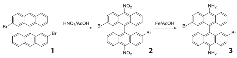
Synthesis of the molecular precursor: To functionalize (3,1) chGNRs with NH2 groups at the edge (named as NH2-chGNRs), we prepared the molecular precursor 2,2’-dibromo-[9,9’-bianthracene]-10,10’-diamine (3, Fig. 2) in two steps from 2,2’-dibromo-9,9’-bianthracene (1). First, selective double nitration of compound 1 led to the synthesis of compound 2, which was isolated with 64% yield. Then, Fe-promoted reduction of bianthracene 2 in acetic acid afforded the GNR precursor 3 with 98% yield (see Supporting Information for details).
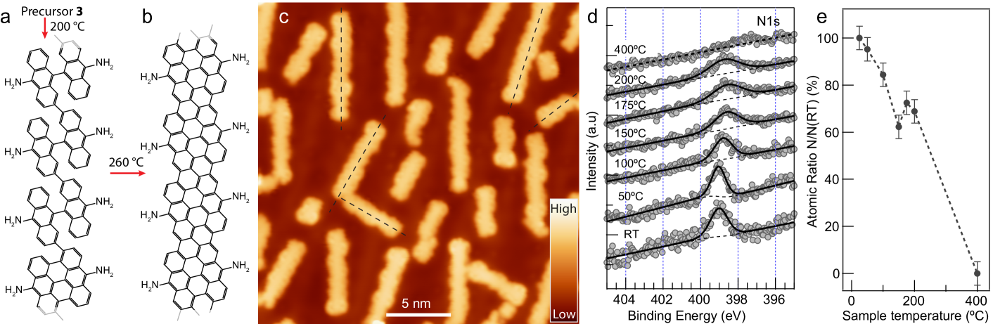
The molecular precursor 3 was sublimated from a Knudsen cell at 212 ∘C onto a clean Au(111) substrate kept at room temperature. The precovered sample was gradually annealed first to 200 ∘C to induce the polymerization of compound 3 through Ullmann-like coupling reactions (Fig. 1(a)). Subsequently, the sample temperature was further raised to 260 ∘C to trigger the cyclodehydrogenation step causing the planarization of the polymers into NH2-chGNRs (Fig. 1(b)). Fig. 1(c) shows a STM overview image of the resulting NH2-chGNRs on a Au(111) surface. The doped chGNRs, as the pristine ones, display preferential orientations on the substrate, but notably shorter than pristine chGNRsDe Oteyza et al. (2016); Merino-Díez et al. (2018), reaching lengths of up to 15 nm. We speculate that the attachment of NH2 groups lowers the diffusion of precursors and thus hinders their polymerization processesBronner et al. (2017). The edges of the resulting NH2-chGNRs are quite inhomogeneous, and their width is larger than pristine chGNRs, suggesting the presence of a certain amount of NH2 groups attached to the edges (see Supporting Information).
To prove the survival of NH2 groups during the on-surface synthesis (OSS) process, we characterized the thermal evolution during the OSS with X-ray photoelectron spectroscopy (XPS) measurements (Fig. 1(d,e)). We find a gradual decrease of the N- XPS peak intensity with increasing annealing temperatures, which completely vanishes at 400∘C. Since the cyclodehydrogenation step completing the synthesis of chGNRs occurs at temperatures slightly above 200∘C De Oteyza et al. (2016); Merino-Díez et al. (2018), the XPS results suggest that about 60% of the NH2 groups survive the OSS.
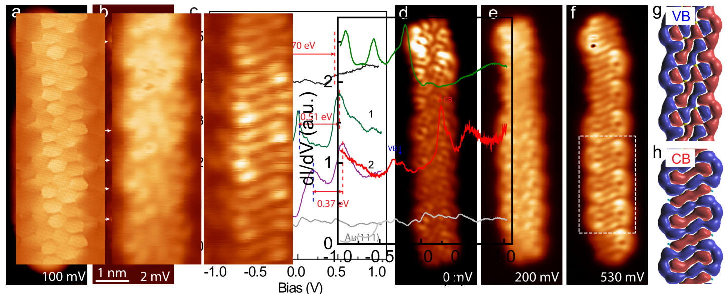
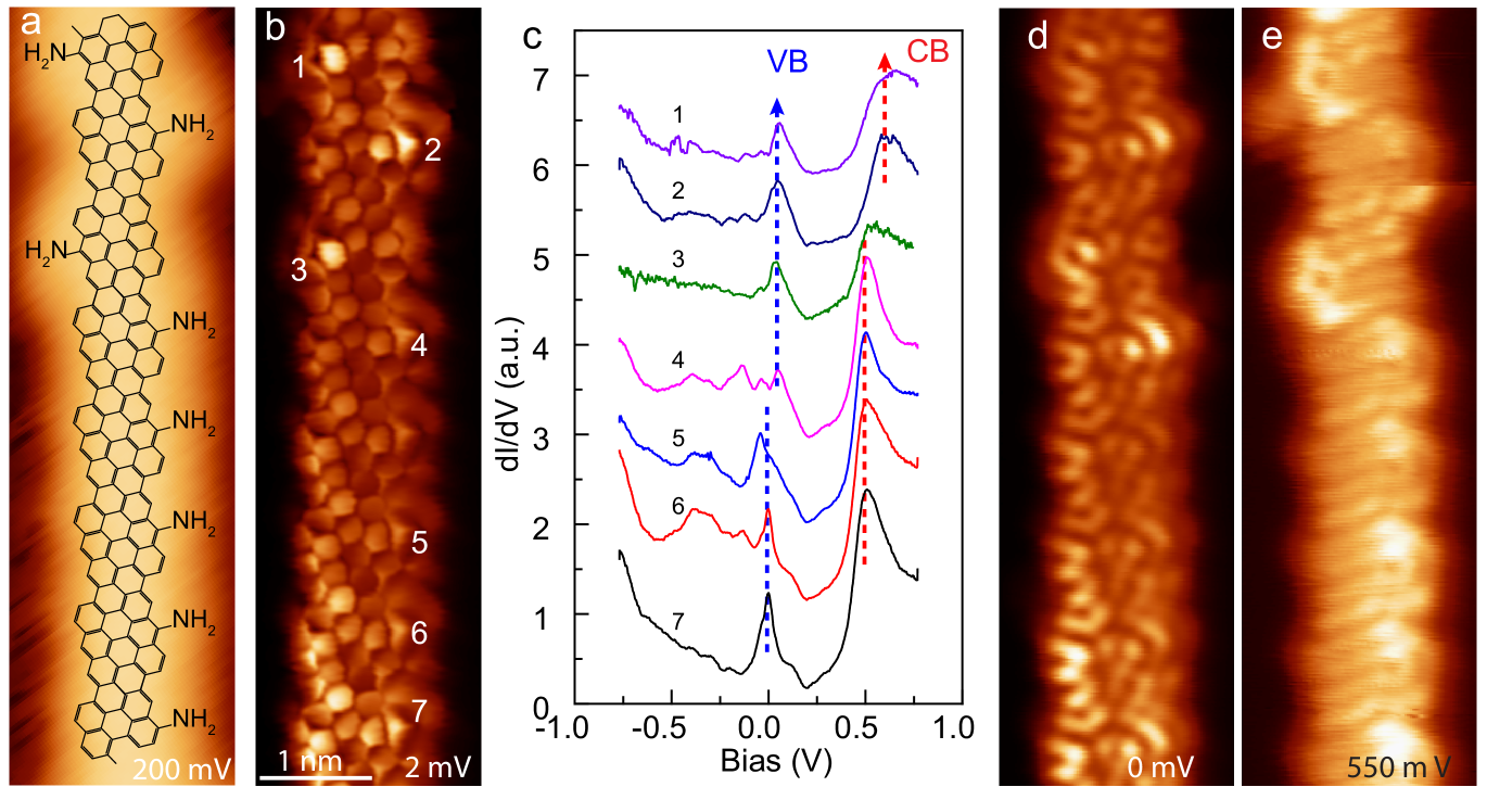
Fig. 3(a) shows the constant current STM image of a NH2-chGNR which shows irregular edges. The superimposed chemical structure of the NH2-chGNR indicates the loss of some NH2 groups on the edges. To further prove this, we acquired high resolution STM image of the same GNR with a CO-terminated tipGross et al. (2009); Kichin et al. (2011), as shown in Fig. 3(b). The high resolution image resolves the backbone structure of the ribbon as composed of zig-zag edged segments Merino-Díez et al. (2018), and also shows distinctive triangular features at many of the zigzag edges (indicated by arrows in Fig. 3(b)), which we attribute to surviving NH2 groups as shown in Fig. 3(a). The carbon atoms with missing NH2 groups are then passivated by hydrogen atoms during the on-surface reactionsTalirz et al. (2013). Most of the chGNR segments maintain at least one of the NH2 edge groups attached at one side, and some appear with both of them intact. From such high resolution images on more than 80 GNRs, we extract that about 75% of the NH2 groups survived the on-surface reaction. This value is even larger than the one obtained from the XPS results, most probably due to the different annealing processes (see methods). This is a remarkable doping value when compared with the chemical doping surviving the reaction for cyano (CN) functionalization of 7-AGNRsCarbonell-Sanromà et al. (2017b). We note that edge functionalization in such chiral ribbons is so efficient due to the lower temperatures required for their complete synthesis, in contrast with e.g. 7-AGNRs, which need more than 300∘C for their OSS.
Electronic structure of doped chGNRs: NH2 groups have an electron donating character. To study their effect on the electronic properties of the chiral nanoribbons, we recorded differential conductance plots (dI/dV) directly over singly and doubly NH2-doped segments, and compare them with measurements on pristine chGNRs. As shown in Fig. 3(c), the characteristic 0.7 eV energy gap of pristine chGNRs Merino-Díez et al. (2018) is severely modified on the NH2-substituted segments, which exhibit instead two pronounced peaks, indicated by red and blue dashed lines in Fig. 3(c). Over the singly doped segment, one of the peaks is pinned at the Fermi level (zero bias), while the other appears at 500 mV above. On the doubly-doped units, both peaks appear slightly shifted upwards, and their separation noticeably decreases.
To elucidate the origin of the peaks, we acquired dI/dV maps at bias values corresponding to the peak energies, which unveil the spatial distribution of these states (Fig. 3(d-f)). dI/dV maps at 0 mV and 200 mV (the bias values of the lower peaks in each case) resemble the VB shape of pristine chGNRs at the singly- and doubly-doped segments, respectively. In contrast, the dI/dV map at 530 mV (in coincidence with the higher-bias peak in both cases) reproduces a wave-front pattern that is characteristic of the CB of pristine chGNRsMerino-Díez et al. (2018). Furthermore, these dI/dV maps show very good agreement with the density functional theory simulations of wave function amplitude of valence and conduction bands along a fully NH2-doped ribbon (Fig. 3(g,h)). Based on these observations, we ascribe the two dI/dV peaks to the VB and CB onsets of NH2-chGNRs, respectively.
Compared to pristine chGNRs, both VB and CB onsets of the NH2-doped segments appear shifted upwards, and the VB-CB energy gap is significantly reduced (as labelled in Fig. 3(c)). This upwards shift of the frontier bands can be attributed to a significant hole-doping introduced by the edge NH2 groups, whose effect increases with the number of groups surviving in each segment. Particularly striking is the alignment of the VB onset with the Fermi-level for the singly-doped segments, and its further shift to 0.18 eV when a second NH2 is present, unveiling a substantial band depopulation by edge-doping, and underpinning of chGNRs on Au(111).
Intriguingly, the number of NH2-dopants per unit is not the only parameter tuning the bands’ onset, but also their relative arrangement. This was concluded from the inspection of ribbons with only one edge group per unit. In order to induce the more diluted functionalization observed in Fig. 4, we annealed the sample to a slightly higher temperature during the OSS process (e.g. to 280 ∘C for the data shown in Fig. 4). The result was a reduction of the edge functionalization to about 50%, with a nice preference to maintain one group per unit cell rather than two or none. We found that NH2 groups could either line-up along the same edge (e.g. as between segments 4 and 7 in Fig. 4(a,b), or alternate at opposite sides (as between 1 and 4 in Fig. 4(a,b)), what had an apparent impact in their corresponding band alignment. While the dI/dV spectra (Fig. 4(c)) and maps (Fig. 4(d) and (e)) appear in every case similar to the ones shown in Fig. 3, both VB and CB peaks in regions with alternating NH2 groups (spectra 1 to 4 in Fig. 4(c)) appear shifted upwards by 50 meV with respect to the regions where NH2 groups line up in the same side (spectra 5 to 7).
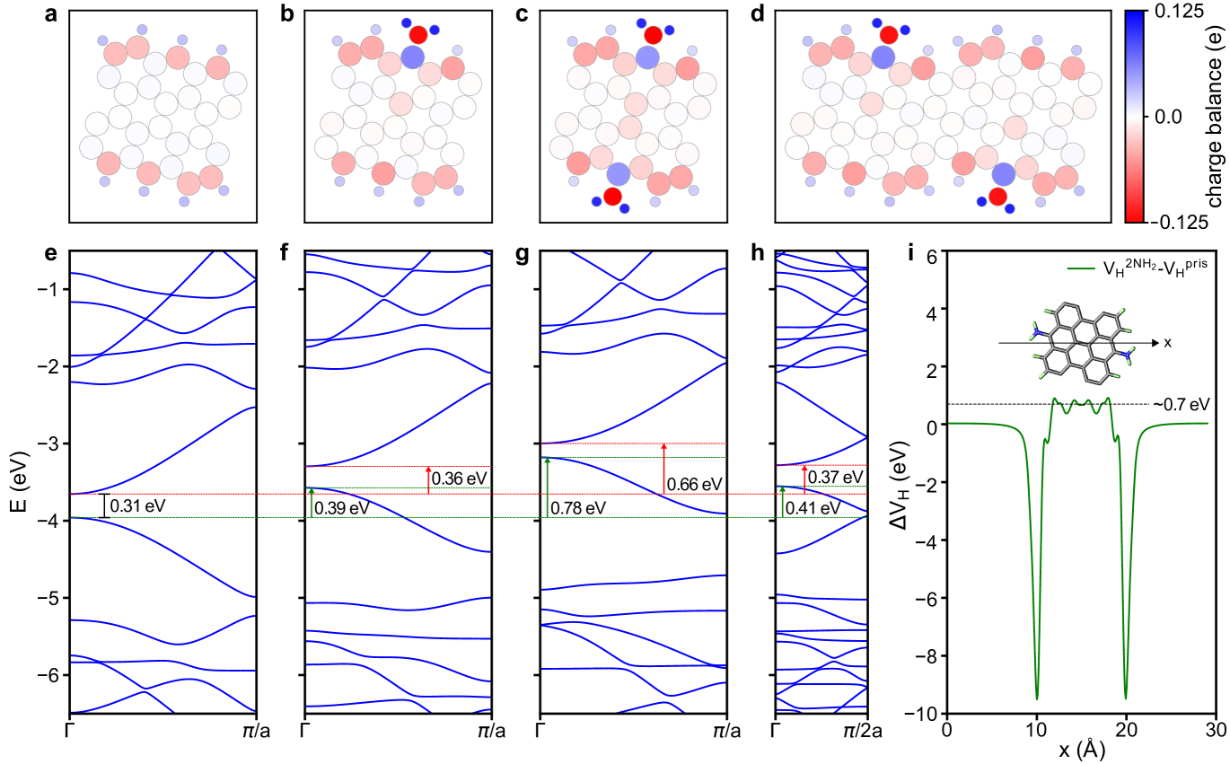
Theoretical analysis: Further insight into the doping effects of NH2 groups is obtained by DFT calculations of free-standing nanoribbons (see Methods for details on the simulations). In particular, we studied the band structure of infinite (3,1) chGNRs with the edge structures shown in Fig. 5(a-d), namely, the three experimentally observed scenarios: NH2 groups only on one side (1a-NH2-chGNR), on both sides (2-NH2-chGNR), and in alternating positions (1b-NH2-chGNR), as well as the pristine chGNRs for comparison. The corresponding band structures (Fig. 5(e-h)) show that, in all three cases, the presence of NH2 groups at the edge induces an upward shift of the bands with respect to the pristine case. For the singly-doped cases, the VB band shifts about 0.4 eV. When both edges are doped (2-NH2-chGNR), the upwards shift of the VB is almost doubled (0.78 eV), showing that it scales with the number of dopants.
The band structure also reproduces the clear reduction of the VB-CB energy gap with doping seen in the experiment. In particular, we find that the band gap of the singly-doped GNR decrease 10% with respect to the pristine case, while this reduction amounts to 40% for the doubly-doped species. A similar band gap closure with doping in GNRs was attributed to the extension of the -conjugated electron cloud outside the graphene backbone with the addition of the functional groups Carbonell-Sanromà et al. (2017b). On the other hand, we note that the calculated shifts of the frontier bands are larger than the measured ones, and the gap reduction obtained numerically is smaller than in experiments. These discrepancies might be related to screening effects of the metallic substrate, which are not included in our calculations. Yet, our DFT results are in very good qualitative agreement with the experimental measurements.
To explain the origin of the band shifts, we studied the charge redistribution induced by the NH2 edge groups in the chGNRs. As seen from the Hirshfeld plots in Fig. 5(a-d), the NH2 groups redistribute their charges by the covalent bonding to the edge and induce a small electron depletion at the connecting C and accumulation right at the middle of the ribbon. Interestingly, the lower electron density at the edge coincides with the higher current of these rings in the chGNR backbone (Fig. 3(a) and Fig. 4(a))). We speculate that the higher current signal over the edge phenyl rings bonding to NH2 and lower current over the neighbour rings at the center are related to the calculated electron depletion and accumulation, respectively, and to their influence on the effective tunneling.
As a result of the charge redistribution, a strong edge dipole of 3.6 D per unit cell pointing outwards from the ribbon backbone emerges around the NH2 group. Considering the edge dipole per unit cell of the pristine chGNR (2.1 D), and since both CH and NH2 dipoles are pointing in the same direction, we can estimate that each NH2 group contributes with a dipole of about 2.0 D. This edge dipole increases the internal electrostatic potential, and consequently, shifts their occupied bands upwards. For example, for the 2-NH2-chGNR case (Fig. 5(i)), we find that the 0.7 eV higher electrostatic potential is the main responsible of the 0.7 eV up-shift of its VB. The result is that the NH2 groups endow their electron-donating character to the ribbons. On the Au(111) surface, this leads to a substantial hole-doping from the metal, reaching the point where the VB is partially depopulated. We note that this scenario differs from the previously reported case of armchair GNRs doped with CN groups. CN induced a dipole pointing opposite, i.e. towards the ribbon’s backbone, leading to a total dipole smaller in magnitude and opposite in sign to the one presented here. This caused a small band down shift, contrary to the situation found here, but the semiconducting character remained unchanged Carbonell-Sanromà et al. (2017b).
I Conclusions
Our results demonstrate a chemical-gating strategy of graphene nanoribbons, by which incorporating electron donating chemical groups to the edge of GNRs causes a significant hole-doping on a Au(111) surface. As found previously with CN groups Carbonell-Sanromà et al. (2017b), the GNR inherits the electron affinity of the attached chemical group. Favored by the lower reaction temperatures for the synthesis of chiral GNRs, we found that a large fraction of NH2 groups survived the on-surface reaction, and caused the depopulation of its VB, signaling its charging. Based on DFT simulations, we explained the enhanced electron-donating character of the doped ribbons as arising from a substantial charge redistribution induced by the dopant. From these results, we foresee that combination of edge terminations with different electron affinity character can be a promising route to engineer hybrid GNRs with active electronic properties.
We acknowledge financial support from: i) AEI/FEDER-EU through grants no. MAT2016-78293-C6, FIS2017-83780-P (AEI/FEDER,EU), the Maria de Maeztu unit of excellence MDM-2016-0618, and the Severo Ochoa program (ICN2) SEV-2017-0706; ii) the European Research Council (grant agreement no. 635919); iii) the Xunta de Galicia (Centro singular de investigación de Galicia, accreditation 2016−2019, ED431G/09); iv) the EU project SPRING (863098); v) the European Regional Development Fund (ERDF) under the program Interreg V-A España-Francia-Andorra (Contract No. EFA 194/16 TNI), vi) the CERCA Program/Generalitat de Catalunya, and vii) the Gobierno Vasco-UPV/EHU (project IT1246-19).
II Methods/Experimental
The experiments were performed on a low temperature (5 K) STM under ultrahigh vacuum (UHV) conditions. The Au(111) substrate was cleaned in UHV by repeated cycles of Ne+ ion sputtering and subsequent annealing to 460 ∘C. The molecular precursor was sublimated at 210 ∘C from a Knudsen cell onto the clean Au(111) substrate kept at room temperature. Then the sample was first annealed at 200 ∘C for 15 minutes and then annealed at 260 ∘C for 5 minutes. A tungsten tip functionalized with a CO molecule was used for high resolution images. The high resolution images were acquired in constant height mode, at very small voltages, and junction resistances of typically 20 M. The signal was recorded using a lock-in amplifier with a bias modulation of mV at 760 Hz.
XPS measurements were carried out using a Specs PHOIBOS 150 hemispherical energy analyzer using a monochromatic X-ray source (Al K line with an energy of 1486.6 eV and 400 W) and energy referenced to the Fermi level. Surface cleanliness was confirmed with x-ray photoelectron spectroscopy (XPS) prior molecules deposition. Organic molecules were sublimated in UHV from a home-made Knudsen-cell type evaporator with an alumina crucible, with the sample kept at room temperature. Then the sample was step-wisely annealed to 400 ∘C. The sample was kept at each temperature step in Fig. 2d for 10 minutes for the XPS measurements.
Computational Procedures
The optimized geometry and electronic structure of free-standing pristine and NH2 doped ch-GNRS were calculated using density functional theory, as implemented in the SIESTA code.Soler et al. (2002) The nanoribbons were relaxed until forces on all atoms were 0.01 eV/Å, and the dispersion interactions were taken into account by the nonlocal optB88-vdW functional.Klimeš et al. (2009) The basis set consisted of double- plus polarization (DZP) orbitals for all species, with an energy shift parameter of 0.01 Ry. A 11100 Monkhorst-Pack mesh was used for the k-point sampling of the Brillouin zone, where the 100 k-points are taken along the direction of the ribbon. A cutoff of 300 Ry was used for the real-space grid integrations. The atomic population analysis was performed using the Hirshfeld scheme for partitioning the electron density.Hirshfeld (1977) The dipole moments were calculated integrating the valence electron density at each point times the distance of that point to the ribbon backbone (central axis). The corresponding neutralizing contribution due to the nuclei is also added. More details can be find in Carbonell-Sanromà et al. 2017b.
References
- Llinas et al. (2017) J. P. Llinas, A. Fairbrother, G. Borin Barin, W. Shi, K. Lee, S. Wu, B. Yong Choi, R. Braganza, J. Lear, N. Kau, W. Choi, C. Chen, Z. Pedramrazi, T. Dumslaff, A. Narita, X. Feng, K. Müllen, F. Fischer, A. Zettl, P. Ruffieux, E. Yablonovitch, M. Crommie, R. Fasel, and J. Bokor, Nat. Commun. 8, 633 (2017).
- Han et al. (2014a) W. Han, R. K. Kawakami, M. Gmitra, and J. Fabian, Nat. Nanotechnol. 9, 794 (2014a).
- Moreno et al. (2018) C. Moreno, M. Vilas-Varela, B. Kretz, A. Garcia-Lekue, M. V. Costache, M. Paradinas, M. Panighel, G. Ceballos, S. O. Valenzuela, D. Peña, and A. Mugarza, Science 360, 199 (2018).
- Yazyev (2013) O. V. Yazyev, Acc. Chem. Res 46, 2319 (2013).
- Kargar and Lee (2009) A. Kargar and C. Lee, 2009 9th IEEE Conference on Nanotechnology (IEEE-NANO) 8, 243 (2009).
- Schwierz (2010) F. Schwierz, Nat. Nanotechnol 5, 487 (2010).
- Bennett et al. (2013) P. B. Bennett, Z. Pedramrazi, A. Madani, Y. C. Chen, D. G. De Oteyza, C. Chen, F. R. Fischer, M. F. Crommie, and J. Bokor, Appl. Phys. Lett 103, 1 (2013), arXiv:1310.0495 .
- Talirz et al. (2016) L. Talirz, P. Ruffieux, and R. Fasel, Adv. Mater. 28, 6222 (2016).
- Clair and De Oteyza (2019) S. Clair and D. G. De Oteyza, Chem. Rev 119, 4717 (2019).
- Cai et al. (2010) J. Cai, P. Ruffieux, R. Jaafar, M. Bieri, T. Braun, S. Blankenburg, M. Muoth, A. P. Seitsonen, M. Saleh, X. Feng, K. Mullen, and R. Fasel, Nature 466, 470 (2010).
- Chen et al. (2013) Y.-C. Chen, D. G. De Oteyza, Z. Pedramrazi, C. Chen, F. R. Fischer, and M. F. Crommie, ACS Nano 7, 6123 (2013).
- Abdurakhmanova et al. (2014) N. Abdurakhmanova, N. Amsharov, S. Stepanow, M. Jansen, K. Kern, and K. Amsharov, Carbon 77, 1187 (2014).
- Basagni et al. (2015) A. Basagni, F. Sedona, C. A. Pignedoli, M. Cattelan, L. Nicolas, M. Casarin, and M. Sambi, J. Am. Chem. Soc 137, 1802 (2015).
- Kimouche et al. (2015) A. Kimouche, M. M. Ervasti, R. Drost, S. Halonen, A. Harju, P. M. Joensuu, J. Sainio, and P. Liljeroth, Nat. Commun 6, 10177 (2015).
- Zhang et al. (2015) H. Zhang, H. Lin, K. Sun, L. Chen, Y. Zagranyarski, N. Aghdassi, S. Duhm, Q. Li, D. Zhong, Y. Li, K. Müllen, H. Fuchs, and L. Chi, J. Am. Chem. Soc 137, 4022 (2015).
- Talirz et al. (2017) L. Talirz, H. Söde, T. Dumslaff, S. Wang, J. R. Sanchez-Valencia, J. Liu, P. Shinde, C. A. Pignedoli, L. Liang, V. Meunier, N. C. Plumb, M. Shi, X. Feng, A. Narita, K. Müllen, R. Fasel, and P. Ruffieux, ACS Nano 11, 1380 (2017).
- Merino-Díez et al. (2017) N. Merino-Díez, A. Garcia-Lekue, E. Carbonell-Sanromà, J. Li, M. Corso, L. Colazzo, F. Sedona, D. Sánchez-Portal, J. I. Pascual, and D. G. De Oteyza, ACS Nano 11, 11661 (2017).
- Han et al. (2014b) P. Han, K. Akagi, F. Federici Canova, H. Mutoh, S. Shiraki, K. Iwaya, P. S. Weiss, N. Asao, and T. Hitosugi, ACS Nano 8, 9181 (2014b).
- Liu et al. (2015) J. Liu, B.-W. Li, Y.-Z. Tan, A. Giannakopoulos, C. Sanchez-Sanchez, D. Beljonne, P. Ruffieux, R. Fasel, X. Feng, and K. Müllen, J. Am. Chem. Soc 137, 6097 (2015).
- Ruffieux et al. (2016) P. Ruffieux, S. Wang, B. Yang, C. Sánchez-Sánchez, J. Liu, T. Dienel, L. Talirz, P. Shinde, C. A. Pignedoli, D. Passerone, T. Dumslaff, X. Feng, K. Müllen, and R. Fasel, Nature 531, 489 (2016).
- De Oteyza et al. (2016) D. G. De Oteyza, A. García-Lekue, M. Vilas-Varela, N. Merino-Díez, E. Carbonell-Sanromà, M. Corso, G. Vasseur, C. Rogero, E. Guitián, J. I. Pascual, J. E. Ortega, Y. Wakayama, and D. Peña, ACS Nano 10, 9000 (2016).
- Rizzo et al. (2018) D. J. Rizzo, G. Veber, T. Cao, C. Bronner, T. Chen, F. Zhao, H. Rodriguez, S. G. Louie, M. F. Crommie, and F. R. Fischer, Nature 560, 204 (2018), arXiv:1805.06470 .
- Gröning et al. (2018) O. Gröning, S. Wang, X. Yao, C. A. Pignedoli, G. Borin Barin, C. Daniels, A. Cupo, V. Meunier, X. Feng, A. Narita, K. Müllen, P. Ruffieux, and R. Fasel, Nature 560, 209 (2018), arXiv:1805.06635 .
- Merino-Díez et al. (2018) N. Merino-Díez, J. Li, A. Garcia-Lekue, G. Vasseur, M. Vilas-Varela, E. Carbonell-Sanromà, M. Corso, J. E. Ortega, D. Peña, J. I. Pascual, and D. G. de Oteyza, J. Phys. Chem. Lett 9, 25 (2018).
- Moreno et al. (2019) C. Moreno, M. Panighel, M. Vilas-Varela, G. Sauthier, M. Tenorio, G. Ceballos, D. Peña, and A. Mugarza, Chem. Mater 31, 331 (2019), https://doi.org/10.1021/acs.chemmater.8b03094 .
- Cai et al. (2014) J. Cai, C. A. Pignedoli, L. Talirz, P. Ruffieux, H. Söde, L. Liang, V. Meunier, R. Berger, R. Li, X. Feng, K. Müllen, and R. Fasel, Nat. Nanotechnol 9, 896 (2014).
- Chen et al. (2015) Y.-C. Chen, T. Cao, C. Chen, Z. Pedramrazi, D. Haberer, de OteyzaDimas G., F. R. Fischer, S. G. Louie, and M. F. Crommie, Nat. Nanotechnol 10, 156 (2015).
- Carbonell-Sanromà et al. (2017a) E. Carbonell-Sanromà, P. Brandimarte, R. Balog, M. Corso, S. Kawai, A. Garcia-Lekue, S. Saito, S. Yamaguchi, E. Meyer, D. Sánchez-Portal, and J. I. Pascual, Nano Lett. 17, 50 (2017a).
- Wang et al. (2017) S. Wang, N. Kharche, E. Costa Girão, X. Feng, K. Müllen, V. Meunier, R. Fasel, and P. Ruffieux, Nano Lett. 17, 4277 (2017).
- Li et al. (2018) J. Li, N. Merino-Díez, E. Carbonell-Sanromà, M. Vilas-Varela, D. G. De Oteyza, D. Peña, M. Corso, and J. I. Pascual, Sci. Adv 4, eaaq0582 (2018).
- Su et al. (2018) X. Su, Z. Xue, G. Li, and P. Yu, Nano Lett. 18, 5744 (2018).
- Li et al. (2019) J. Li, S. Sanz, M. Corso, D. J. Choi, D. Peña, T. Frederiksen, and J. I. Pascual, Nat. Commun. 10, 200 (2019).
- Krull et al. (2013) C. Krull, R. Robles, A. Mugarza, and P. Gambardella, Nat. Mater. 12, 337 (2013).
- Bronner et al. (2013) C. Bronner, S. Stremlau, M. Gille, F. Brauße, A. Haase, S. Hecht, and P. Tegeder, Angew. Chem 52, 4422 (2013).
- Kawai et al. (2015) S. Kawai, S. Saito, S. Osumi, S. Yamaguchi, A. S. Foster, P. Spijker, and E. Meyer, Nat. Commun 6, 8098 (2015).
- Cloke et al. (2015) R. R. Cloke, T. Marangoni, G. D. Nguyen, T. Joshi, D. J. Rizzo, C. Bronner, T. Cao, S. G. Louie, M. F. Crommie, and F. R. Fischer, J. Am. Chem. Soc 137, 8872 (2015).
- Nguyen et al. (2016) G. D. Nguyen, F. M. Toma, T. Cao, Z. Pedramrazi, C. Chen, D. J. Rizzo, T. Joshi, C. Bronner, Y. C. Chen, M. Favaro, S. G. Louie, F. R. Fischer, and M. F. Crommie, J. Phys. Chem. C 120, 2684 (2016).
- Durr et al. (2018) R. A. Durr, D. Haberer, Y.-L. Lee, R. Blackwell, A. M. Kalayjian, T. Marangoni, J. Ihm, S. G. Louie, and F. R. Fischer, J. Am. Chem. Soc 140, 807 (2018).
- Carbonell-Sanromà et al. (2018) E. Carbonell-Sanromà, A. Garcia-Lekue, M. Corso, G. Vasseur, P. Brandimarte, J. Lobo-Checa, D. G. de Oteyza, J. Li, S. Kawai, S. Saito, S. Yamaguchi, J. E. Ortega, D. Sánchez-Portal, and J. I. Pascual, J. Phys. Chem. C 122, 16092 (2018).
- Wang et al. (2018) X. Y. Wang, J. I. Urgel, G. B. Barin, K. Eimre, M. Di Giovannantonio, A. Milani, M. Tommasini, C. A. Pignedoli, P. Ruffieux, X. Feng, R. Fasel, K. Müllen, and A. Narita, J. Am. Chem. Soc 140, 9104 (2018).
- Kawai et al. (2018) S. Kawai, S. Nakatsuka, T. Hatakeyama, R. Pawlak, T. Meier, J. Tracey, E. Meyer, and A. S. Foster, Sci. Adv. 4, 1 (2018).
- Carbonell-Sanromà et al. (2017b) E. Carbonell-Sanromà, J. Hieulle, M. Vilas-Varela, P. Brandimarte, M. Iraola, A. Barragán, J. Li, M. Abadia, M. Corso, D. Sánchez-Portal, D. Peña, and J. I. Pascual, ACS Nano 11, 7355 (2017b), arXiv:1705.07045 .
- Cao et al. (2017) T. Cao, F. Zhao, and S. G. Louie, Phys. Rev. Lett. 119, 076401 (2017).
- Bronner et al. (2017) C. Bronner, T. Marangoni, D. J. Rizzo, R. A. Durr, J. H. Jørgensen, F. R. Fischer, and M. F. Crommie, J. Phys. Chem. C 121, 18490 (2017).
- Gross et al. (2009) L. Gross, F. Mohn, N. Moll, P. Liljeroth, and G. Meyer, Science 325, 1110 (2009).
- Kichin et al. (2011) G. Kichin, C. Weiss, C. Wagner, F. S. Tautz, and R. Temirov, J. Am. Chem. Soc 133, 16847 (2011).
- Talirz et al. (2013) L. Talirz, H. Söde, J. Cai, P. Ruffieux, S. Blankenburg, R. Jafaar, R. Berger, X. Feng, K. Müllen, D. Passerone, R. Fasel, and C. A. Pignedoli, J. Am. Chem. Soc. 135, 2060 (2013).
- Soler et al. (2002) J. M. Soler, E. Artacho, J. D. Gale, A. García, J. Junquera, P. Ordejón, and D. Sánchez-Portal, J. Phys. Condens. Matter 14, 2745 (2002).
- Klimeš et al. (2009) J. Klimeš, D. R. Bowler, and A. Michaelides, J. Phys. Condens. Matter 22, 022201 (2009).
- Hirshfeld (1977) F. Hirshfeld, Theor. Chim. Acta 44, 129 (1977).