Au-decorated black TiO2 produced via laser ablation in liquid
Abstract
Rational combination of plasmonic and all-dielectric concepts within unique hybrid nanomaterials provides promising route toward devices with ultimate performance and extended modalities. However, spectral matching of plasmonic and Mie-type resonances for such nanostructures can only be achieved for their dissimilar characteristic sizes, thus making the resulting hybrid nanostructure geometry complex for practical realization and large-scale replication. Here, we produced unique amorphous TiO2 nanospheres simultaneously decorated and doped with Au nanoclusters via single-step nanosecond-laser ablation of commercially available TiO2 nanopowders dispersed in aqueous HAuCl4. The fabricated hybrids demonstrate remarkable light-absorbing properties (averaged value 96%) in the visible and near-IR spectral range mediated by bandgap reduction of the laser-processed amorphous TiO2, as well as plasmon resonances of the decorating Au nanoclusters, which was confirmed by combining optical spectroscopy, advanced electron energy loss spectroscopy, transmission electron microscopy and electromagnetic modeling. Excellent light-absorbing and plasmonic properties of the produced hybrids were implemented to demonstrate catalytically passive SERS biosensor for identification of analytes at trace concentrations and solar steam generator that permitted to increase water evaporation rate by 2.5 times compared with that of pure water under identical one-sun irradiation conditions.
I Introduction
Collective resonant oscillations of conduction electrons (also known as localized surface plasmon resonance (LSPR)) in noble-metal nanoparticles (NPs) provide a common and reliable way to control electromagnetic fields at nanoscale. Such oscillations facilitate resonant absorption of incident energy that can be converted to strongly enhanced electromagnetic fields around the NP surface or dissipate via nonradiative damping to induce localized heating. Both electromagnetic and photothermal effects have found numerous practical applications in photovoltaics, solar energy conversion, biomedicine, sensing, etc Giannini et al. (2011); Atwater and Polman (2011); Baffou et al. (2020). Alternatively, NPs made of various materials with high refractive index (high-) and low dissipative losses in the visible and near-IR spectral range (for example, Si, Ge and TiO2) emerged recently as alternative platform for nanoscale light management Kuznetsov et al. (2016). In particular, such nanomaterials support Mie-type resonances that permit to concentrate electromagnetic energy inside the NP bulk giving rise to efficient photothermal conversion and highly enhanced nonlinear optical effects Alessandri and Lombardi (2016); Makarov et al. (2017a); Zograf et al. (2017); Mitsai et al. (2019); Koshelev et al. (2020).
Rational combination of plasmonic and all-dielectric concepts to design hybrid nanostructures is expected to provide a promising route toward advanced nanomaterials with optimized optical response and extended operation range Jiang et al. (2014); Zuev et al. (2016); Timpu et al. (2017); Makarov et al. (2017b); Milichko et al. (2018). Meanwhile, spectral matching of LSPRs and Mie-type resonances of noble-metal and high- NPs is crucial for optimal performance. Unfortunately, in the visible and near-IR spectral ranges such resonant matching can be achieved for dissimilar characteristic dimensions of both types of NPs (in particular, 100-500 nm for dielectric NPs and less than 50 nm - for plasmonic ones). This makes the NP geometry quite complex for practical realization, even with time- and money-consuming lithography-based techniques.
Laser ablation in liquids (LAL) has emerged as a promising high-performance and green approach for nanomaterial preparation Amendola and Meneghetti (2009); Zeng et al. (2012); Zhang et al. (2017a). When compared with wet-chemistry methods, LAL represents a simple and environmentally friendly technology that can be carried out under normal environmental conditions without external stimuli. Intense pulsed laser radiation generates extremely high local pressures, temperatures and quenching rates, thus providing experimental conditions for production of nanostructures with different phase composition (including unique meta-stable phases) Vailionis et al. (2011), complex chemical composition and morphology Zhang et al. (2017b); Shih et al. (2018); Alexander et al. (2019); Tymoczko et al. (2019). However, so far only a few studies reported on LAL-generated hybrid nanomaterials where plasmonic and dielectric counterparts were combined within practically relevant design Liu et al. (2015); Saraeva et al. (2018); Mintcheva et al. (2020), yet without rigorous assessment of their nanophotonic properties and practical applications.
In this paper, unique amorphous TiO2 spherical-shaped NPs simultaneously decorated and doped with Au nanoclusters were produced via single-step ablation of commercially available TiO2 nanopowders in presence of HAuCl4 with nanosecond (ns) laser pulses. The fabricated hybrids demonstrated remarkable broadband light-absorbing properties (averaged value 96%) in the visible and near-IR spectral ranges mediated by bandgap reduction of the laser-processed amorphous TiO2, as well as LSPRs of the decorating Au nanoclusters, which was confirmed by combining optical spectroscopy, advanced EELS/TEM and electromagnetic modeling. When placed on a back reflecting mirror, the produced Au@TiO2 NPs demonstrated excellent SERS performance resulted from the coupled Mie resonance of TiO2 spheres and the LSPRs of their decorating Au nanoclusters. Isolated Au@TiO2 NPs were found to provide background-free chemically non-perturbing optical identification of various analytes at initial concentrations down to 10-8 M. Au@TiO2 NPs with their strong broadband optical absorption make them promising for photothermal conversion of the solar energy. As a proof-of-concept, by using a commercial cellulose membrane functionalized with solar-energy absorbing Au@TiO2, we realized a lab-scale water steam generator which permitted to increase evaporation rate by 2.5 times compared with that of pure water.
II Materials and Methods
II.1 Fabrication of Au@TiO2 nanoparticles.
Two types of commercially available TiO2 nanopowders (anatase powder from Wako Chemicals, 99.99 % pure, and P25 from Degussa, 99.5 % pure) with average particle sizes 125 and 21 nm, respectively, were used as supplied. First, NPs were dispersed in deionized water by means of ultrasonication to achieve 0.001% solution. Then, the suspension (7.5 ml) was transferred to a quartz cuvette (3 x 3 x 6 cm3) and a certain amount of aqueous tetrachloroauric acid (HAuCl4, concentration of 10-3 M) was added. Finally, the suspension was magnetically stirred and irradiated with focused nanosecond laser pulses (Quantel Ultra 50, with 7 ns, 532 nm and 20-Hz as pulse width, wavelength and repetition rate, respectively) for a certain period of time at laser fluence of 20 J/cm2.
II.2 Characterization.
Scanning electron microscopy. SEM images of Au@TiO2 NPs were obtained at accelerating voltage of 5 kV and beam current of 50 pA (Helios NanoLab 450S, Thermo Fisher, USA). Backscattered electron detector was used to achieve atomic number-sensitive contrast for highlighting Au particles.
To obtain information about the internal structure and composition of the Au@TiO2 system, the cross-sections of NPs produced by focused ion beam (FIB) milling (beam current of 43 pA at an accelerating voltage of 30 kV) were studied via SEM imaging. A platinum layer was locally deposited atop of Au@TiO2 nanospheres to protect their surface structures during FIB milling (see Supporting Information).
More detailed characterization of composition and inner structure was carried out using transmission electron microscopy (TEM) combined with electron tomography. For TEM studies, particles were ultrasonicated in acetone for 5 min and then 30 L droplet was placed on copper grid with lacy carbon. TEM/scanning TEM (STEM) imaging, electron tomography and STEM-EDX (energy dispersive x-ray spectroscopy, EDAX) chemical mapping experiments were performed at an acceleration voltage of 300 kV (Titan 60-300, Thermo Fisher, USA). The microscope was equipped with a monochromated X-FEG and spectrometer Quantum GIF (Gatan, USA) which allowed performing electron energy loss spectroscopy (EELS) with energy resolution better than 50 meV. Probing of the surface plasmons around Au NPs was done by means of high-resolution EELS technique combined with STEM image acquisition. In this case, the microscope was tuned at a beam energy of 60 keV in STEM mode. The spectrum images were recorded in the low loss region, including zero-loss peak. At each pixel of HAADF stem image, an EELS spectrum was stored with the length of 2048 pixels and energy dispersion 0.01 eV.
Three-dimensional particle morphology was characterized by the electron tomography technique using high angular annular dark-field STEM (HAADF-STEM) imaging mode. The HAADF-STEM regime provides contrast that is strongly dependent on the atomic number (?Z2) in which Au NPs appear much brighter at HAADF-STEM images. Tomographic tilt series were acquired automatically at angles between ?74o and +74o at 2o tilt step. Images were taken with a FEI Tomography 4.0 software in automatic mode; the dwell time for acquisition was set to 2 ?s for the images of 2048 x 2048 pixels with the pixel size of 1.3 nm. The fiducial-less tilt-series alignment and tomographic reconstructions with simultaneous iterative reconstruction (SIRT) techniques were done using in-house Digital Micrograph (Gatan, USA) scripts Rajabalinia et al. (2019). Reconstructed volumes had a voxel size of 5.2 nm 3. The intensity-based segmentation with a local threshold criterion and manual supervision was used. Depending on the intensity value of pixels, they were assumed as belonging to the Au nanoparticles (bright) or TiO2 sphere (grey). Segmentation of Au and TiO2 phases, subsequent 3-D rendering, and statistical calculations were done by using FEI Avizo 8.1 software.
Optical and Raman spectroscopy. Absorption coefficient A=1-R was measured in 200-1700 nm spectral range with an integrating sphere spectrophotometer (Cary Varian 5000) at a spectral resolution of 1 nm. Halogen and deuterium lamps were used as radiation sources for Vis-NIR and UV range, respectively. The bandgap value Eg was determined by the position of the fundamental absorption edge according to Tauc equation:
| (1) |
where is a Planck’s constant, is the oscillation frequency of electromagnetic waves, B is a constant showing slope of the linear fit, while F(R) = (1 - R)2/2R represents Kubelka-Munk s function. The value of the exponent denotes the nature of the interband electronic transitions - for direct allowed transitions is equal to n=1/2. The basic procedure for Tauc analysis is to acquire optical absorbance data that cover a range of energies below and above the bandgap transition. Eg value was determined by extrapolating the linear part of the Tauc curve ()1/n on the axis.
Raman spectra of pristine TiO2 NPs, as well as produced and post-annealed AuTiO2 hybrids, were acquired with commercial Raman microscope (Alpha WiTec) at 532 nm (2.33 eV) pump. Similar device was used to probe the SERS performance of AuTiO2 NPs. The Au@TiO2 NPs were functionalized with several types of practically relevant analytes, namely organic dyes (Rhodamine 6G and Acridine orange), medical drags (Diphendramine hydrochloride) and histological marker molecules (4 ,6-diamidino-2-phenylindole dihydrochloride, DAPI). The NPs were added into the 10-8M analyte solutions and stored there for 2 h. Then, suspensions were drop-cast onto a bulk Ag mirror. SERS signal was obtained only from isolated NPs on the substrate and averaged over 50 similar measurements for each analyte to account for random distribution of Au nanoclusters.
FDTD modeling. Finite-difference time domain calculations (Lumerical Solutions Ltd.) were undertaken to assess plasmonic properties of the Au@TiO2 hybrids. The structures were excited with linearly polarized broadband source. Exact 3D models of the NPs were reconstructed from the corresponding STEM images produced during the EELS studies. The dispersion functions for Au and TiO2 were obtained from Palik Palik (1998).
II.3 Modelling of radiation-induced heating of Au@TiO2 particles.
Theoretical description of nanoparticle heating (induced by either solar or laser radiation) was carried out numerically with a commercial software package (Comsol Multiphysics). A numerical model was first constructed for a plane wave irradiation of a bare and Au-decorated ellipsoidal TiO2 NP (the diameter along the long axis is 21 nm) in water. Distribution of electromagnetic fields in the simulated volume excited by a single laser pulse (532 nm wavelength and 25 mJ pulse energy) was calculated by solving wave equation in the frequency domain. Then, the following heat equation in a time domain during the single-pulse irradiation was calculated:
| (2) |
where the right side of this equation describes the heat source with the current density and electric field amplitude , whereas the left side corresponds to temperature evolution in time and space with corresponding parameters: Cp is the heat capacity at constant pressure, is the material density, and is the thermal conductivity. We suppose the refractive indexes are =0.56+3.44i, =1.335, = 2.54+0.0001i. Thermal conductivities for TiO2, gold and water are taken as =7 , =320 and =0.6 . Similar procedure was used to calculate heating efficiency of the Au@TiO2 hybrids of variable diameter in water and on the glass substrate. For these calculations, an excitation source with central wavelength at 550 nm and intensity 0.1 W/cm2 was considered providing reliable approximation of solar irradiation. We define thermal boundary conditions as a heat flux through the computational domain surface , where is the heat transfer coefficient, = 293 K is the external temperature, is the calculated temperature near boundary of the computational domain. We assume that temperature gradient is weak because in real experiment there are a lot of particle nearby. From these considerations we estimate value of equals 0.22 W/(mK).
II.4 Water steam generation.
Nanofluid was prepared by suspending 50 mg of Au@TiO2 in 10 mL of distilled water. Also, similar suspension was filtered through a commercial cellulose membrane (pore diameter of 200 nm) and then dried in vacuum. The produced nanofluid and “black” membrane loaded with Au@TiO2 NPs was placed on the surface of distilled water and irradiated by commercial solar simulator at 0.1 W/cm2. A microbalance was used to detect weight loss of water after evaporation. The evaporation rate observed in both cases was compared with that of distilled water. Heating process was visualized with calibrated IR camera. All experiments were performed at 25 oC and relative humidity of 40 %.
.
III Results and Discussions
III.1 Au-decorated black TiO2: fabrication and structural properties.
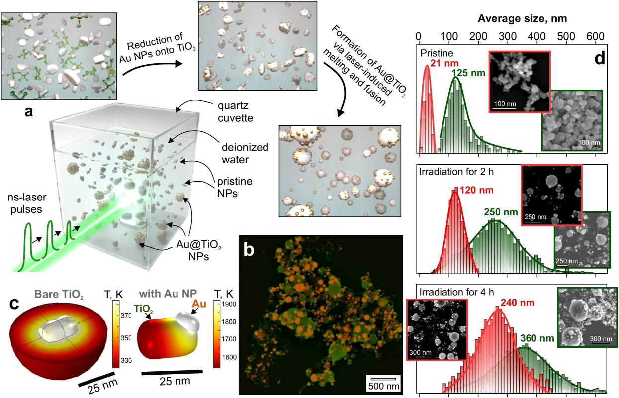
Figure 1a schematically illustrates key processes underlying formation of Au@TiO2 NPs using the LAL technique. In our experiments, aqueous suspension of as-supplied TiO2 NPs (mainly anatase, average size of 21 and 125 nm, respectively) mixed with aqueous solution of HAuCl4 was irradiated by ns-laser pulses for a few hours resulting in formation of spherical TiO2 decorated with multiple nano-sized Au clusters (Fig. 1b). Typically, the average size of resulting Au@TiO2 hybrids is always larger than the size of as-supplied NPs (for both types of starting nanopowders). Accordingly, the observed increase in size and spherical shape of produced NPs indicate melting and fusion as key processes involved in their formation.
However, the as-supplied crystalline TiO2 NPs of both types weakly absorb visible light, which results in their single-pulse laser heating that is insufficient to reach the melting point of bulk TiO2 (1900 K, Liu and Chen (2014)). In particular, 21-nm sized TiO2 nanoparticle suspended in water can be homogeneously heated up to only 385 K upon irradiation with a single 7-ns laser pulse at fluence of 20 J/cm2, as confirmed by corresponding numerical modeling (see Fig. 1c and Methods for simulation details). In a sharp contrast, Au nanoclusters can efficiently absorb laser radiation at 532 nm that is close to the wavelength of their localized plasmon resonance. The absorbed radiation is converted to Joule heating by resonantly excited conduction electrons, while the generated heat can be further transferred to surroundings. Similar modeling of the single-pulse laser heating of isolated 21-nm-big TiO2 NP decorated with an Au cluster with its diameter of 5 nm shows that such a hybrid nanostructure can easily reach temperatures exceeding 1900 K at the same irradiation laser fluence (Fig. 1d).
To further support this idea, LAL experiments were performed with a similar suspensions of pristine TiO2 NPs at the same laser fluence and irradiation time but without addition of HAuCl4. Careful SEM analysis of obtained nanomaterials indicated no modification/melting of pristine NPs revealing the key role of Au nanoclusters decorating TiO2 NPs. The formation of such Au NPs can occur even without laser-irradiation and is expected to be preferentially facilitated on TiO2 NPs via surface chemistry driven by active sites and laser-induced enhanced temperature near NP’s surface.
As mentioned above, after irradiation for 2 h all as-supplied TiO2 NPs with irregular shapes were successfully transferred to spherically-shaped Au@TiO2 hybrids. For both types of used pristine NPs with their average sizes of 21 and 125 nm, the obtained hybrids had the averaged diameters of 120 and 220 nm, respectively, indicating that fusion of several NPs is crucial (see Fig. 1d). Shorter irradiation time resulted in particular presence of pristine TiO2 NPs in the obtained product. However, irradiation of the TiO2 suspension for 4 h yielded in even larger size of obtained Au@TiO2 hybrids. The number of adsorbed Au nanoclusters and their size were also found to increase with laser irradiation time, as well as with the concentration of HAuCl4 in the irradiated dispersion. From practical application point of view, it is important to control both the average size of the spherical-shaped TiO2 NPs and their decoration degree. We showed that by varying the starting size of pristine TiO2 NPs, irradiation time and HAuCl4 content, both the two parameters could be flexibly controlled. The related information is summarized in Fig. 1d and in the Supporting Information.
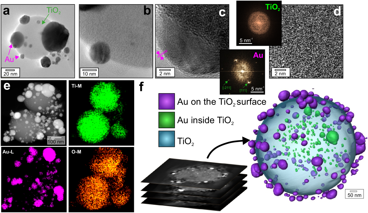
HR-TEM imaging was used to identify crystalline structure of the obtained Au@TiO2 product. TEM images of representative NPs are shown in Fig. 2(a-d), revealing completely disordered lattice in the TiO2 nanosphere and crystalline structure of decorating Au nanoclusters. FFT analysis of the selected HR-TEM images confirmed amorphous nature of the TiO2 nanospheres (insets in Fig. 3c,d). Additionally, Raman spectroscopy was utilized to provide more information regarding the crystallinity of TiO2 statistically averaged over multiple NPs before and after LAL processing. These studies permitted clearly to identify all main anatase Raman bands for both types of as-supplied TiO2 NPs and low-intense bands at 440 and 610 cm-1 that can be attributed to a small amount of nanocrystalline rutile inclusions in the Au@TiO2 product (Supporting information). EDX mapping was used to confirm chemical composition of the Au@TiO2 NPs (Fig. 2e).
The formation mechanism suggests that Au NPs stimulating TiO2 melting can appear not only on the surface of Au@TiO2 product but also in its bulk upon fusion. To clarify this, several FIB cross-sectional cuts of Au@TiO2 were first visualized using SEM revealing high-contrast nanoscaled inclusions that appeared much brighter compared with TiO2 surrounding and can be attributed to Au (see Supporting Information). More detailed studies of the inner structures of Au@TiO2 NPs were carried out via tomographic reconstruction of HAADF-STEM image series (see Methods for details). The reconstructed 3D model of an isolated Au@TiO2 NP is shown in Fig. 2f, where Au nanoclusters located on the surface and inside the TiO2 NP are highlighted by different colors for clarity. Systematic studies of the produced Au@TiO2 hybrids indicated that the inner Au nanoclusters occupy 0.5-1% of the volume of TiO2 nanosphere. Noteworthy, such Au nanoclusters both embedded into TiO2 NPs and simultaneously decorating their surface are reported for the first time.
III.2 Optical and plasmonic properties of Au@TiO2 hybrids
We started by probing the plasmonic response of Au nanoclusters capping amorphous spherical-shaped TiO2 using high-resolution EELS. The corresponding EELS spectrum measured from an isolated Au nanocluster sitting on the surface of a 225-nm-sized spherical TiO2 NP demonstrates a pronounced signal at 2.3 eV (Fig. 3a). Several peaks with their maxima centered at 2.02, 2.145 and 2.365 eV can be resolved indicating the multi-resonant nature of the electron plasma oscillations in the Au nanocluster. Mapping of the intensity of this signal (1.9-2.5 eV) confirmed that it originates from the Au nanocluster surface (Fig. 3b). Noteworthy, the EELS peaks at 5.7 and 10 eV can be attributed to low-loss edges of Ti.
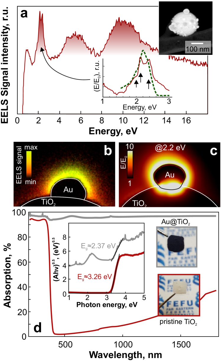
The size of the considered Au nanocluster is comparable with a doubled skin depth of gold ( 30 nm) thus suggesting the dipolar approximation applicable for formal analysis of its plasmon modes. Considering the dielectric permittivity of Au, the localized dipolar plasmon resonance of such a NP in vacuum should appear around 2.35 eV redshifting to 2.1 eV when it attaches a high-refractive-index TiO2 surface. Also, the shape of the obtained Au nanoclusters (grown above or even penetrating into their TiO2 support; see Supporting Information) is typically irregular suggesting a certain splitting of localized plasmon resonances. The resonance seen at 2.365 eV can be attributed to the quadrupolar localized plasmonic modes appeared owing to the substrate-induced redshift of the dipolar one. These inferences are consistent with the supporting FDTD calculations showing the near-field plasmonic spectrum of the slightly elliptical Au nanocluster capping TiO2 NP as well as the normalized amplitude of the electromagnetic near-fields under resonant excitation (at 2.1 eV; Fig. 3a,c).
Statistical EELS studies averaged over various Au clusters found on TiO2 spheres indicate that the Au@TiO2 hybrids support localized plasmons in a rather broad spectral range spanning from 500 to 650 nm. Moreover, closely spaced Au clusters can act as plasmonic oligomers with their resonance shifted to near-IR part of the spectrum (see Supporting Information).
Further, we assessed optical properties Au@TiO2 hybrids in the visible and near-IR part of their spectra (Fig. 3d). For this purpose, the Au@TiO2 suspension (produced by laser irradiation of P25 pristine TiO2 for 4h) was dried on a cover glass slide to form a uniform opaque coating. In a sharp contrast to the similar coating made of both types of pristine TiO2 NPs, the color of the Au@TiO2 nanomaterial appeared black indicating high absorption in the visible spectral range. Corresponding measurements of absorption coefficient (A=1-R; R - diffuse reflectance) of both coatings revealed an order of magnitude larger vis-to-IR absorption by the “black” Au-decorated amorphous TiO2 with respect to its “white” pristine precursors. Based on the obtained diffuse reflectance, we found a considerable decrease of the bandgap Eg value from 3.26 (pristine TiO2) to 2.37 eV (Au@TiO2 hybrids) for direct transitions according to Kubelka-Munk method (see inset in Fig. 3d). Noteworthy, the maximal absorption (97%) mediated by the localized plasmon resonances of the Au nanoclusters capping TiO2 was observed at 2.2 eV that is consistent with the numerical modeling and EELS studies.
III.3 Use of Au@TiO2 hybrids for label-free biosensing and photo-thermal conversion.
The observed remarkable optical and plasmonic properties of the prepared Au@TiO2 hybrids suggest several areas for their potential application. First of all, the plasmon-mediated EM field localized near Au particles make such Au@TiO2 NPs attractive for label-free biosensing based on the surface-enhanced Raman scattering (SERS) effect . As the electromagnetic contribution to the SERS signal is predominant roughly scaling as forth power of the local electromagnetic field amplitude normalized over the pump field amplitude E4/E , maximization of this value gives a general route for boosting SERS performance. Au nanoclusters produce enhanced plasmon-mediated EM fields under resonant visible-light excitation. However, the nanoscale size of such particles results in their low absorption cross-section. As a result, they efficiently interact only with a small fraction of incident pump laser beam with a diffraction-limited lateral size typical for SERS experiments. In the produced Au@TiO2 hybrids, the excitation of decorating Au particles can be improved via additional coupling of the incident field to the Mie resonances supported by TiO2 spheres with diameters comparable with the diffraction-limited (/2) laser spot Checcucci et al. (2018); Milichko et al. (2018). To illustrate this, we performed FDTD calculations showing a 2-fold increase of the local E/E0 amplitude near an isolated Au particle upon its attachment to a 240-nm-sized TiO2 sphere supporting magnetic-type dipolar resonance at plasmon excitation wavelength (2.2 eV). Such improvement permits to expect contribution to SERS electromagnetic enhancement which is at least an order of magnitude larger compared to those from a similar isolated Au particle in a free-space.
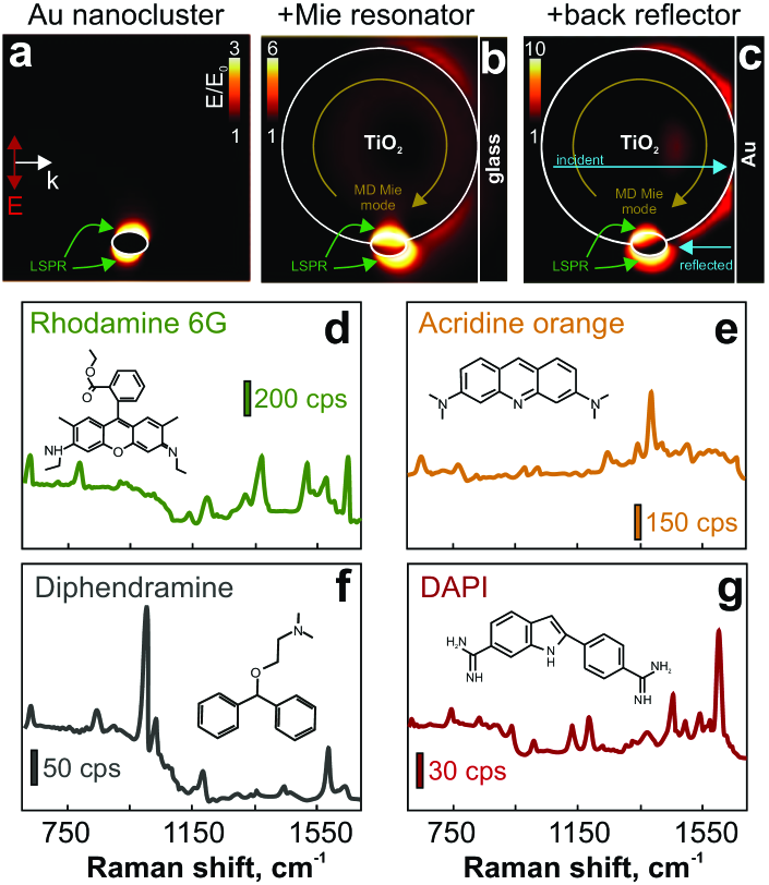
Additionally, the geometry of the prepared Au@TiO2 hybrids permits to apply them with back-reflector mirror . In our case, this concept can be simply realized upon SERS studies by placing Au@TiO2 NPs on a smooth refectory mirror. In this respect, the TiO2 sphere acts as a support for Au nanoclusters providing required gap between plasmonic NPs and reflecting mirror. Corresponding FDTD calculations of the local EM field enhancement gave more than a 3-fold enhancement of E/E0 (compared with the case of a Au particle in a free-space) near the isolated Au nanocluster yielding in further increase of the SERS performance (Fig. 4c). Amorphous structure of TiO2 spheres contributed to the background free SERS studies.
To assess the applicability of Au@TiO2 hybrids for reliable label-free optical identification at trace concentrations, we probed the SERS signal from isolated NPs with an average diameter 24020 nm at 532 nm (2.33 eV) as pumping wavelength. The Au@TiO2 NPs were functionalized with several types of practically relevant analytes, namely organic dyes (Rhodamine 6G and Acridine orange), medical drags (diphendramine hydrochloride) and histological marker molecules (4’,6-diamidino-2-phenylindole dihydrochloride, DAPI). Considering the rather random size distribution and arrangement of decorating Au nanoclusters, for each type of the analyte we statistically averaged the SERS signal over at least 50 NPs performing measurements only from isolated structures. By laser pumping isolated Au@TiO2 NPs stored in the analyte solution for 2h and then drop-cast onto a silver mirror, we found distinct SERS signals from all tested analytes (Fig. 4d). The spectral position of the identified Raman bands were found to be in good agreement with previously reported studies substantiating isolated Au@TiO2 hybrids as simple all-in-one SERS platforms with optimized EM response for reliable fingerprinting at initial concentrations down to 10-8M Pavliuk et al. (2020).
Noteworthy, NPs for SERS studies provide additional flexibility for device designs. For example, quantitative SERS measurements using NPs can be performed in liquids to detect and trace catalytic processes Mitsai et al. (2018). Moreover, the analyte solution loaded with functional NPs can be dried on a substrate with specially designed wetting properties. After solvent evaporation on such surface, the deposition area for both the molecules and functional NPs will be substantially reduced increasing local concentration of the analyte already mixed with SERS-active probes De Angelis et al. (2011); Yang et al. (2016); Pavliuk et al. (2020). Realization of such NP-based sensor active device will become a subject of our forthcoming studies.
Moreover, several studies highlighted the excellent catalytic performance of laser-modified titania again visible-light driven degradation of dye molecules Chen et al. (2015). In this work we used methylene blue and methylene orange organic dye molecules to benchmark photocatalytic activity of the produced Au@TiO2 hybrids (see Supporting information). Remarkably, in comparison with the as-supplied pristine TiO2 (P25 from Degussa) the Au@TiO2 hybrids with amorphous core demonstrate weak activity in photocatalytic degradation of both dye molecules. Noteworthy, weak photocatalytic activity of nanostructres are beneficial in designing SERS substrates for studies where non-invasive and non-perturbing identification of analytes is mandatory Mitsai et al. (2018). It should be noted that post-annealing of the Au@TiO2 product can be used to convert its amorphous core to rutile crystalline phase. Raman spectroscopy indicated appearance of characteristic rutile bands (at 445 and 610 cm-1) in Au@TiO2 hybrids after their annealing at 500o for 2h (see Supporting Information). However, more detailed studies of post-annealing of Au@TiO2 NPs and its effect on crystalline structure and photocatalytic activity of the product are out of the scope of this paper.
Finally, the excellent broadband optical absorption of the prepared Au@TiO2 NPs make them promising for photothermal conversion of the solar energy. In particular, the maximal visible light absorption (97%) of Au@TiO2 nanomaterial mediated by the localized plasmon resonances in Au nanoclusters was observed at 2.2 eV which perfectly matches the maximum of the solar energy spectrum. Our numerical calculations showed that Au@TiO2 NPs with diameters ranging from 300 to 500 nm in the form of water suspensions or continuous coatings on a substrate can be efficiently heated under optical excitation that is close to one-sun irradiation conditions (Fig. 5a). Thus, local heating provided by Au@TiO2 NPs allows for fast liquid-vapor phase transition that can be applied for steam generation or water desalination Zhou et al. (2016); Zhao et al. (2020); Neumann et al. (2013); Jin et al. (2016).
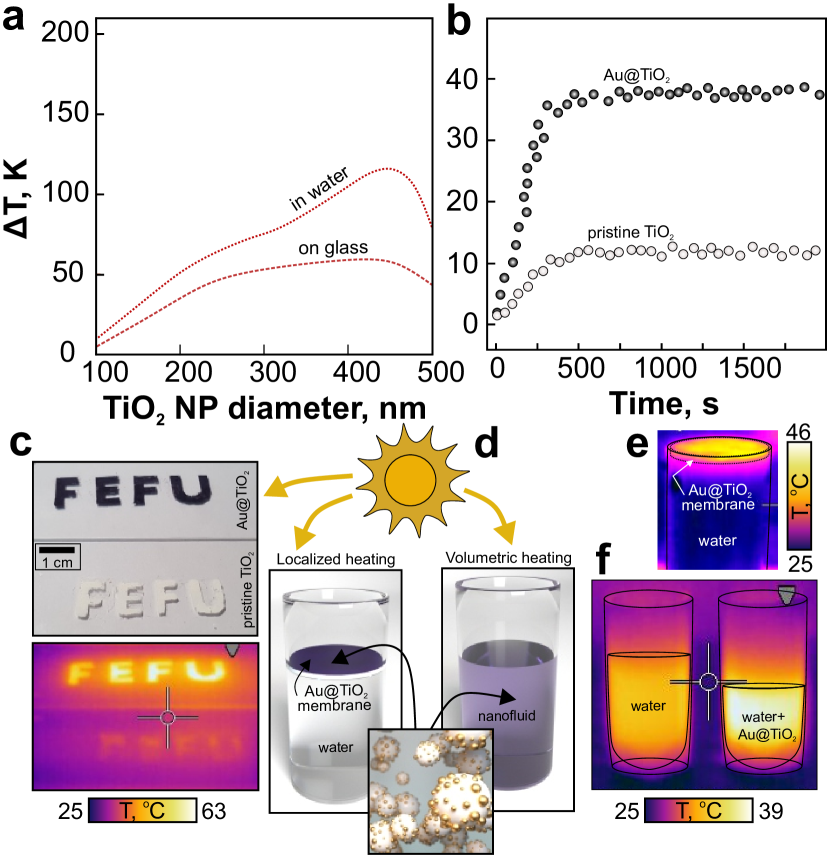
We performed comparative experiments run with custom-built solar simulator heating the black Au@TiO2 product and pristine white TiO2 material arranged to form “FEFU” letters under one-sun irradiation (0.1 W/cm2). These experiments showed that the black Au@TiO2 coating reaches a maximal temperature of about 60oC within 5 min in a sharp contrast with white TiO2 reaching only 35oC as indicated by temperature measurements with a calibrated IR-camera (see Fig. 5b). Further, using the Au@TiO2 product, we realized a lab-scale water steam generator based on both localized and volumetric heating approaches (Fig. 5d). To do this, the weight loss of water during evaporation under one-sun irradiation (at 0.1 W/cm2) was monitored and evaluated for a Au@TiO2-based nanofluid and cellulose-membrane functionalized Au@TiO2 NPs (Fig. 5d). For both approaches, we found that the evaporation rate increases 2-2.5 times compared with that for pristine distilled water, implying the potential applicability of light-absorbing AuTiO2 for solar steam generation and water desalination.
IV Conclusions
Here, we produced unique amorphous TiO2 nanospheres simultaneously decorated and doped with Au nanoclusters via single-step ns-laser ablation of commercially available TiO2 nanopowders dispersed in presence of aqueous HAuCl4. The fabricated hybrids demonstrate remarkable light-absorbing properties (averaged value 96% in the visible and near-IR spectral range mediated by bandgap reduction of the laser-processed amorphous TiO2 as well as LSPRs of the decorating Au nanoclusters that was confirmed by combining optical spectroscopy, advanced EELS/TEM and electromagnetic modeling. Excellent light-absorbing and plasmonic properties of the produced hybrids were implemented to demonstrate catalytically passive SERS biosensing for identification of analytes at trace concentration, as well as solar steam generator that permits to increase water evaporation rate by 2.5 times compared with that of pure water under identical one-sun irradiation conditions. We also envision that the produced Au@TiO2 product will be useful as transport layers in third-generation solar cells, for which our NPs possess excellent optical properties, low cost, and ability to be deposited by scalable wet-chemistry approaches. Similarly, the developed nanomaterials, being mixed with a binder, can be further used for production of highly adhesive ultrablack coatings for optical devices where even weak undesired reflections represent crucial issue.
Conflicts of interest
There are no conflicts to declare.
Acknowledgements
Laser-related experiments were supported by Russian Science Foundation (grant no. 19-79-00214). A.K. and S.M. express their gratitude to the Ministry of Science and Higher Education of the Russian Federation (grants nos. M -3258.2019.8 and MK-3514.2019.2) regarding performed calculations of temperature profiles and light-to-heat conversion efficiency.
References
- Giannini et al. (2011) V. Giannini, A. I. Fernández-Domínguez, S. C. Heck, and S. A. Maier, Chemical Reviews 111, 3888 (2011).
- Atwater and Polman (2011) H. A. Atwater and A. Polman, in Materials for Sustainable Energy: A Collection of Peer-Reviewed Research and Review Articles from Nature Publishing Group (World Scientific, 2011) pp. 1–11.
- Baffou et al. (2020) G. Baffou, F. Cichos, and R. Quidant, Nature Materials , 1 (2020).
- Kuznetsov et al. (2016) A. I. Kuznetsov, A. E. Miroshnichenko, M. L. Brongersma, Y. S. Kivshar, and B. Luk yanchuk, Science 354, aag2472 (2016).
- Alessandri and Lombardi (2016) I. Alessandri and J. R. Lombardi, Chemical Reviews 116, 14921 (2016).
- Makarov et al. (2017a) S. V. Makarov, M. I. Petrov, U. Zywietz, V. Milichko, D. Zuev, N. Lopanitsyna, A. Kuksin, I. Mukhin, G. Zograf, E. Ubyivovk, et al., Nano Letters 17, 3047 (2017a).
- Zograf et al. (2017) G. P. Zograf, M. I. Petrov, D. A. Zuev, P. A. Dmitriev, V. A. Milichko, S. V. Makarov, and P. A. Belov, Nano Letters 17, 2945 (2017).
- Mitsai et al. (2019) E. Mitsai, M. Naffouti, T. David, M. Abbarchi, L. Hassayoun, D. Storozhenko, A. Mironenko, S. Bratskaya, S. Juodkazis, S. Makarov, et al., Nanoscale 11, 11634 (2019).
- Koshelev et al. (2020) K. Koshelev, S. Kruk, E. Melik-Gaykazyan, J.-H. Choi, A. Bogdanov, H.-G. Park, and Y. Kivshar, Science 367, 288 (2020).
- Jiang et al. (2014) R. Jiang, B. Li, C. Fang, and J. Wang, Advanced Materials 26, 5274 (2014).
- Zuev et al. (2016) D. A. Zuev, S. V. Makarov, I. S. Mukhin, V. A. Milichko, S. V. Starikov, I. A. Morozov, I. I. Shishkin, A. E. Krasnok, and P. A. Belov, Advanced Materials 28, 3087 (2016).
- Timpu et al. (2017) F. Timpu, N. R. Hendricks, M. Petrov, S. Ni, C. Renaut, H. Wolf, L. Isa, Y. Kivshar, and R. Grange, Nano Letters 17, 5381 (2017).
- Makarov et al. (2017b) S. V. Makarov, A. S. Zalogina, M. Tajik, D. A. Zuev, M. V. Rybin, A. A. Kuchmizhak, S. Juodkazis, and Y. Kivshar, Laser & Photonics Reviews 11, 1700108 (2017b).
- Milichko et al. (2018) V. A. Milichko, D. A. Zuev, D. G. Baranov, G. P. Zograf, K. Volodina, A. A. Krasilin, I. S. Mukhin, P. A. Dmitriev, V. V. Vinogradov, S. V. Makarov, et al., Laser & Photonics Reviews 12, 1700227 (2018).
- Amendola and Meneghetti (2009) V. Amendola and M. Meneghetti, Physical Chemistry Chemical Physics 11, 3805 (2009).
- Zeng et al. (2012) H. Zeng, X.-W. Du, S. C. Singh, S. A. Kulinich, S. Yang, J. He, and W. Cai, Advanced Functional Materials 22, 1333 (2012).
- Zhang et al. (2017a) D. Zhang, B. Go?kce, and S. Barcikowski, Chemical Reviews 117, 3990 (2017a).
- Vailionis et al. (2011) A. Vailionis, E. G. Gamaly, V. Mizeikis, W. Yang, A. V. Rode, and S. Juodkazis, Nature Communications 2, 1 (2011).
- Zhang et al. (2017b) D. Zhang, Z. Ma, M. Spasova, A. E. Yelsukova, S. Lu, M. Farle, U. Wiedwald, and B. Gökce, Particle & Particle Systems Characterization 34, 1600225 (2017b).
- Shih et al. (2018) C.-Y. Shih, R. Streubel, J. Heberle, A. Letzel, M. V. Shugaev, C. Wu, M. Schmidt, B. Gökce, S. Barcikowski, and L. V. Zhigilei, Nanoscale 10, 6900 (2018).
- Alexander et al. (2019) D. T. Alexander, D. Forrer, E. Rossi, E. Lidorikis, S. Agnoli, G. D. Bernasconi, J. Butet, O. J. Martin, and V. Amendola, Nano Letters 19, 5754 (2019).
- Tymoczko et al. (2019) A. Tymoczko, M. Kamp, C. Rehbock, L. Kienle, E. Cattaruzza, S. Barcikowski, and V. Amendola, Nanoscale Horizons 4, 1326 (2019).
- Liu et al. (2015) P. Liu, H. Chen, H. Wang, J. Yan, Z. Lin, and G. Yang, The Journal of Physical Chemistry C 119, 1234 (2015).
- Saraeva et al. (2018) I. N. Saraeva, N. Van Luong, S. I. Kudryashov, A. A. Rudenko, R. A. Khmelnitskiy, A. L. Shakhmin, A. Y. Kharin, A. A. Ionin, D. A. Zayarny, P. Van Duong, et al., Journal of Photochemistry and Photobiology A: Chemistry 360, 125 (2018).
- Mintcheva et al. (2020) N. Mintcheva, P. Srinivasan, J. B. B. Rayappan, A. A. Kuchmizhak, S. Gurbatov, and S. A. Kulinich, Applied Surface Science 507, 145169 (2020).
- Rajabalinia et al. (2019) N. Rajabalinia, S. Hamzehlou, E. Modin, A. Chuvilin, J. R. Leiza, and J. M. Asua, Macromolecules 52, 5298 (2019).
- Palik (1998) E. D. Palik, Handbook of optical constants of solids, Vol. 3 (Academic press, 1998).
- Liu and Chen (2014) L. Liu and X. Chen, Chemical Reviews 114, 9890 (2014).
- Checcucci et al. (2018) S. Checcucci, T. Bottein, J.-B. Claude, T. Wood, M. Putero, L. Favre, M. Gurioli, M. Abbarchi, and D. Grosso, Advanced Functional Materials 28, 1801958 (2018).
- Pavliuk et al. (2020) G. Pavliuk, D. Pavlov, E. Mitsai, O. Vitrik, A. Mironenko, A. Zakharenko, S. A. Kulinich, S. Juodkazis, S. Bratskaya, A. Zhizhchenko, et al., Nanomaterials 10, 49 (2020).
- Mitsai et al. (2018) E. Mitsai, A. Kuchmizhak, E. Pustovalov, A. Sergeev, A. Mironenko, S. Bratskaya, D. Linklater, A. Balčytis, E. Ivanova, and S. Juodkazis, Nanoscale 10, 9780 (2018).
- De Angelis et al. (2011) F. De Angelis, F. Gentile, F. Mecarini, G. Das, M. Moretti, P. Candeloro, M. Coluccio, G. Cojoc, A. Accardo, C. Liberale, et al., Nature Photonics 5, 682 (2011).
- Yang et al. (2016) S. Yang, X. Dai, B. B. Stogin, and T.-S. Wong, Proceedings of the National Academy of Sciences 113, 268 (2016).
- Chen et al. (2015) X. Chen, D. Zhao, K. Liu, C. Wang, L. Liu, B. Li, Z. Zhang, and D. Shen, ACS Applied Materials & Interfaces 7, 16070 (2015).
- Zhou et al. (2016) L. Zhou, Y. Tan, J. Wang, W. Xu, Y. Yuan, W. Cai, S. Zhu, and J. Zhu, Nature Photonics 10, 393 (2016).
- Zhao et al. (2020) F. Zhao, Y. Guo, X. Zhou, W. Shi, and G. Yu, Nature Reviews Materials , 1 (2020).
- Neumann et al. (2013) O. Neumann, A. S. Urban, J. Day, S. Lal, P. Nordlander, and N. J. Halas, ACS Nano 7, 42 (2013).
- Jin et al. (2016) H. Jin, G. Lin, L. Bai, A. Zeiny, and D. Wen, Nano Energy 28, 397 (2016).