Two-orbital degeneracy lifted state as a local precursor to a metal-insulator transition
Abstract
The recent discovery of a local fluctuating orbital-degeneracy-lifted (ODL) state in CuIr2S4 as a high temperature precursor to the metal-insulator transition (MIT) opens the door to a possible widespread presence of precursor states in scarcely studied high-temperature regimes of transition-metal-based quantum materials. Although in CuIr2S4 the ODL state comprises one orbital per Ir, there is no fundamental reason to exclude multi-orbital ODL states in general. The transition-metal spinel MgTi2O4 exhibits a MIT on cooling below T 250 K, accompanied by long-range Ti orbital ordering and spin singlet dimerization with associated average symmetry reduction to tetragonal. It shares with CuIr2S4 the pyrochlore transition metal sublattice with active orbitals. This, together with its different orbital filling ( versus ) make it a candidate for hosting a multi-orbital ODL precursor state. By combining x-ray and neutron pair distribution function (PDF) analyses to track the evolution of the local atomic structure across the MIT we find that local tetragonality already exists, in the metallic globally cubic phase at high temperature. Local distortions are observed up to at least 500 K, the highest temperature assessed in this study. Significantly, the high temperature local state revealed by PDF is not continuously connected to the orbitally ordered band insulator ground state, and so the transition cannot be characterized as a trivial order-disorder type. The shortest Ti-Ti bond lengths corresponding to spin singlet dimers expand abruptly on warming across the transition, but they are still shorter than those seen in the cubic average structure. These seemingly contradictory observations can be understood within the model of a local fluctuating two-orbital ODL precursor state. The ODL state in MgTi2O4 has a correlation length of about 1 nm at high temperature. We discuss that this extended character of the local distortions is consistent with the two-orbital nature of the ODL state imposed by the charge filling and the bond charge repulsion.
I Introduction
The AB2X4 transition-metal spinels, with B cations forming a geometrically frustrated pyrochlore lattice SickafusStructureSpinel1999 , exhibit a wealth of interesting electronic and magnetic properties, such as spin dimerization RadaelliFormationisomorphicIr32002b ; SchmidtSpinSingletFormation2004 , spin-lattice instability MatsudaSpinlatticeinstability2007 , orbital ordering RadaelliOrbitalorderingtransitionmetal2005a , charge ordering WrightLongRangeCharge2001 , and metal-insulator transitions NagataMetalinsulatortransitionspineltype1998b ; IsobeObservationPhaseTransition2002 ; ItoPressureInducedSuperconductorInsulatorTransition2003 . For instance, the CuIr2S4 thiospinel system exhibits a transition from paramagnetic metal to diamagnetic insulator on cooling FurubayashiStructuralMagneticStudies1994c ; MatsunoPhotoemissionstudymetalinsulator1997b ; MatsumotoMetalinsulatortransitionsuperconductivity1999a , the formation of structural isomorphic octamers IshibashiXraydiffractionstudy2001a ; RadaelliFormationisomorphicIr32002b , and anomalous electrical properties BurkovAnomalousresistivitythermopower2000b ; TakuboIngapstateeffect2008b . In particular, CuIr2S4 has broken symmetries in its ground state accompanied by formation of charge order, orbital order, and the creation of magnetic spin singlets on dimerized Ir4+-Ir4+ pairs RadaelliFormationisomorphicIr32002b . These dimers were shown to disappear on warming through the transition, but preformed local symmetry broken states were seen at temperatures well above the global long-range ordering structure transition BozinLocalorbitaldegeneracy2019c . Thus, on cooling, a precursor state exists that has broken local symmetry but is high symmetry over long range. This behavior was shown to be driven by breaking of -electron orbital degeneracies, resulting in a local fluctuating orbital-degeneracy-lifted (ODL) state distinct from spin singlet dimer. This local state results from direct Ir orbital overlap promoted by the topology of the crystal structure. In the regime of partial filling and high crystal symmetry that imposes the degeneracy of the orbital manifold, a molecular-orbital-like state is formed, accompanied by local structure distortion. Many of the interesting physical properties of CuIr2S4 could be explained by the short-range-ordered ODL mechanism, such as the non-conventional conduction in the high-temperature metallic phase BurkovAnomalousresistivitythermopower2000b , and the apparently contradictory observation of the destruction of the dimers at the phase transition but the persistence of poor metallic response above the transition BozinDetailedMappingLocal2011c .
Similar local symmetry-broken ODL states preformed at high temperature have been observed in other non-spinel -electron systems such as the FeSe superconductor KochRoomtemperaturelocal2019d ; FrandsenQuantitativecharacterizationshortrange2019a . Even the well studied physics of the perovskite lanthanum manganites (LaMnO3) could be interpreted in the same way qiu;prl05 , arguing that the ODL phenomenon may be widespread among the myriad of materials with partially filled -electron manifolds. To explore this hypothesis it is important to seek it out systematically and characterize other materials that have the potential of revealing both commonalities and novel aspects that give more insights into the general ODL phenomenon. For instance, there is no fundamental reason for the ODL states to be exclusively comprised of one orbital per transition metal, yet multi-orbital ODL states have so far not been identified. Here we show that such multi-orbital ODL state exists in a related spinel, MgTi2O4, and that this is a consequence of the electron configuration decorating the pyrochlore lattice.
MgTi2O4 shares many features with CuIr2S4 KhomskiiOrbitallyInducedPeierls2005a . They are both cubic spinels at high temperature with a transition metal ion pyrochlore sublattice of corner-shared tetrahedra. In both materials the crystal field splits the -orbitals into a triplet and an doublet with the orbitals partially occupied and the orbitals empty. They both exhibit a temperature-dependent metal-insulator transition (MIT) NagataMetalinsulatortransitionthiospinel1994a ; IsobeObservationPhaseTransition2002 , the origin of which has been attributed to an orbital-selective Peierls mechanism KhomskiiOrbitallyInducedPeierls2005a . They both exhibit a global symmetry lowering, from cubic to tetragonal, at the MIT on cooling. They both have anomalous electrical resistivity behavior in the high-temperature metallic phase ZhouOpticalstudyMgTi2O42006 ; BurkovAnomalousresistivitythermopower2000b . The symmetry lowering at the MIT is accompanied by a dimerization of transition metal ions that results in alternating short and long metal-metal bonds, and a resulting tetramerization CroftMetalinsulatortransitionmathrmCuIr2003a , along linear chains of ions on the pyrochlore sublattice KhomskiiOrbitallyInducedPeierls2005a . The short bonds are associated with spin singlet dimer formation RadaelliFormationisomorphicIr32002b ; SchmidtSpinSingletFormation2004 . The charge filling is also electron-hole symmetric between the systems with Ti3+ having one electron in the manifold, whilst Ir4+ has one hole, although the nominal charge of Ir in CuIr2S4 is 3.5+ which would place half a hole per Ir in the anti-bonding band on average in this compound.
Despite the similarities, there are also notable differences. The Ti valence electrons reside in 3 orbitals whereas for Ir they are 5, which are more extended and should result in larger bandwidth. Indeed, the average separation between the transition metals on the undistorted tetrahedral pyrochlore sublattices of their cubic structures follow this expectation: Ti-Ti separation is shorter (3.0 Å) than Ir-Ir separation (3.5 Å). Also, experimentally, in MgTi2O4 the tetragonal distortion is shown to be compressive () below the MIT SchmidtSpinSingletFormation2004 while it is tensile () FurubayashiStructuralMagneticStudies1994c in CuIr2S4. Dimers form helical superstructures in MgTi2O4, whereas they form octameric “molecules” in CuIr2S4, thereby lowering the symmetry further to triclinic. Both materials tetramerize below the MIT; however, CuIr2S4 has a 3+-3+-4+-4+ charge ordering (CO) that accompanies an orbital ordering (OO) BozinLocalorbitaldegeneracy2019c , whereas a uniform 3+ charge on Ti rules out CO in MgTi2O4. With both similarities – orbitals on pyrochlore lattice, formation of dimerized singlets – and differences to CuIr2S4 – lack of charge order, electron rather than hole states – MgTi2O4 provides a natural next step in a deeper, broader mapping of ODL phenomena. As we show below, this includes the emergence of a multi-orbital ODL state.
Neutron PDF (nPDF) analysis on MgTi2O4 has been performed previously TorigoeNanoscaleicetypestructural2018 to study the spin singlet dimers, suggesting that they do persist to high temperature. However, due to the weak and negative neutron scattering length of Ti, and appreciable overlap with substantially stronger oxygen signal, nPDF itself cannot fully reveal how the local structure behaves with temperature. Here we have applied a combined x-ray and neutron analysis to understand the full picture. We find unambiguously that the Ti-Ti dimers do disassemble on warming through MIT. However, the local structure does not agree with the average structure even at high temperature. In analogy with CuIr2S4, partially filled transition metal orbital manifolds of MgTi2O4, which are triply degenerate in the average cubic symmetry, utilize their favorable overlaps fostered by the pyrochlore sublattice topology to form an ODL state. Its structural signatures are observed up to at least 500 K ( 2Ts), the highest temperature measured. The spatial extent of the local structural response is found to be greater than that observed in CuIr2S4, consistent with the proposed two-orbital character of the ODL state in MgTi2O4.
II Methods
Sample preparation & characterization.— TiO2, Ti metal, and an excess of MgO were mixed and reacted using a spark plasma sintering technique in a graphite crucible. Synthesis at 1100 o C was complete in 15 minutes. The sample was reground and fired a second time under similar conditions. Powder X-ray diffraction analysis of the product showed well-crystallized MgTi2O4 spinel accompanied by an extremely small concentration of Ti2O3 as a second phase. Magnetization measurements were conducted using SQUID magnetometer on a specimen with mass of 6.2 mg. The data show a pronounced low temperature Curie tail which was subtracted. The Curie-Weiss fit to the low temperature data yielded a Weiss temperature of -0.45 K, consistent with isolated spins, and a Curie constant of 0.023 emuK/mol which corresponds to % by mole of putative Ti3+ spin-1/2 impurities
The PDF method.—The local structure was studied using the atomic pair distribution function (PDF) technique egami;b;utbp12 ; billi;b;itoch18 . The PDF analysis of x-ray and neutron powder diffraction datasets has been demonstrated to be an excellent tool for revealing local-structural distortions in many systems BozinLocalorbitaldegeneracy2019c ; KochRoomtemperaturelocal2019d ; billi;prl96 ; qiu;prl05 ; YoungApplicationspairdistribution2011a ; Keencrystallographycorrelateddisorder2015a ; LavedaStructurepropertyinsights2018 . The PDF gives the scaled probability of finding two atoms in a material a distance apart and is related to the density of atom pairs in the material. It does not presume periodicity so goes well beyond just well ordered crystals egami;b;utbp12 ; billi;b;itoch18 . The experimental PDF, denoted , is the truncated Fourier transform of the reduced total scattering structure function farro;aca09 , :
| (1) |
where is the magnitude of the scattering momentum transfer. The total scattering structure function, , is extracted from the Bragg and diffuse components of x-ray, neutron or electron powder diffraction intensity. For elastic scattering, , where is the wavelength of the probe and is the scattering angle. In practice, values of and are determined by the experimental setup and is often reduced below the experimental maximum to eliminate noisy data from the PDF since the signal to noise ratio becomes unfavorable in the high- region egami;b;utbp12 .
X-ray PDF experiment.— The synchrotron x-ray total scattering measurements were carried out at the PDF beamline (28-ID-1) at the National Synchrotron Light Source II (NSLS-II) at Brookhaven National Laboratory (BNL) using the rapid acquisition PDF method (RAPDF) chupa;jac03 . The MgTi2O4 powder sample was loaded in a 1 mm diameter polymide capillary and measured from 90 K to 500 K on warming using a flowing nitrogen cryostream provided by Oxford Cryosystems 700 Series Cryocooler. The experimental setup was calibrated by measuring the crystalline Ni as a standard material. A two-dimensional (2D) PerkinElmer area detector was mounted behind the samples perpendicular to the primary beam path with a sample-to-detector distance of 227.7466 mm. The incident x-ray wavelength was 0.1668 Å. The PDF instrument resolution effects are accounted for by two parameters in modeling, and proff;jac99 ; farro;jpcm07 . For x-ray PDF measurement, these were determined as Å-1 and Å-1 by fitting the x-ray PDF from a well crystallized sample of Ni collected under the same experimental conditions.
In order to verify the data reproducibility, an additional set of x-ray data (in 90 K to 300 K temperature range on warming) was collected at the XPD beamline (28-ID-2) at the NSLS-II at BNL using a similar RAPDF setup but with x-ray wavelength of 0.1901 Å and sample-to-detector distance of 251.1493 mm. The corresponding instrument resolution parameters were determined to be Å-1 and Å-1, implying similar instrument resolution effects across the two sets of measurements.
The collected x-ray data frames were summed, corrected for detector and polarization effects, and masked to remove outlier pixels before being integrated along arcs of constant momentum transfer , to produce 1D powder diffraction patterns using the pyFAI program Ashiotisfastazimuthalintegration2015a . Standardized corrections and normalizations were applied to the data to obtain the reduced total scattering structure function, , which was Fourier transformed to obtain the PDF, using PDFgetX3 juhas;jac13 within xPDFsuite YangxPDFsuiteendtoendsoftware2015 . The maximum range of data used in the Fourier transform was chosen to be Å-1, so as to give the best trade-off between statistical noise and real-space resolution.
Neutron PDF experiment.— The time-of-flight (TOF) neutron total scattering measurements were conducted at the NOMAD beamline (BL-1B) NeuefeindNanoscaleOrderedMAterials2012 at the Spallation Neutron Source (SNS) at Oak Ridge National Laboratory. The MgTi2O4 powder sample was loaded into a 3 mm diameter silica capillary mounted on a sample shifter, and data were collected from 100 K to 500 K on warming using an Ar flow cryostream. The neutron PDF instrument resolution parameters were determined as Å-1 and Å-1 by fitting the neutron PDF of a NIST standard 640d crystalline silicon sample.
The neutron data were reduced and transformed to the PDF with Å-1 using the automated data reduction scripts available at the NOMAD beamline.
Structural modeling.— The PDF modeling programs PDFgui and DiffPy-CMI were used for PDF structure refinements farro;jpcm07 ; juhas;aca15 . In these refinements (Å2) is the isotropic atomic displacement parameter (ADP) and the ADPs of the same type of atoms are constrained to be the same; (Å2) is a parameter that describes correlated atomic motions jeong;jpc99 . The PDF instrument parameters and determined by fitting the PDF from the well crystallized standard sample under the same experimental conditions are fixed in the structural refinements on MgTi2O4 dataset.
To estimate the lengthscale of local structural correlations, a dependent fit is performed on select high temperature x-ray data (300 K, 400 K, and 500 K). During the fit, the average high-temperature cubic MgTi2O4 model is used, fixing the upper limit of fit range as Å, but changing the lower limit from Å in 0.2 Å -steps. Each fit range uses the same initial parameter values and term is not applied since the data fitting range does not consistently include the low- region in all these refinements.
The Rietveld refinement on the neutron TOF Bragg data was implemented in the GSAS-II software TobyGSASIIgenesismodern2013c . The instrument parameters were refined to the standard silicon data collected under the same experimental conditions and then fixed in the MgTi2O4 Rietveld refinements. The sequential refinement option was used to refine the temperature series dataset collected at detector bank from 100 K to 500 K in a systematic manner.
Structural models.— Two candidate MgTi2O4 models were fit against the experimental data. In the cubic MgTi2O4 model (space group: ), the atoms sit at the following Wyckoff positions: Mg at 8a (0.125,0.125,0.125), Ti at 16d (0.5, 0.5, 0.5), and O at 32e (, , ). The initial lattice parameters and atomic positions are Å and O at (0.25920, 0.25920, 0.25920) SchmidtSpinSingletFormation2004 . The Ti pyrochlore sublattice is shown in Fig. 1(a), indicating that all the Ti-Ti bonds are of equal length, reflecting regular Ti4 tetrahedra.
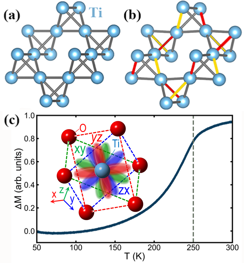
In the tetragonal MgTi2O4 model (space group: ), the atoms sit at the following Wyckoff positions: Mg at 4a (,,0), Ti at 8b (, , ), O1 at 8b (, , ), and O2 at 8b (, , ). The initial lattice parameters and atomic positions are Å, Å, Mg at (0.7448, 0.7448, 0), Ti at (-0.0089, 0.2499, -0.1332), O1 at (0.4824 0.2468 0.1212), and O2 at (0.2405, 0.0257, 0.8824) SchmidtSpinSingletFormation2004 . The corresponding distorted Ti sublattice is presented in Fig. 1(b), showing that one Ti-Ti bond gets shorter (indicated in red) and one gets longer (in yellow) out of the six Ti-Ti bonds of each Ti4 tetrahedron. The lattice parameters of these two models have the relationship of , where the subscripts and refer to the cubic and tetragonal models, respectively.
III Vanishing spin singlet dimers
III.1 Canonical behavior
The magnetization, , of the sample up to 300 K is shown in Fig. 1(c). A low temperature Curie-Weiss like component has been subtracted to account for the effect of magnetic impurities contributing to the signal at low temperature. The onset of a broad dimerization transition is observed at around 250 K, which is close to the literature reported MIT temperature of 260 K IsobeObservationPhaseTransition2002 ; VasilievSpecificheatmagnetic2006 and implies canonical behavior of our sample.
We next establish that the average structural behavior of our sample agrees with other observations in the literature IsobeObservationPhaseTransition2002 ; SchmidtSpinSingletFormation2004 ; TalanovTheorystructuralphase2015 by carrying out Rietveld refinements fitting tetragonal and cubic MgTi2O4 models to the neutron time-of-flight (TOF) Bragg data measured from 100 K to 500 K. The refinement results from select fits are reproduced in Table 1 presented in Appendix A, and a representative fit, of the tetragonal model to the 100 K dataset, is shown in Fig. 2(a).
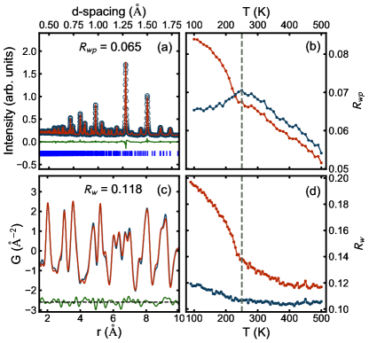
The tetragonal distortion, given by , is small at all temperatures and the data are not of high enough resolution to directly observe distinct peaks that would indicate any tetragonal distortion. However, the temperature dependence of the weighted profile agreement factor, , of the two models is shown in Fig. 2(b), which clearly implicates the tetragonal model as preferred at low-temperature, but not at high temperature, above around 230-250 K, which is close the literature reported MIT temperature 260 K IsobeObservationPhaseTransition2002 .
III.2 Local structure behavior
We investigate the behavior of the local structure by performing a PDF structural refinement of the x-ray total scattering PDF data from 90 K to 500 K. Fits were carried out over the data range of Å and Å and the results are qualitatively the same. A representative fit of the tetragonal model to the 100 K dataset is shown in Fig. 2(c). This time, the temperature dependence of the goodness-of-fit parameter, , of the tetragonal and cubic models (Fig. 2(d)) indicates that the tetragonal model fits the local structure better than the cubic model at all temperatures. In the local structure the tetragonal distortion is always evident.
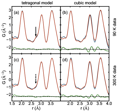
To see visually in the PDF how the cubic model fails we consider the low and high-temperature PDFs and fits with the cubic and tetragonal models, shown in Fig. 3.
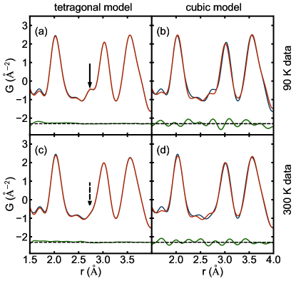
The low temperature PDFs (below the MIT) are shown in the top row ((a) and (b)) and the high temperature (above the MIT) in the lower row ((c) and (d)). The tetragonal model fit is shown in the first column ((a) and (c)) and the cubic fit is shown in the second column ((b) and (d)). At low temperature, as expected, the tetragonal model (Fig. 3(a)) fits much better than the cubic model (Fig. 3(b)). At 300 K we have already established that the global structure is cubic; however, again we see that in these local structural fits the tetragonal model (Fig. 3(c)) is superior to that of the cubic model (Fig. 3(d)), with smaller oscillations in the difference curve. To emphasize our argument, we note that the difference curves are similar when data collected at distinct temperatures are fit using identical symmetry constraints. Specifically, careful inspection of the difference curves in Fig. 3(b) and (d) reveals that the positions of residual maxima and minima are nearly identical, although their amplitudes are smaller in Fig. 3(d). Since this difference signal represents the inability of the cubic model to fit the data, it is further support to the idea that a similar tetragonally distorted structure is present in the low- region at 90 K and at 300 K, but smaller in amplitude at 300 K. To validate that the results are reproducible, Fig. 3 represents x-ray PDF data collected at the PDF beamline, and data collected from the XPD beamline is shown in Fig. 8 presented in Appendix A, which reproduces the same result as discussed above.
In the cubic MgTi2O4 structure unit cell, all six Ti-Ti bonds have the same length, 3.008 Å, whereas in the tetragonal MgTi2O4 model, one of the six Ti-Ti distances is shortened (2.853 Å), forming dimers, and one Ti-Ti bond is longer (3.157 Å) TalanovTheorystructuralphase2015 . The dimer contact is considerably shorter than the 2.917 Å found in titanium metal Hullcrystalstructurescommon1922 , indicating a strong covalent interaction in MgTi2O4. From the analysis of the average structure, it was understood that the MIT was accompanied by the formation of Ti-Ti structural dimers. Above we showed the observation from the PDF of local tetragonality at high temperature which may indicate that local dimers survive above the MIT. This would seem to be in qualitative agreement with a prior observation from a neutron PDF study that reported the persistence of Ti-Ti dimers up to high temperature TorigoeNanoscaleicetypestructural2018 . In this picture the dimers survive locally but become disordered at the transition where the structure becomes globally cubic on warming. However, our PDF analysis clearly shows that the large amplitude structural dimers actually do disappear at the MIT, as described below, and that the local tetragonality that we observe at high temperature in the PDF has a more subtle origin as we develop in greater detail below.
III.3 Orbital degeneracy lifting or spin dimerization?
To establish the disappearance of the structural dimers at the phase transition, we simulated x-ray PDFs of the cubic (no dimers) and tetragonal (dimers) models. These are plotted as the red and blue curves, respectively, in Fig. 4 (a).
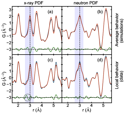
A number of the PDF peaks are affected on the transition through the MIT, with the largest change observed on the peak at around 3.0 Å, which contains the shortest Ti-Ti distances. Based on the change in crystal structure, the expected change in the PDF results in the characteristic M-shaped signature in the difference curve seen in Fig. 4 (a) which comes from the disappearance of the long and short Ti-Ti bonds associated with the dimer, leaving all the Ti-Ti distances the same. This can be compared to the difference in the measured PDF data as MgTi2O4 crosses the MIT, shown in Fig. 4(c) at 300 K (red) and 100 K (blue). First, we see a significant change in the relevant 3.0 Å peak when moving through the MIT. This change is inconsistent with the conclusion from the previous neutron PDF study that the dimers are retained, unaltered, to high temperature in the local structure TorigoeNanoscaleicetypestructural2018 , necessitating an order-disorder scenario in which the local structure across the transition is unchanged, with small or no change in the PDF. Instead we see a significant change in the x-ray PDF at the phase transition, with PDF intensity moving from the short position towards that of the average Ti-Ti distance, as indicated by the grey circle in Fig. 4(c).
It is clear from careful inspection that the observed and simulated difference curves across the transition (Fig. 4(c) and (a), respectively) are significantly different. PDF peak intensity redistribution associated with the dimer removal is seen in the differential curve as a transfer of intensity from 2.71 Å to 2.89 Å, i.e., on the low- side of the average Ti-Ti distance at 3.01 Å marked by the vertical dashed line in the figure. This implies that the very short Ti-Ti ion dimers disappear at the phase transition, but the associated PDF intensity is shifted to a position that is still shorter than that of the average Ti-Ti distance: shortened Ti-Ti contacts in fact do exist at high temperature above the MIT but they are not the original spin singlet dimers. Our results are consistent with the disappearance of dimers at the phase transition on warming, but a persistence of a local tetragonality, smaller in magnitude than that associated with the the dimer phase. This behavior is shown on an expanded scale in Fig. 5(b) where PDFs measured at multiple temperatures are compared, focusing on the 3 Å peak.
The feature in the difference curve that shows the shift of the intensity associated with the Ti-Ti short bonds from 2.71 Å to the longer position (2.89 Å) is shown below shaded green (for the loss of short bonds) and purple (for the gain in longer bonds). This result is qualitatively supported by the PDF structural modeling over the range of Å, shown in Fig. 5(e) and (f), where the short Ti-Ti bond shifts from 2.83 Å to 2.88 Å over the same temperature range, but never converges to the average cubic value of 3.01 Å at 300 K.
The full temperature dependence of the dimer disappearance may then be extracted by integrating the shaded regions in the difference curve and is shown in Fig. 5(c), where we plot the integrated area shown shaded in green in the difference curve vs. temperature. In this case the difference is always taken with respect to the 500 K dataset. The intensity decreases gradually until around 200 K, where it rapidly falls off. The rate of fall-off in this intensity then slows again above 250 K. There are two principal contributions to the differential signal below the transition: the changes in the local structure and the thermal broadening effects. Thermal broadening effects in the differential are gradual and typically small, particularly from one data point to the next corresponding to the trends observed below 200 K and above 250 K. On the other hand, the signal in the differential coming from the dimers is considerably larger, roughly proportional to the big step seen at the transition, and dissipates rapidly as the temperature passes through the MIT range. This is a model-independent way of observing how the local dimer disappears on warming.
III.4 X-ray PDF versus neutron PDF
We may seek an explanation for why we see the dimers disappear at the transition from our x-ray PDF measurements, whereas this was not evident in the earlier neutron PDF study TorigoeNanoscaleicetypestructural2018 . Observing the local Ti dimer is complicated in the neutron case by the relatively weak and negative neutron scattering length of titanium, and an appreciable overlap of titanium contributions with strong oxygen contributions. To show this, we perform the same comparison that we just made for the x-ray PDFs, but on neutron data. The neutron simulations are shown in Fig. 4(b) and the neutron experimental PDFs in Fig. 4(d). It is clear that the signals in the difference curve, both for the simulations based on the average structure and the data PDFs themselves, are much smaller than for the x-ray case. This is because the most important signal in MgTi2O4 is coming from the Ti ions that are relatively strong scatterers in the x-ray case but not in the neutron measurement. Specifically, in the x-ray case, Ti scatters 2.75 times stronger than O and 1.8 times stronger than Mg, whereas for neutrons the scattering of Ti is 1.7 times weaker than that of O and 1.6 times weaker than that of Mg. In addition, the neutron PDF intensity in the range of interest is dominated by the contributions from O-O pairs constituting TiO6 octahedra, evidenced in the data as the additional shoulder intensity features around the 3 Å peak (Fig. 4(d)), as compared to the x-ray case where such features are largely absent (Fig. 4(c)). This may explain the different interpretation in the earlier neutron PDF study TorigoeNanoscaleicetypestructural2018 . However, the x-ray data unambiguously show the disappearance of the full-amplitude spin singlet Ti-Ti dimers.
III.5 Detection of the ODL state
This behavior, where sizeable distortions associated with ordered spin singlet dimers evolve into smaller local distortions with spin singlets disassembled, is reminiscent of that observed in the CuIr2S4 system. In that case a local symmetry broken orbital-degeneracy-lifted (ODL) state was observed up to the highest temperatures studied BozinLocalorbitaldegeneracy2019c . On cooling these local distortions of broken symmetry ordered into a long-range orbitally ordered (LROO) state. Only below the LROO transition did charges disproportionate, forming a charge density wave accompanied by a Peierls distortion and the formation of Ir-Ir dimers with very short Ir-Ir bonds. We believe our observations in MgTi2O4 suggest a similar kind of ODL behavior, which we explore to a greater extent below, though different in detail because of the different charge filling.
As discussed above, we can rule out that the local structure is changing in the same way as the average structure. The expected changes in the PDF at low- due to average structure changes at the MIT make all the low- peaks sharper for , as shown in Fig. 4(a). However, the data do not show this (Fig. 4(c)). There is a small change in the local structure, evidenced by the feature in the difference curve around 2.9 Å, indicated by the grey circle in Fig. 4(c), but it is smaller than the average structure change at the MIT. As discussed above, the short Ti-Ti dimers ( Å shoulder) go away on warming by a shift to a longer bond (around Å), but this “longer” bond is still shorter than the average 3.01 Å Ti-Ti bond distance expected from the cubic average model. These two behaviors exactly mimic the ODL state found in CuIr2S4 BozinLocalorbitaldegeneracy2019c .
As is evident in Fig. 2(d), the tetragonal model fits the local structure better than the cubic model at all temperatures to 500 K, the highest measurement temperature. As in CuIr2S4 BozinLocalorbitaldegeneracy2019c the dimers disappear at the MIT transition on warming, but the local symmetry broken state with, presumably, fluctuating short Ti-Ti bonds is present to high temperature.
III.6 Spatial extent of the ODL state
It is of interest to explore whether the fluctuating short Ti-Ti bonds at temperature above the MIT correlate with each other, and how this varies with temperature. To extract the correlation length of the local fluctuating symmetry broken states, dependent PDF fits were performed on selected high-temperature datasets (300 K, 400 K, and 500 K). In these models the high-temperature average cubic model was fit over a range from to a fixed Å, where was allowed to vary from Å. When is large the fit is over just the high- region of the PDF and will retrieve the average structure and the cubic fit will be good. As extends to lower values, progressively more of the local structure that has a tetragonal distortion is included in the fit and the agreement of the cubic model becomes degraded. The resulting is shown in Fig. 5(d). The cubic fits are good over the entire region, Å, with very little variation in indicated by the horizontal grey band in the figure, but rapidly degrade below this length-scale. This suggests that the symmetry broken local distortions have a correlation length of around 1 nm but that this correlation length does not vary significantly in the temperature range above 300 K. This agrees well with the previously reported length-scale of local tetragonality based on neutron PDF analysis TorigoeNanoscaleicetypestructural2018 .
IV Two-orbital ODL state
IV.1 The ODL regime
The driving force behind the high temperature local symmetry breaking in CuIr2S4 was shown to be of electronic origin involving orbital degeneracy lifting BozinLocalorbitaldegeneracy2019c . Significantly, the isostructural and isoelectronic sister compound, CuIr2Se4, remains metallic down to the lowest temperature and does not show local symmetry breaking orbital-degeneracy-lifted (ODL) effects. This intriguingly suggests that the ODL state could be a prerequisite for the MIT BozinLocalorbitaldegeneracy2019c in these spinels. Here we argue that also in MgTi2O4 local ODL effects are present and produce the local symmetry breaking in the high temperature region that we report here. By analogy with CuIr2S4, the ODL states are precursor states to the spin singlet dimerization and MIT.
The long range pattern of ordered spin singlet dimers differs in detail in the two systems, being octamers in CuIr2S4 RadaelliFormationisomorphicIr32002b and helices in MgTi2O4 SchmidtSpinSingletFormation2004 , and is presumably dictated by delicate energetics DiMatteoValencebondcrystallattice2005 ; RahamanTetramerorbitalordering2019 . Although intriguing, these intricate aspects of long range order are not of concern here. Our interest lies with internal dimer structure, the relationship between the dimer state and the ODL state, and the electron-hole symmetry that links the two systems KhomskiiOrbitallyInducedPeierls2005a .
Prior to pursuing the analogy between the CuIr2S4 and MgTi2O4, we appraise the two systems from the perspective of a regime under which the ODL state can form in a transition metal system BozinLocalorbitaldegeneracy2019c . Three criteria need to be fulfilled: (i) filling – the transition metal in the system possesses partially filled orbitals, (ii) symmetry – the high crystallographic point symmetry of the system imposes the orbital degeneracy, and (iii) topology – the structural topology promotes adequate highly directional orbital overlaps. In practice, materialization of the ODL state could further be impacted by the existence of other competing degeneracy lifting channels that may be available to the system, such as the relativistic spin orbit coupling, the effects of crystal field, etc. Eventually, formation of the ODL local broken symmetry state is accompanied by associated local structure distortion. Both CuIr2S4 and MgTi2O4 systems meet the criteria: partial filling (Ir is , Ti is ), degeneracy promoting high symmetry structure (cubic spinel structure imposes 3-fold degeneracy of the manifolds in both materials), and favorable structural topology (edge shared IrS6 and TiO6 octahedra fostering direct overlap). In both systems short nearest neighbor transition metal contacts are observed in the high temperature metallic regime, that are distinct from the very short spin singlet dimer bonds observed in the insulating state at low temperature.
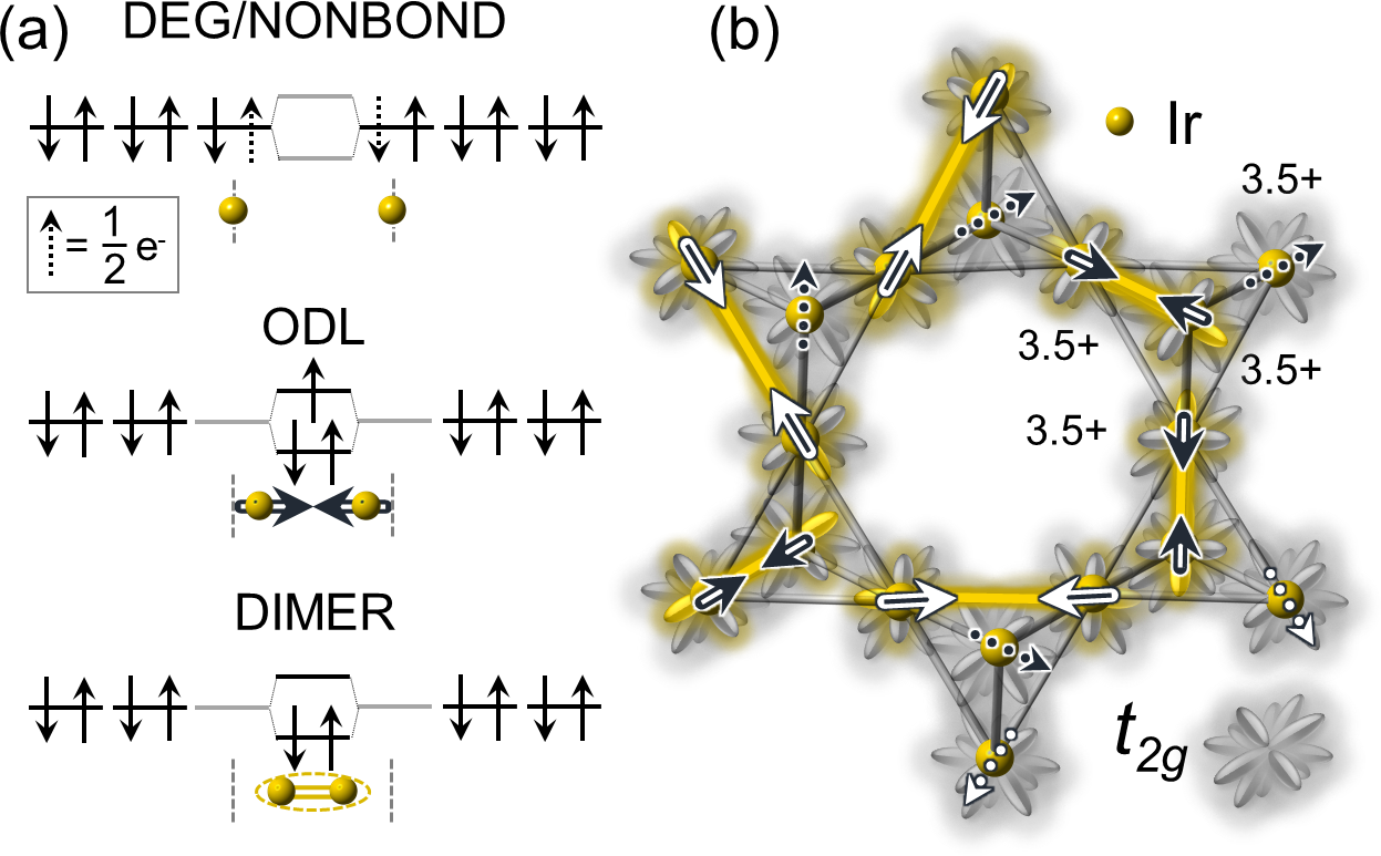
In what follows we first review the ODL state and the dimerization mechanism within the local picture of CuIr2S4, summarized in Fig. 6. We then utilize the analogies between the two systems to put forward a scenario portraying the high temperature state and the dimerization mechanism in MgTi2O4, illustrated in Fig. 7.
IV.2 Single-orbital ODL state
In CuIr2S4 the dimers involve two Ir4+ ions in configuration with hole character BozinLocalorbitaldegeneracy2019c . In the localized electron picture, the ODL state is based on molecular orbital (MO) concepts. In this, two neighboring transition metal ions with partially filled degenerate orbitals form a MO state with shared electrons (holes) that lifts the degeneracy and lowers the system energy. In the high temperature regime of CuIr2S4 iridium nominally has a half-integer valence (3.5+) MatsunoPhotoemissionstudymetalinsulator1997b ; YagasakiHoppingConductivityCuIr2S42006b . This means that each Ir3.5+ on the pyrochlore sublattice is in a nominal state corresponding to half a hole per three degenerate orbitals (Fig. 6(a), top). The sublattice geometry fosters direct overlaps of orbitals from the manifolds (Fig. 6(b)), which, in turn allows neighboring Ir pairs to form a bound state sharing a single hole in the antibonding MO BozinLocalorbitaldegeneracy2019c (middle panel of Fig. 6(a)).
Importantly, for any choice of two neighboring Ir on a pyrochlore lattice only one member of the three orbitals overlap (e.g. xy with xy, etc.) along the Ir4 tetrahedral edges. Due to the specifics of filling (0.5 holes/Ir) each Ir participates in exactly one such paired state at a time. We call this a one-orbital ODL (1O-ODL) state. The ODL state is hence comprised of two atomic orbitals, one from each Ir in the pair, with on average 1.5 electrons (0.5 holes) per Ir, resulting in MO with 3 electrons and one hole, as shown in Fig. 6(a), with a net spin of 1/2. This configuration results in the observed contraction of the Ir-Ir separation in the local structure vis-à-vis that expected if orbital degeneracy is retained. Since each iridium has 6 Ir neighbors to pair with, the ODL state fluctuates spatially and, presumably, temporally among (xy, xy), (yz, yz), and (zx, zx) variants, which results in an undistorted cubic structure on average BozinLocalorbitaldegeneracy2019c . One such configuration is illustrated in Fig. 6(b), with strong short-range correlations governed by the “one ODL state per Ir” and the Coulomb bond charge repulsion principles, resulting in a single ODL state per Ir4 tetrahedron. Pursuant to this, the ODL state in CuIr2S4 follows the “two in – two out” ice rules ander;pr56 , which will be addressed later.
In this localized picture, the ODL state is also a precursor for the spin singlet dimer. The dimer state is attained by removing an excess electron from the antibonding MO of the ODL state, thus stabilizing the bond, as shown in the bottom panel of Fig. 6(a). The process involves charge transfer between two ODL Ir3.5+-Ir3.5+ pairs, one of which becomes dimerized Ir4+-Ir4+ by losing an electron (or gaining a hole, hence hole dimer) and the other becomes non-ODL (and non-dimer) Ir3+-Ir3+ by gaining an electron (- configuration).
IV.3 Two-orbital ODL state
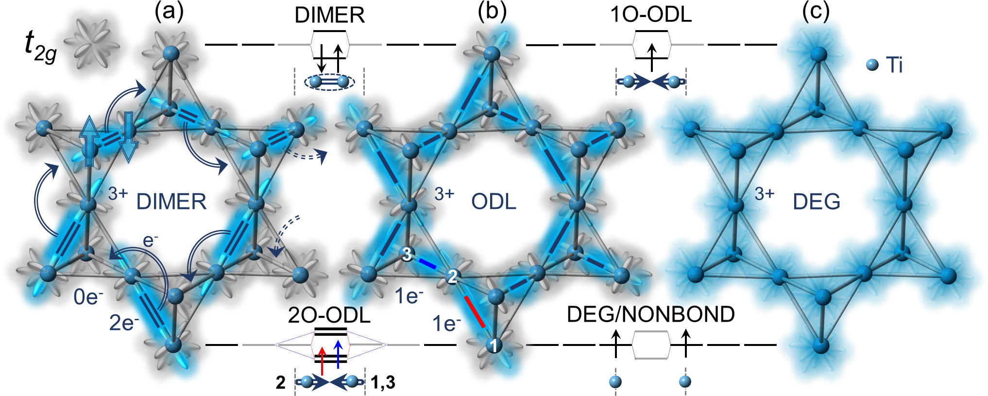
We now turn to MgTi2O4 starting from the local view of spin singlet dimers. Dimerization in MgTi2O4, depicted using the Ti manifold representation, is shown in Fig. 7(a), overlaying a fragment of pyrochlore sublattice as seen in the model. In MgTi2O4 the dimers involve two Ti3+ in configuration with a single electron in manifold, hence Ti3+-Ti3+ dimers inevitably have electron character by construction. These result in short Ti-Ti dimerized contacts observed in the tetragonal structure at low temperature. Each dimer carries 2 of net charge. Since each Ti participates in a dimer, and since there is exactly one dimer per Ti4 tetrahedron DiMatteoValencebondcrystallattice2005 ; TorigoeNanoscaleicetypestructural2018 , nominal charge count results in 1/Ti site. This is consistent with no CO being observed experimentally in MgTi2O4, in contrast to CuIr2S4, implying that all Ti sites are equivalent in this regard SchmidtSpinSingletFormation2004 . From the average structure perspective and in the localized picture, supported by the experimentally observed paramagnetism and poor metallic conduction, above the MIT the dimers could be seen to disassemble in such a way as to statistically distribute 1 of charge evenly across the three degenerate orbitals, resulting in a cubic structure with all Ti-Ti nearest neighbor contacts equivalent, as schematically presented in Fig. 7(c). This implies that, despite nominal charge equivalence of all Ti sites and no site CO observed, some charge transfer, presumably involving bond charge, still has to take place at the transition. This then inevitably implies that the ground state has to involve the bond charge order which coincides with, and is hence indistinguishable from, the observed dimer order in the diffraction measurements.
The implication of charge being equally distributed across the triply degenerate manifold of Ti in a manner depicted in Fig. 7(c), presented also in associated energy diagram (bottom right inset to the Figure), would be that the pyrochlore sublattice is comprised of regular Ti4 tetrahedra with equidistant Ti-Ti contacts corresponding to equal bond charge, as described within the cubic spinel model. However, this is not what is observed experimentally. The observations based on the PDF analyses clearly demonstrate that the pyrochlore sublattice is comprised of locally distorted Ti4 tetrahedra with a distribution of distances, suggesting that the bond charge remains inequivalent above the MIT. One possibility is that the dimers indeed persist in the metallic regime, as hinted at in the earlier neutron study TorigoeNanoscaleicetypestructural2018 . However, such an interpretation would be inconsistent with magnetization measurements of MgTi2O4 that establish the disappearance of spin singlets above the MIT. It would further be inconsistent with the elongation of the short Ti-Ti dimer contacts evidenced in our x-ray PDF analysis, which implies disappearance of spin singlet dimers at MIT even locally. In analogy with CuIr2S4, it is plausible that the dimer state gets replaced locally by an ODL-type state in the high temperature metallic phase. Observation of heterogeneous local Ti-Ti contacts corresponding to inequivalent bond charge is consistent with an ODL-like state in MgTi2O4. Since the local structures at high temperature and in the ground state are distinct, the MIT in MgTi2O4 cannot be assigned to a trivial order-disorder type.
Importantly, as careful assessment shows, the ODL state in MgTi2O4 cannot be exactly mapped onto the ODL state seen in CuIr2S4, since the charge filling is different in the two systems (1 /Ti in MgTi2O4, 0.5 holes/Ir in CuIr2S4). In the latter case two Ir neighbors can reduce the energy of the system by forming a 1O-ODL state accommodating a common hole, as described in the previous section. This results in a 3-electron state shown in the middle panel of Fig. 6(a). Given that the destruction of the spin singlet dimer in MgTi2O4 involves removal of one of the electrons from the dimer state sketched in the energy diagram in the top left inset to Fig. 7, consistent with destabilization and elongation of the short Ti-Ti bond, in the ODL picture the one electron left behind would indeed result in an electron-hole antipode of the 1O-ODL state seen in CuIr2S4, as shown in the top right inset to Fig. 7. The issue arises with the placement of the extra electron from the dimer. In case of CuIr2S4, and due to filling, the removal of one of the two holes constituting a dimer results in generation of two 1O-ODL states on two different pairs of Ir. There, since only 50% of Ir participates in dimerization, in terms of filling one should think of this process as a replacement of –4+–4+–3+–3+– charge tetramer with a pair of –3.5+–3.5+– ODL states achieved by hole redistribution. Since there is no site charge disproportionation in MgTi2O4 and since each Ti is involved in dimerization, the dimer density per formula unit is twice as large in MgTi2O4 as in CuIr2S4 and each Ti has to participate in two independent 1O-ODL states simultaneously. Notably, the geometry of orbital overlaps imposes a constraint that the two 1O-ODL states for each Ti have to be assembled with two different Ti neighbors, as illustrated in Fig. 7(a),(b). There, for example, the dimer in Fig. 7(a) involving Ti labeled “2” in Fig. 7(b) disassembles at MIT to make two 1O-ODL states, one with the Ti neighbor labeled “1” and another with the neighbor labeled “3”. This results in two short Ti-Ti contacts that are both longer than the dimer distance, but shorter than the average Ti-Ti separation in the cubic structure. We call this a two-orbital ODL state (2O-ODL) given that two atomic orbitals of a single Ti ion are utilized. Such 2O-ODL state is hence comprised of a superposition of two 1O-ODL states of different variety (e.g. (xy, xy) and (yz, yz), etc.) that point in different directions and lie along different edges of the pyrochlore sublattice. These ODL distances are marked as thick lines color coded as red and blue in Fig. 7(b), and the corresponding state is schematically shown on the energy diagram in the bottom left inset to the Figure, where the matching color coding of the electron spins signifies that they belong to different 1O-ODL states.
The existence of 2O-ODL state in MgTi2O4 is corroborated by another experimentally observed difference between the two systems: Spatial extent of the local order associated with the metallic regime of MgTi2O4 is observably larger than that seen in CuIr2S4 BozinLocalorbitaldegeneracy2019c . The extended character of the local structural distortions associated with the 2O-ODL state in MgTi2O4 are expected for the following reasons. First, due to filling the two ingredient 1O-ODL prong states in the 2O-ODL super-state cannot both be of the same type (e.g. if one is (xy, xy), the other can only be (yz, yz) or (zx, zx)) which would tend to increase local structural correlations. Second, the bond charge on different 1O-ODL bonds is of the same sign, resulting in the Coulomb repulsion which would also maximize the span of the 2O-ODL state itself. The observed local distortions over the length scale of 1 nm, spanning approximately three Ti4 tetrahedra TorigoeNanoscaleicetypestructural2018 , are therefore consistent with the presence of the 2O-ODL state in MgTi2O4.
The energetic benefit of the 2O-ODL state over the local spin singlet dimer state is not apparent and remains elusive. The local 2O-ODL and disordered dimer states would both increase the system entropy and, in turn, the entropic contribution to energy would stabilize corresponding short range ordered state at elevated temperature. On the other hand, some vestigial magnetic correlations could be expected in the high temperature ODL regime. Another possibility then is that the principal stabilization of the 2O-ODL state stems from its presumed magnetism. The character of local spin correlations within the 2O-ODL state cannot be established by the analysis carried out in this work. In the 2O-ODL diagram shown in Fig. 7 the two spins are arbitrarily drawn as parallel to avoid any confusion with the dimer state, but their relationship in fact has not been established experimentally. In fact, the magnetic response of MgTi2O4 above the MIT, Fig. 1(c), is neither Pauli-like nor Curie-Weiss-like. Rather it resembles that of charge density wave systems at high temperature BenchimolAnisotropymagneticsusceptibility1978 , the regime associated with a pseudogap in the electronic density of states BorisenkoPseudogapChargeDensity2008b . In these systems magnetic susceptibility behavior at TTs was attributed to fluctuations of the charge density wave amplitude JohnstonThermodynamicsChargeDensityWaves1984 ; ZhangIntertwineddensitywaves2020 . It is thus tempting to speculate that in MgTi2O4 the observed magnetic response may similarly be due to ODL fluctuations. It would therefore be of appreciable interest to explore this aspect of the 2O-ODL state by techniques sensitive to local magnetism, such as SR McKenziepositivemuonmSR2013 and magnetic PDF FrandsenMagneticpairdistribution2014 , which would be particularly informative in that regard.
While the PDF probe used here provides the information on instantaneous atomic structure, and as such does not differentiate between static and dynamic disorder, the ODL state in these systems is expected to be dynamic. Spatiotemporal fluctuations then average out to perceived undistorted cubic average structure as observed crystallographically. Notably, the resistivity just above the MIT in CuIr2S4 is about 2 mcm and linearly increasing with temperature BurkovAnomalousresistivitythermopower2000b , ascribed to bipolaronic hopping mechanism YagasakiHoppingConductivityCuIr2S42006b ; TakuboIngapstateeffect2008b , whereas in the metallic regime of MgTi2O4 electric resistivity is not only substantially higher, but it decreases with increasing temperature in an insulator-like manner IsobeObservationPhaseTransition2002 . This stark difference in the observed electronic transport could be considered as an important indicator, albeit indirect, of the underlying difference reflecting the 1O-ODL and 2O-ODL characters of the high temperature states in these two systems, respectively.
Although the Ir dimers in CuIr2S4 are strictly speaking equivalent to the Ti dimers in MgTi2O4, the mechanism of their local formation from the ODL state is electron-hole symmetric and in that sense the dimers in these two systems could be considered as having a different flavor derived from their origin. Formation of dimers in CuIr2S4 requires transfer of holes from one half of the available population of ODL states to the other, and the ODLs receptors of a hole become dimers, accounting for only 50 % of Ir being dimerized. In contrast, the dimers in MgTi2O4 assemble from the ODL states by a virtue of electron transfer, where 1O-ODL states that receive electrons become dimers with all Ti participating in dimerization. While in both systems the process involves bond charge disproportionation, in CuIr2S4 this consequentially results in the observed site charge disproportionation and subsequent charge order, which is presumably imposed by the specifics of the filling and reflected in the dimer density per formula unit.
IV.4 Consequences for ice-type nanoscale fluctuations
Presence of 2O-ODL state has another important consequence for MgTi2O4. Each Ti4 tetrahedron inevitably hosts two 1O-ODL states. Due to the Coulomb repulsion of the bond charge we would expect the two states to be placed on the opposite skew edges of each tetrahedron, although other constellations cannot be excluded. On the other hand, and irrespective of the details of their distribution, multiple 1O-ODLs on one Ti4 tetrahedron would cause distortions that are incompatible with the ice-type structural fluctuations in the MgTi2O4 system such as those suggested in the previous study of the local structure of MgTi2O4 TorigoeNanoscaleicetypestructural2018 . Given that the spin singlet dimer distortion in the ground state of MgTi2O4 SchmidtSpinSingletFormation2004 follows the “two in – two out” ice rules ander;pr56 on each individual Ti4 tetrahedron in the structure, the proposition that the local Ti atomic displacements have the same configuration in the cubic phase TorigoeNanoscaleicetypestructural2018 presumably originates in part from the order-disorder type view of the MIT in MgTi2O4 that would be implicated by the survival of dimer-like distortions in the high temperature phase. Our analysis does not support this picture. However, based on the considerations described above, CuIr2S4 may possibly be a better candidate for exhibiting the distortions of the “two in – two out” type in the disordered ODL regime, as illustrated by mapping shown in Fig. 6(b). Exploring this matter further both experimentally and theoretically should provide a more detailed understanding of these systems. Single crystal diffuse scattering based methods Weberthreedimensionalpairdistribution2012b ; KrogstadReciprocalspaceimaging2020 ; DavenportFragile3DOrder2019 ; RothModelfreereconstructionmagnetic2018b ; RothSolvingdisorderedstructure2019b and dynamical mean-field and advanced first-principles approaches PramudyaNearlyfrozenCoulomb2011 ; MahmoudianGlassyDynamicsGeometrically2015 ; WangTetragonalFeSePolymorphous2020 would be particularly useful in that regard.
V Conclusion
Here we applied joint x-ray and neutron pair distribution function analysis on the dimerized MgTi2O4 spinel, a candidate system for hosting multi-orbital orbital-degeneracy-lifted (ODL) state, to track the evolution of its local atomic structure across its localized-to-itinerant electronic transition. Consistent with recent reports, the local structure does not agree with the average structure above the MIT temperature of 250 K and deep in the metallic cubic regime. However, in stark contrast to previous findings TorigoeNanoscaleicetypestructural2018 , we provide unambiguous evidence that spin singlet dimers are vanishing at the MIT. The shortest Ti-Ti distance corresponding to spin singlet dimers experiences a discontinuous elongation locally on warming through the MIT but remains shorter than that prescribed by the cubic average structure. The local distortion in the metallic regime is quantitatively and qualitatively different than that observed in association with the spin singlet state, implying that MIT is not a trivial order-disorder type transition. The distortion characterizes the entire metallic regime, and persists up to at least 500 K (2Ts). The observed behavior is a fingerprint of the local symmetry broken ODL state observed in the related CuIr2S4 system. The correlation length of local distortions associated with the ODL state in MgTi2O4 is about 1 nm, which is double that seen in CuIr2S4, implying two-orbital character of the ODL state. The observations exemplify that high temperature electronic precursor states that govern emergent complex low temperature behaviors in quantum materials can indeed have a multi-orbital degeneracy lifting character.
Acknowledgements.
Work at Brookhaven National Laboratory was supported by the U.S. Department of Energy (US DOE), Office of Science, Office of Basic Energy Sciences under contract DE-SC0012704. LY and MGT acknowledge support from the ORNL Graduate Opportunity (GO) program, which was funded by the Neutron Science Directorate, with support from the Scientific User Facilities Division, Office of Basic Energy Science, US DOE. Work in the Materials Science Division at Argonne National Laboratory (sample synthesis and characterization) was sponsored by the US DOE Office of Science, Basic Energy Sciences, Materials Science and Engineering Division. X-ray PDF measurements were conducted at 28-ID-1 and 28-ID-2 beamlines of the National Synchrotron Light Source II, a US DOE Office of Science User Facility operated for the DOE Office of Science by Brookhaven National Laboratory. Neutron diffraction experiments were carried out at the NOMAD beamline of the Spallation Neutron Source, Oak Ridge National Laboratory, which was sponsored by the Scientific User Facilities Division, Office of Basic Energy Science, US DOE.Appendix A Supplemental Results
Structural parameters obtained from Rietveld refinements of the tetragonal and cubic models fit to 100 K and 500 K neutron time-of-flight powder diffraction data, respectively, are summarized in Table 1. These confirm the expected average structure behavior of our sample and are in good quantitative agreement with the values reported previously SchmidtSpinSingletFormation2004 .
The data collected in a separate x-ray PDF experiment carried out at a different beamline under the experimental conditions similar to those of the main x-ray PDF experiment are shown in Fig 8. The PDF analysis of these data reproduces the observations from the main experiment presented in Fig 3.
| Temperature (K) | 100 | 500 |
|---|---|---|
| Model | tetragonal | cubic |
| (Å) | ||
| (Å) | - | |
| Mg | - | |
| Ti (,,) | (-0.0112,0.2451,-0.1414) | - |
| O1 (,,) | (0.4795,0.2480,0.1182) | (0.2595,0.2595,0.2595) |
| O2 (,,) | (0.2410,0.0219,0.8805) | - |
| Mg (Å2) | ||
| Ti (Å2) | ||
| O (Å2) |
References
- (1) K. E. Sickafus, J. M. Wills, and N. W. Grimes, J. Am. Ceram. Soc. 82, 3279 (1999).
- (2) P. G. Radaelli, Y. Horibe, M. J. Gutmann, H. Ishibashi, C. H. Chen, R. M. Ibberson, Y. Koyama, Y.-S. Hor, V. Kiryukhin, and S.-W. Cheong, Nature 416, 155 (2002).
- (3) M. Schmidt, W. Ratcliff, P. G. Radaelli, K. Refson, N. M. Harrison, and S. W. Cheong, Phys. Rev. Lett. 92, 056402 (2004).
- (4) M. Matsuda, H. Ueda, A. Kikkawa, Y. Tanaka, K. Katsumata, Y. Narumi, T. Inami, Y. Ueda, and S.-H. Lee, Nature Phys. 3, 397 (2007).
- (5) P. G. Radaelli, New J. Phys. 7, 53 (2005).
- (6) J. P. Wright, J. P. Attfield, and P. G. Radaelli, Phys. Rev. Lett. 87, 266401 (2001).
- (7) S. Nagata, N. Matsumoto, Y. Kato, T. Furubayashi, T. Matsumoto, J. P. Sanchez, and P. Vulliet, Phys. Rev. B 58, 6844 (1998).
- (8) M. Isobe and Y. Ueda, J. Phys. Soc. Jpn. 71, 1848 (2002).
- (9) M. Ito, J. Hori, H. Kurisaki, H. Okada, A. J. P. Kuroki, N. Ogita, M. Udagawa, H. Fujii, F. Nakamura, T. Fujita, and T. Suzuki, Phys. Rev. Lett. 91, 077001 (2003).
- (10) T. Furubayashi, T. Matsumoto, T. Hagino, and S. Nagata, J. Phys. Soc. Jpn. 63, 3333 (1994).
- (11) J. Matsuno, T. Mizokawa, A. Fujimori, D. A. Zatsepin, V. R. Galakhov, E. Z. Kurmaev, Y. Kato, and S. Nagata, Phys. Rev. B 55, R15979 (1997).
- (12) N. Matsumoto, R. Endoh, S. Nagata, T. Furubayashi, and T. Matsumoto, Phys. Rev. B 60, 5258 (1999).
- (13) H. Ishibashi, T. Sakai, and K. Nakahigashi, J. Magn. Magn. Mater. 226-230, 233 (2001).
- (14) A. T. Burkov, T. Nakama, M. Hedo, K. Shintani, K. Yagasaki, N. Matsumoto, and S. Nagata, Phys. Rev. B 61, 10049 (2000).
- (15) K. Takubo, T. Mizokawa, N. Matsumoto, and S. Nagata, Phys. Rev. B 78, 245117 (2008).
- (16) E. S. Bozin, W. G. Yin, R. J. Koch, M. Abeykoon, Y. S. Hor, H. Zheng, H. C. Lei, C. Petrovic, J. F. Mitchell, and S. J. L. Billinge, Nat. Commun. 10, 1 (2019).
- (17) E. S. Bozin, A. S. Masadeh, Y. S. Hor, J. F. Mitchell, and S. J. L. Billinge, Phys. Rev. Lett. 106, 045501 (2011).
- (18) R. J. Koch, T. Konstantinova, M. Abeykoon, A. Wang, C. Petrovic, Y. Zhu, E. S. Bozin, and S. J. L. Billinge, Phys. Rev. B 100, 020501 (2019).
- (19) B. A. Frandsen, Q. Wang, S. Wu, J. Zhao, and R. J. Birgeneau, Phys. Rev. B 100, 020504 (2019).
- (20) X. Qiu, T. Proffen, J. F. Mitchell, and S. J. L. Billinge, Phys. Rev. Lett. 94, 177203 (2005).
- (21) D. I. Khomskii and T. Mizokawa, Phys. Rev. Lett. 94, 156402 (2005).
- (22) S. Nagata, T. Hagino, Y. Seki, and T. Bitoh, Physica B 194-196, 1077 (1994).
- (23) J. Zhou, G. Li, J. L. Luo, Y. C. Ma, D. Wu, B. P. Zhu, Z. Tang, J. Shi, and N. L. Wang, Phys. Rev. B 74, 245102 (2006).
- (24) M. Croft, W. Caliebe, H. Woo, T. A. Tyson, D. Sills, Y. S. Hor, S.-W. Cheong, V. Kiryukhin, and S.-J. Oh, Phys. Rev. B 67, 201102 (2003).
- (25) S. Torigoe, T. Hattori, K. Kodama, T. Honda, H. Sagayama, K. Ikeda, T. Otomo, H. Nitani, H. Abe, H. Murakawa, H. Sakai, and N. Hanasaki, Phys. Rev. B 98, 134443 (2018).
- (26) T. Egami and S. J. L. Billinge, Underneath the Bragg peaks: structural analysis of complex materials, Elsevier, Amsterdam, 2nd edition, 2012.
- (27) S. J. L. Billinge, in International Tables of Crystallography, edited by C. Gilmore et al., volume H, Chester, UK, 2019, International Union of Crystallography.
- (28) S. J. L. Billinge, R. G. DiFrancesco, G. H. Kwei, J. J. Neumeier, and J. D. Thompson, Phys. Rev. Lett. 77, 715 (1996).
- (29) C. A. Young and A. L. Goodwin, J. Mater. Chem. 21, 6464 (2011).
- (30) D. A. Keen and A. L. Goodwin, Nature 521, 303 (2015).
- (31) J. V. Laveda, B. Johnston, G. W. Paterson, P. J. Baker, M. G. Tucker, H. Y. Playford, K. M. Ø. Jensen, S. J. L. Billinge, and S. A. Corr, J. Mater. Chem. A 6, 127 (2018).
- (32) C. L. Farrow and S. J. L. Billinge, Acta Crystallogr. A 65, 232 (2009).
- (33) P. J. Chupas, X. Qiu, J. C. Hanson, P. L. Lee, C. P. Grey, and S. J. L. Billinge, J. Appl. Crystallogr. 36, 1342 (2003).
- (34) T. Proffen and S. J. L. Billinge, J. Appl. Crystallogr. 32, 572 (1999).
- (35) C. L. Farrow, P. Juhás, J. Liu, D. Bryndin, E. S. Božin, J. Bloch, T. Proffen, and S. J. L. Billinge, J. Phys: Condens. Mat. 19, 335219 (2007).
- (36) G. Ashiotis, A. Deschildre, Z. Nawaz, J. P. Wright, D. Karkoulis, F. E. Picca, and J. Kieffer, J. Appl. Crystallogr. 48, 510 (2015).
- (37) P. Juhás, T. Davis, C. L. Farrow, and S. J. L. Billinge, J. Appl. Crystallogr. 46, 560 (2013).
- (38) X. Yang, P. Juhás, C. L. Farrow, and S. J. L. Billinge, arXiv:1402.3163 (2015).
- (39) J. Neuefeind, M. Feygenson, J. Carruth, R. Hoffmann, and K. K. Chipley, Nucl. Instrum. Methods Phys. Res. B 287, 68 (2012).
- (40) P. Juhás, C. L. Farrow, X. Yang, K. R. Knox, and S. J. L. Billinge, Acta Crystallogr. A 71, 562 (2015).
- (41) I. Jeong, T. Proffen, F. Mohiuddin-Jacobs, and S. J. L. Billinge, J. Phys. Chem. A 103, 921 (1999).
- (42) B. H. Toby and R. B. Von Dreele, J. Appl. Crystallogr. 46, 544 (2013).
- (43) A. N. Vasiliev, M. M. Markina, M. Isobe, and Y. Ueda, J. Magn. Magn. Mater. 300, e375 (2006).
- (44) V. M. Talanov, V. B. Shirokov, V. V. Ivanov, and M. V. Talanov, Crystallogr. Rep. 60, 101 (2015).
- (45) A. W. Hull, J. Franklin Inst. 193, 189 (1922).
- (46) S. Di Matteo, G. Jackeli, and N. B. Perkins, Phys. Rev. B 72, 024431 (2005).
- (47) A. Rahaman, M. Chakraborty, T. Paramanik, R. K. Maurya, S. Mahana, R. Bindu, D. Topwal, P. Mahadevan, and D. Choudhury, Phys. Rev. B 100, 115162 (2019).
- (48) K. Yagasaki, T. Nakama, M. Hedo, Y. Uwatoko, Y. Shimoji, S. Notsu, K. Uchima, N. Matsumoto, S. Nagata, H. Okada, H. Fujii, H. Yoshida, H. M. Kimura, Y. Yamaguchi, and A. T. Burkov, J. Phys. Soc. Jpn. 75, 074706 (2006).
- (49) P. W. Anderson, Phys. Rev. 102, 1008 (1956).
- (50) J. L. Benchimol, F. T. Hedgcock, and F. J. Di Salvo, Solid State Commun. 25, 677 (1978).
- (51) S. V. Borisenko, A. A. Kordyuk, A. N. Yaresko, V. B. Zabolotnyy, D. S. Inosov, R. Schuster, B. Büchner, R. Weber, R. Follath, L. Patthey, and H. Berger, Phys. Rev. Lett. 100, 196402 (2008).
- (52) D. C. Johnston, Phys. Rev. Lett. 52, 2049 (1984).
- (53) J. Zhang, D. Phelan, A. S. Botana, Y.-S. Chen, H. Zheng, M. Krogstad, S. G. Wang, Y. Qiu, J. A. Rodriguez-Rivera, R. Osborn, S. Rosenkranz, M. R. Norman, and J. F. Mitchell, arXiv:2004.07897 (2020).
- (54) I. McKenzie, Annu. Rep. Prog. Chem., Sect. C: Phys. Chem. 109, 65 (2013).
- (55) B. Frandsen, X. Yang, and S. J. L. Billinge, Acta Crystallogr. A 70, 3 (2014).
- (56) T. Weber and A. Simonov, Z. Kristallogr. Cryst. Mater. 227, 238 (2012).
- (57) M. J. Krogstad, S. Rosenkranz, J. M. Wozniak, G. Jennings, J. P. C. Ruff, J. T. Vaughey, and R. Osborn, Nat. Mater. 19, 63 (2020).
- (58) M. A. Davenport, M. J. Krogstad, L. M. Whitt, C. Hu, T. C. Douglas, N. Ni, S. Rosenkranz, R. Osborn, and J. M. Allred, arXiv:1909.12704 (2019).
- (59) N. Roth, A. F. May, F. Ye, B. C. Chakoumakos, and B. B. Iversen, IUCrJ 5, 410 (2018).
- (60) N. Roth and B. B. Iversen, Acta Crystallogr. A 75, 465 (2019).
- (61) Y. Pramudya, H. Terletska, S. Pankov, E. Manousakis, and V. Dobrosavljević, Phys. Rev. B 84, 125120 (2011).
- (62) S. Mahmoudian, L. Rademaker, A. Ralko, S. Fratini, and V. Dobrosavljević, Phys. Rev. Lett. 115, 025701 (2015).
- (63) Z. Wang, X. Zhao, R. Koch, S. J. L. Billinge, and A. Zunger, arXiv:1911.02670 (2020).