2Institute of Neuroinformatics UZH/ETHZ, Zurich, Switzerland,
*Corresponding author: ecksteinn@janelia.hhmi.org
Microtubule Tracking in Electron Microscopy Volumes
Abstract
We present a method for microtubule tracking in electron microscopy volumes. Our method first identifies a sparse set of voxels that likely belong to microtubules. Similar to prior work, we then enumerate potential edges between these voxels, which we represent in a candidate graph. Tracks of microtubules are found by selecting nodes and edges in the candidate graph by solving a constrained optimization problem incorporating biological priors on microtubule structure. For this, we present a novel integer linear programming formulation, which results in speed-ups of three orders of magnitude and an increase of 53% in accuracy compared to prior art (evaluated on three volumes of Drosophila neural tissue). We also propose a scheme to solve the optimization problem in a block-wise fashion, which allows distributed tracking and is necessary to process very large electron microscopy volumes. Finally, we release a benchmark dataset for microtubule tracking, here used for training, testing and validation, consisting of eight 30 x 1000 x 1000 voxel blocks () of densely annotated microtubules in the CREMI data set (https://github.com/nilsec/micron).
1 Introduction

Microtubules are part of the cytoskeleton of a cell and crucial for a variety of cellular processes such as structural integrity and intracellular transport of cargo [15]. They are of particular interest for the connectomics community, as they directly follow the morphology of neurons. Tracking of microtubules therefore provides additional structural information that can potentially be leveraged for guided proof-reading of neuron segmentation and aid in the identification of neural subcompartments such as backbones and twigs [17].
Manual tracking of microtubules faces the same limitations as neuron segmentation and synapse annotations. The resolution needed to discern individual structures of interest like neural arbors, synapses, and microtubules can only be achieved with high resolution electron microscopy (EM), which results in large datasets (several hundred terabytes) even for small model organisms like Drosophila melanogaster [22]. With datasets of these sizes, a purely manual analysis becomes impractical. Consequently, the field of connectomics sparked a surge of automatic methods to segment neurons (for recent advances see [7, 11, 13, 14]), annotate synapses [2, 12, 19, 3, 9, 10, 6], and identify other structures of biological relevance such as microtubules [4] or mitochondria [21, 5, 6]. Large scale automatic reconstruction of microtubules is a particularly challenging problem. With an outer diameter of , microtubules are close to the resolution limit of serial section EM111Resolution is around for ssTEM, and for FIB-SEM [20].. Especially in anisotropic EM volumes, the appearance of microtubules changes drastically depending on their angle of incidence to the imaging plane. Furthermore, they are often locally indistinguishable from other cell organelles (like endoplasmic reticulum) or noise.
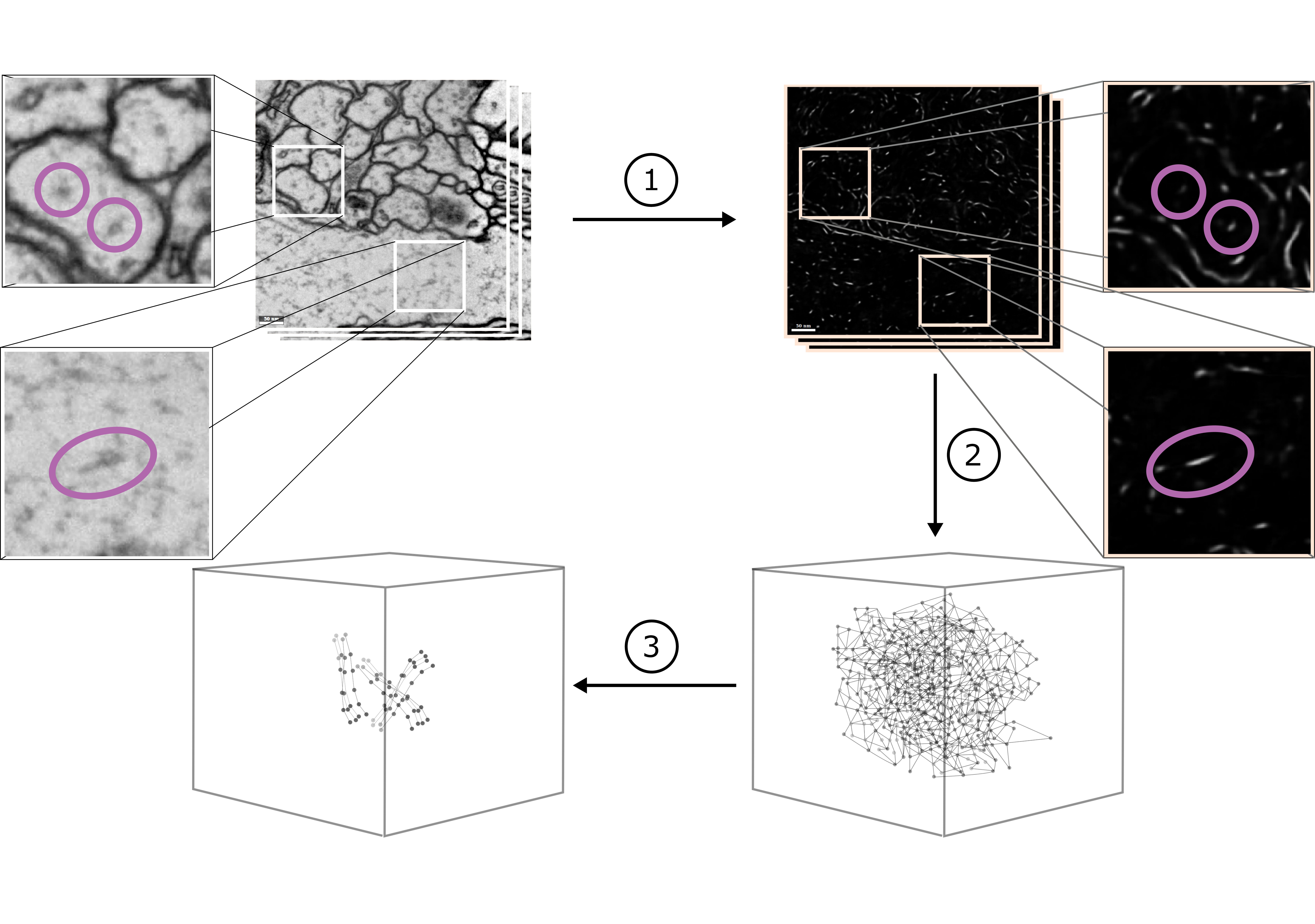
Our method for microtubule tracking is based on the formulation proposed
in [4], with significant improvements in terms of efficiency and accuracy.
Similar to [4], we first predict a score for each voxel to be
part of a microtubule. We then identify promising candidate points and
possible links between them in a candidate graph as nodes and edges. Finally,
we solve a constraint optimization problem incorporating biological priors to
find a subset of edges that constitute microtubule tracks (for an overview
see Fig. 1).
Our four main contributions are as follows:
1. We propose a new integer linear program (ILP) formulation, which decreases
the time needed to solve the constraint optimization by several orders of
magnitude.
2. We devise a scheme to solve the resulting optimization problem in a
block-wise fashion in linear time, and thus are able to process real-world
sized volumes.
3. Our formulation allows tracking of microtubules in arbitrary orientations
in anisotropic volumes by introducing a non-maxima suppression (NMS) based
candidate extraction method.
4. We improve the voxel-level classifier by training a 3D
UNet [16, 7] on skeleton annotations, leading to more
accurate microtubule scores.
We evaluate our method on a new benchmark comprising of densely traced microtubules, demonstrating a 53% increase in accuracy ( F1 score) compared to the prior state of the art. Source code and datasets are publicly available at https://github.com/nilsec/micron.
2 Method
2.1 Predictions
Starting from the raw EM input data, we train a 3D UNet [16] to predict a microtubule score for each voxel. We generate microtubule scores for training from manually annotated skeletons by interpolating between skeleton markers on a voxel grid followed by Gaussian smoothing. In addition, we train the network to predict spatial gradients of the microtubule score up to second order. This is motivated by the idea that the spatial gradient encodes the local shape of a predicted object. Since microtubule segments have locally line-like shapes this auxiliary task potentially regularises microtubule score predictions.
2.2 Candidate Extraction
Given the predicted microtubule score we perform candidate extraction via two NMS passes, to guarantee that two successive candidates of a single microtubule track are not farther apart than the distance threshold we will use to connect two candidates with each other. In a first pass, we perform NMS and thresholding with a stride equal to the NMS window size, guaranteeing at least one candidate per NMS window if the maximum is above the threshold. This strategy is problematic if the local maximum lies on the boarder or corner of a NMS window as this produces multiple, in the worst case eight, candidates that are direct neighbors of each other. We remove this redundancy by performing a second NMS pass on the already extracted maxima, providing us with the final set of microtubule segment candidate detections .
2.3 Constrained Optimization
Following [4], we represent each candidate microtubule segment as a node in a graph with an associated position . A priori we do not know which microtubule segments belong together and form a microtubule. Thus, we connect all microtubule candidates with each other that are below a certain distance threshold . More formally, we introduce an undirected graph , where is the set of microtubule candidate segments augmented with a special node and is the set of possible links between them. The special node is used to mark the beginning or end of a microtubule track and is connected to all candidates in . We further define a set of all directly connected triplets on .
As observed in [4], we can make use of the fact that microtubules do not branch and have limited curvature [8]. We encode these priors as constraints and costs respectively, and solve the resulting optimization problem with an ILP. As outlined in Fig. 2, and in contrast to [4], we formulate consistency and ”no-branch” constraints on triplets of connected nodes only, leading to an orders of magnitude improvement in ILP solve time (see Fig. 4). To this end, we introduce a binary indicator variable for each and define selection costs for each triplet by propagating costs on nodes and on edges as follows:
| (1) |
where is the cost for beginning/ending a track and is the prior on node selection. measures the distance between candidates and , whereas accumulates the predicted evidence for microtubules on all voxels on a line connecting and . measures deviations of a 180 degree angle between two pairs of edges, and thus introduces a cost on curvature. The values are free parameters of the method and found via grid search on a validation dataset.
2.4 Blockwise Processing
In order to be able to apply the constraint optimization to arbitrary sized volumes, we decompose the candidate graph spatially into a set of blocks . For each block , we define a constant-size context region , which encloses the block and is chosen to be large enough such that decisions outside the context region are unlikely to change the ILP solution inside the block. We next identify sets of blocks that are pairwise conflict free, where we define two blocks and to be in conflict if overlaps with . All blocks of a subset can then be distributed and processed in parallel. The corresponding ILP for each block is solved within , however, assignments of the binary indicators are only stored for indicators corresponding to nodes in . To obtain consistent solutions across block boundaries, existing indicator assignments from previous runs of conflicting blocks are acknowledged by adding additional constraints to the block ILP. See supplement for an illustration.
2.5 Evaluation
To evaluate reconstructed tracks against groundtruth, we resample both reconstruction and groundtruth tracks equidistantly and match nodes based on distance using Hungarian matching. Results are reported in terms of precision and recall on edges, which we consider correct if they connect two matched nodes that are matched to the same track.
3 Results


3.1 Dataset
We densely annotated microtubules in eight 1.2x4x4 (30x1000x1000 voxel) volumes of EM data in all six CREMI 222MICCAI Challenge on Circuit Reconstruction in EM Images, https://cremi.org. volumes A, B, C, A+, B+, C+ using Knossos [1] and split the data in training (A+, B+, C+), validation (, ) and test (A, B, C) sets.
3.2 Comparison
NMS_* models refer to the model described in the methods section, where NMS_SM uses a 3D UNet predicting microtubule score only, NMS_GRAD additionally predicts spatial gradients of the microtubule score up to second order and NMS_RFC uses a random forest classifier (RFC) instead of a 3D UNet.
For each, we first select the best performing UNet architecture (for NMS_RFC we interactively train an RFC using Ilastik [18]) and NMS
candidate extraction threshold in terms of recovered candidates on the
validation datasets, followed by a grid search over the distance threshold
and ILP parameters for 150 different parameter combinations.
For the NMS candidate extraction we use a window size of 1x10x10 voxels for the
first NMS pass to offset the anisotropic resolution of 40x4x4 nm.
For the second NMS pass we use a window size of 1x3x3 voxels, removing double
detections.
Baseline refers to an adaptation333We use our ILP formulation,
which was necessary to process larger volumes. of the method
in [4], that uses an RFC for
prediction, z section-wise connected component (CC) analysis on the thresholded
microtubule scores for candidate extraction, and a fixed orientation estimate
for each microtubule candidate pointing in the z direction444Orientation
estimate used in [4] (direct communication with authors)..
For the baseline we interactively train two (for microtubules of
different angles of incidence on the imaging plane) RFCs on training volumes A,
B, C using Ilastik [18]. We find the threshold for CC
candidate extraction, distance threshold and ILP parameters via grid
search over 242 parameter configurations on the validation set. For an overview
see Table 1.
3.3 Test Results
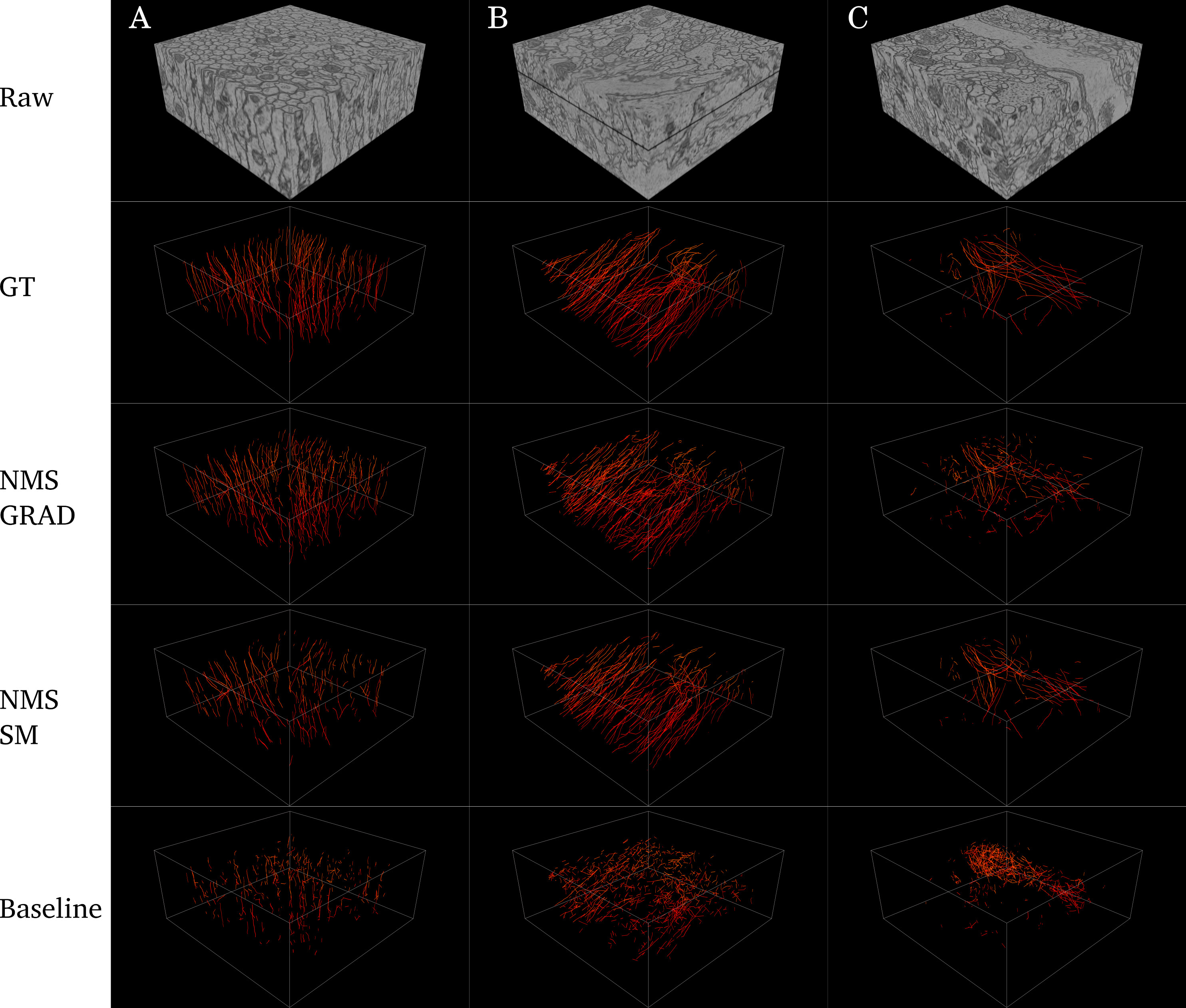
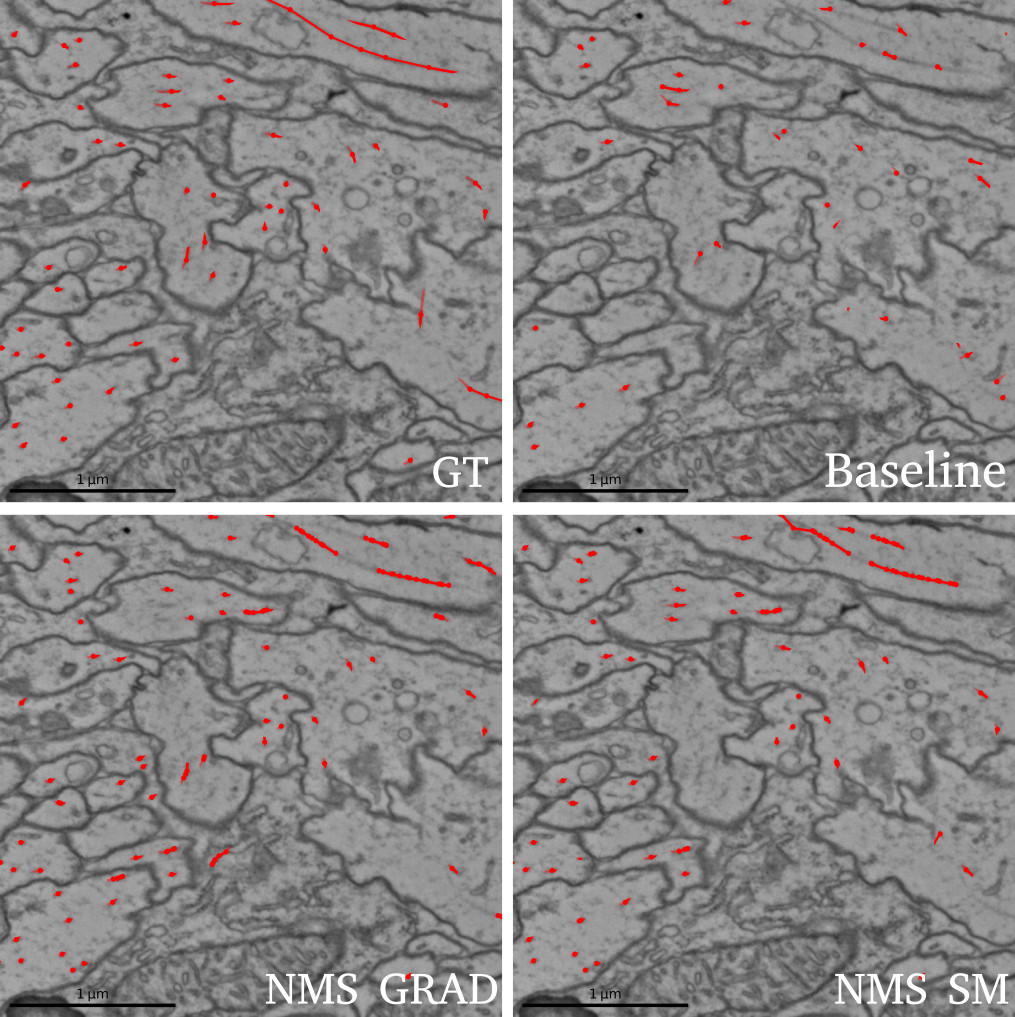
Fig. 4 shows that both variants of our proposed model outperform the prior state of the art [4] substantially. Averaged over test data sets A,B,C, we demonstrate a 53% increase in accuracy for NMS_GRAD. Table 1 further shows test best F1 scores for each individual dataset. In accordance with the qualitative results shown in Fig. 3, NMS_GRAD performs substantially better for test set A while NMS_SM is more accurate for volumes B and C. Ablation experiments show that CC candidate extraction leads to overall less accurate reconstructions. Exchanging the UNet with an RFC while retaining NMS candidate extraction seriously harms performance, resulting in large numbers of false positive detections. For extended qualitative results, including reconstruction of microtubules in the Calyx, a 76 x 52 x 64 region of the Drosophila Melanogaster brain, see supplement.
| Model | Prediction | Cand. Extr. | Edge Score | A | B | C | Avg |
|---|---|---|---|---|---|---|---|
| NMS_GRAD | UNet+GRAD | NMS | Evidence | 0.784 | 0.827 | 0.757 | 0.789 |
| NMS_SM | UNet | NMS | Evidence | 0.711 | 0.828 | 0.785 | 0.775 |
| Baseline | RFC | CC | Orientation | 0.454 | 0.547 | 0.549 | 0.517 |
| CC_GRAD | UNet+GRAD | CC | Evidence | 0.660 | 0.723 | 0.537 | 0.640 |
| NMS_RFC | RFC | NMS | Evidence | 0.366 | 0.375 | 0.302 | 0.348 |
4 Discussion
Although some of our improvements in accuracy can be attributed to the use of a deep learning classifier, the presented method relies mostly on an effective way of incorporating biological priors in the form of constraint optimization. In particular our ablation studies (CC_GRAD) show that the strided NMS-based candidate extraction method positively impacts accuracy: Since a single microtubule could potentially extend far in the x-y imaging plane, it is not sufficient to represent candidates in one plane by a single node, as done in [4]. The strided NMS detections homogenize the candidate graph and likely allow transferring our method to datasets of different resolutions. A potential downside is poor precision when combined with extremely noisy microtubule score predictions (see NMS_RFC). In this case NMS on a grid extracts too many candidate segments, and besides structural priors, the only remaining cost we use to extract final microtubule tracks is directly derived from the (noisy) predicted microtubule score (see equation (1)). Note that the baseline does not suffer as much from noisy microtubule scores, because it uses a fixed orientation prior and is thus limited to a subset of microtubules in any given volume. Finally, the reformulation of the ILP and the block-wise processing scheme result in a dramatic speed-up and the ability to perform distributed, consistent tracking, which is required to process petabyte-sized datasets.
5 Acknowledgements
We thank Tri Nguyen and Caroline Malin-Mayor for code contribution; Arlo Sheridan for helpful discussions and Albert Cardona for his contagious enthusiasm and support. This work was supported by Howard Hughes Medical Institute and Swiss National Science Foundation (SNF grant 205321L 160133).
References
- [1] Boergens, K.M., Berning, M., Bocklisch, T., Bräunlein, D., Drawitsch, F., Frohnhofen, J., Herold, T., Otto, P., Rzepka, N., Werkmeister, T., et al.: webknossos: efficient online 3d data annotation for connectomics. nature methods 14(7), 691–694 (2017)
- [2] Buhmann, J., Krause, R., Ceballos Lentini, R., Eckstein, N., Cook, M., Turaga, S., Funke, J.: Synaptic partner prediction from point annotations in insect brains. Medical Image Computing and Computer Assisted Intervention – MICCAI 2018 11071 (09 2018)
- [3] Buhmann, J., Sheridan, A., Gerhard, S., Krause, R., Nguyen, T., Heinrich, L., Schlegel, P., Lee, W.C.A., Wilson, R., Saalfeld, S., Jefferis, G., Bock, D., Turaga, S., Cook, M., Funke, J.: Automatic detection of synaptic partners in a whole-brain drosophila em dataset. bioRxiv (2019). https://doi.org/10.1101/2019.12.12.874172, https://www.biorxiv.org/content/early/2019/12/13/2019.12.12.874172
- [4] Buhmann, J.M., Gerhard, S., Cook, M., Funke, J.: Tracking of microtubules in anisotropic volumes of neural tissue. In: 2016 IEEE 13th International Symposium on Biomedical Imaging (ISBI) (2016). https://doi.org/10.1109/isbi.2016.7493275, http://dx.doi.org/10.1109/ISBI.2016.7493275
- [5] Cheng, H.C., Varshney, A.: Volume segmentation using convolutional neural networks with limited training data. In: 2017 IEEE international conference on image processing (ICIP). pp. 590–594. IEEE (2017)
- [6] Dorkenwald, S., Schubert, P.J., Killinger, M.F., Urban, G., Mikula, S., Svara, F., Kornfeld, J.: Automated synaptic connectivity inference for volume electron microscopy. Nature methods 14(4), 435–442 (2017)
- [7] Funke, J., Tschopp, F.D., Grisaitis, W., Sheridan, A., Singh, C., Saalfeld, S., Turaga, S.C.: Large scale image segmentation with structured loss based deep learning for connectome reconstruction. IEEE Transactions on Pattern Analysis and Machine Intelligence pp. 1–1 (2018). https://doi.org/10.1109/TPAMI.2018.2835450
- [8] Gittes, F., Mickey, B., Nettleton, J., Howard, J.: Flexural rigidity of microtubules and actin filaments measured from thermal fluctuations in shape. The Journal of cell biology 120(4), 923–934 (1993)
- [9] Heinrich, L., Funke, J., Pape, C., Nunez-Iglesias, J., Saalfeld, S.: Synaptic cleft segmentation in non-isotropic volume electron microscopy of the complete drosophila brain. In: International Conference on Medical Image Computing and Computer-Assisted Intervention. pp. 317–325. Springer (2018)
- [10] Huang, G.B., Scheffer, L.K., Plaza, S.M.: Fully-automatic synapse prediction and validation on a large data set. Frontiers in neural circuits 12, 87 (2018)
- [11] Januszewski, M., Kornfeld, J., Li, P.H., Pope, A., Blakely, T., Lindsey, L., Maitin-Shepard, J., Tyka, M., Denk, W., Jain, V.: High-precision automated reconstruction of neurons with flood-filling networks. Nature methods p. 1 (2018)
- [12] Kreshuk, A., Funke, J., Cardona, A., Hamprecht, F.A.: Who is talking to whom: Synaptic partner detection in anisotropic volumes of insect brain. In: Navab, N., Hornegger, J., Wells, W.M., Frangi, A. (eds.) Medical Image Computing and Computer-Assisted Intervention – MICCAI 2015. pp. 661–668. Springer International Publishing, Cham (2015)
- [13] Lee, K., Lu, R., Luther, K., Seung, H.S.: Learning Dense Voxel Embeddings for 3D Neuron Reconstruction. arXiv e-prints arXiv:1909.09872 (Sep 2019)
- [14] Lee, K., Zung, J., Li, P., Jain, V., Seung, H.S.: Superhuman accuracy on the snemi3d connectomics challenge. arXiv preprint arXiv:1706.00120 (2017)
- [15] Nogales, E.: Structural insights into microtubule function. Annual Review of Biochemistry 69(1), 277–302 (2000)
- [16] Ronneberger, O., Fischer, P., Brox, T.: U-net: Convolutional networks for biomedical image segmentation. In: International Conference on Medical image computing and computer-assisted intervention. pp. 234–241. Springer (2015)
- [17] Schneider-Mizell, C.M., Gerhard, S., Longair, M., Kazimiers, T., Li, F., Zwart, M.F., Champion, A., Midgley, F.M., Fetter, R.D., Saalfeld, S., et al.: Quantitative neuroanatomy for connectomics in drosophila. eLife 5, e12059 (2016)
- [18] Sommer, C., Straehle, C.N., Koethe, U., Hamprecht, F.A., et al.: Ilastik: Interactive learning and segmentation toolkit. In: ISBI. vol. 2, p. 8 (2011)
- [19] Staffler, B., Berning, M., Boergens, K.M., Gour, A., van der Smagt, P., Helmstaedter, M.: Synem, automated synapse detection for connectomics. Elife 6, e26414 (2017)
- [20] Takemura, S.y., Xu, C.S., Lu, Z., Rivlin, P.K., Parag, T., Olbris, D.J., Plaza, S., Zhao, T., Katz, W.T., Umayam, L., et al.: Synaptic circuits and their variations within different columns in the visual system of drosophila. Proceedings of the National Academy of Sciences 112(44), 13711–13716 (2015)
- [21] Xiao, C., Chen, X., Li, W., Li, L., Wang, L., Xie, Q., Han, H.: Automatic mitochondria segmentation for em data using a 3d supervised convolutional network. Frontiers in Neuroanatomy 12, 92 (2018). https://doi.org/10.3389/fnana.2018.00092, https://www.frontiersin.org/article/10.3389/fnana.2018.00092
- [22] Zheng, Z., Lauritzen, J.S., Perlman, E., Robinson, C.G., Nichols, M., Milkie, D., Torrens, O., Price, J., Fisher, C.B., Sharifi, N., et al.: A complete electron microscopy volume of the brain of adult drosophila melanogaster. Cell 174(3), 730–743 (2018)