Optical floating zone crystal growth of rare-earth disilicates, Si2O7 ( = Er, Ho, and Tm)
Abstract
The wealth of structural phases seen in the rare-earth disilicate compounds promises an equally rich range of interesting magnetic properties. We report on the crystal growth by the optical floating zone method of members of the rare-earth disilicate family, Si2O7 (with R = Er, Ho, and Tm). Through a systematic study, we have optimised the growth conditions for Er2Si2O7. We have grown, for the first time using the floating zone method, crystal boules of Ho2Si2O7 and Tm2Si2O7 compounds. We show that the difficulties encountered in the synthesis of polycrystalline and single crystal samples are due to the similar thermal stability ranges of different rare-earth silicate compounds in the temperature-composition phase diagrams of the -Si-O systems. The addition of a small amount of SiO2 excess allowed the amount of impurity phases present in the powder samples to be minimised. The phase composition analysis of the powder X-ray diffraction data collected on the as-grown boules revealed that they were of single phase, except in the case of thulium disilicate, which comprised of two phases. All growths resulted in multi-grain boules, from which sizeable single crystals could be isolated. The optimum conditions used for the synthesis and crystal growth of polycrystalline and single crystal Si2O7 materials are reported. Specific heat measurements of erbium and thulium disilicate compounds confirm an antiferromagnetic phase transition below = 1.8 K for D-type Er2Si2O7 and a Schottky anomaly centred around 3.5 K in C-type Tm2Si2O7, suggesting the onset of short-range magnetic correlations. Magnetic susceptibility data of E-type Ho2Si2O7 reveals an antiferromagnetic ordering of the Ho spins below = 2.3 K.
keywords:
Crystal growth, Optical floating zone method, Rare-earth disilicate, Er2Si2O7, Ho2Si2O7, Tm2Si2O7, Frustrated magnetism.TBC (Thermal barrier coating), EBC (Environmental barrier coating), CVT (Chemical vapour transport), FZ (Floating zone), QDM (Quantum dimer magnet), BEC (Bose-Einstein condensate), GOF (Goodness of fit).
1 Introduction
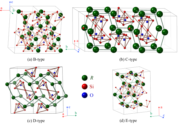
Rare-earth disilicates, Si2O7, where R is a rare-earth element, were studied in the past due to the polymorphism exhibited by these compounds and their physical properties. At ambient pressure, Si2O7 compounds can crystallise in seven different structural types 1, 2, 3. The seven polymorphs, conventionally referred to as A, B (or ), C (or ), D (or ), E (or ), F, G, have, respectively, tetragonal (41), triclinic (), monoclinic (2/), monoclinic (21/), orthorhombic ( or ), triclinic (), and monoclinic (21/) crystal structure 2. At room temperature and ambient pressure, Si2O7 compounds can be stabilised in one of three of these structures, depending on the ionic radius of the rare-earth element, . Rare-earth disilicate compounds containing large rare-earth elements (with La Sm) crystallise in the tetragonal A-type structure. Compounds Si2O7 that incorporate smaller rare-earth ions (where Eu Tm) adopt a triclinic B-type structure. The smallest rare-earth elements, Lu and Yb, form disilicate compounds crystallising in the monoclinic C-type structure. At high temperature (950 1500 ∘C), rare-earth disilicate compounds Si2O7 (where La Tm) undergo one or more structural phase transitions. The number of phase transitions they undergo and their transition temperatures depend on the nature of the rare-earth ion 2. A detailed diagram describing the thermal area of stability of the polymorphs, as well as the characteristics of each crystallographic structure, for each rare-earth disilicate compound, can be found in the early work of Felsche 2. Two new polymorphs, K and L, with monoclinic (21/) and triclinic () crystal symmetries, were later synthesised under pressure 4.
Si2O7 compounds are widely investigated for their luminescent and optical properties, with potential applications as crystal scintillators and in the detection of and X-rays 5, 6, 7, 8, 9. Recently, rare-earth disilicates were identified as promising candidates for thermal barrier coating/environmental barrier coating (TBC/EBC) systems, due to their low thermal conductivities 10, 11, 12, 13, 14, 15.
Other properties, such as the magnetic behaviour of Si2O7 compounds, have not been investigated in great detail, partly due to the complexity of the structural phase diagram. The successful growth of ytterbium and erbium disilicate crystals using optical FZ method 16 has resulted in a resurgence of interest in the magnetic ground states of these compounds 17, 18. Yb2Si2O7 is the first Yb3+ based material that exhibits a quantum dimer magnet (QDM) state, consisting of nearest neighbour spin dimers, with magnetic field-induced order reminiscent of a Bose-Einstein condensate (BEC) phase. Nevertheless, ytterbium disilicate is one of the very few Si2O7 compounds that crystallises in only one structural type 2, whereas for the other members of the rare-earth disilicate compounds stabilising in two or more crystallographic structures, extensive studies of the magnetic properties are scarce in the literature.
The growth of Si2O7 crystals opens up a route to further investigation of these materials, with the potential to unearth some very interesting magnetic properties, similar to what has been observed in Yb2Si2O7. There have been many attempts to grow crystals of the rare-earth disilicate compounds. Typical routes used to prepare Si2O7 crystals include the Verneuil 19, flux 20, 21, 22, 23, 24, 25, Czochralski 8, chemical vapour transport (CVT) 26, floating zone (FZ) 6, 16 and micro-pulling-down (-PD) 27 techniques. The FZ method of crystal growth has been widely employed in the past for the growth of oxides, and not only is it the ideal technique to produce large, high quality single crystals, but it is also one of the most appropriate methods due to its advantages compared to the conventional techniques of crystal growth 28, 29, 30. The main benefits of employing the standard FZ technique for the growth of crystals are: (a) the high purity of the crystals grown thanks to the absence of a container (crucible) and solvent (flux), (b) the relatively large size of the crystal boules obtained compared with other techniques, (c) the good crystalline quality and uniformity of the physical properties throughout the crystals, (d) this method can be used for growing refractory materials.
This has motivated us to embark upon the study of the highly polymorphic rare-earth disilicate compounds. In the present work, we investigate three rare-earth disilicate compounds, Si2O7, where R = Er, Ho, and Tm, with focus on the crystal growth of these materials using the optical FZ method. Tm2Si2O7 is dimorphic with temperature, whereas Er2Si2O7 and Ho2Si2O7 display a high degree of polymorphism 2, 31, 32, 24, 33, 9, 25. A list of the various polymorphs of the disilicate compounds under investigation in this study along with the temperature ranges of their formation/stability, and their respective crystal structures and melting points is given in Table 1.
Rare-earth disilicate compounds have previously been grown in crystal form using the flux method 20, 21, 22, 23, 24, 25, however, the temperature regions where only one crystallographic phase forms are narrow and small differences (of 50 to C) in the soak temperature could result in a yield of crystals of two different polymorphs or in a different structural phase to the one desired 31, 33. Previous studies 34, 35 of other materials that undergo thermally-induced structural phase transitions have proven that the stabilisation of a specific structural type is feasible in FZ grown crystals. The crystal growth of the high temperature stable polymorphs of rare-earth disilicate compounds is therefore made possible by the FZ method, especially for those structural types where the melting points (1700 1800 ∘C) are higher than the temperatures of the structural phase transitions (950 1500 ∘C) 2.
| Si2O7 | Polymorph | Space | Thermal stability | Melting |
| group | range | point () | ||
| Er2Si2O7 | B-type | 1025 ∘C | C | |
| C-type | 2/ | 1025 1400 ∘C | ||
| D-type | 21/ | 1350 ∘C | ||
| Ho2Si2O7 | B-type | 1200 ∘C | C | |
| C-type | 2/ | 1200 1275 ∘C | ||
| D-type | 21/ | 1275 1500 ∘C | ||
| E-type | 1500 ∘C | |||
| Tm2Si2O7 | B-type | 950 ∘C | C | |
| C-type | 2/ | 950 ∘C |
Erbium disilicate exists in three polymorphs (see Table 1), a triclinic () low temperature phase (referred to as B-type), a monoclinic (2/) structure (C-type), and a high temperature monoclinic (21/) arrangement (D-type) 2, 31, 32. C-type Er2Si2O7 is reported to order antiferromagnetically below = 2.50(5) K 22, 24. The structural modification stable at high temperature, D-type, is one of the most studied polymorphs of Er2Si2O7, from a magnetic point of view 36, 37. Furthermore, C D-type structural phase transition occurs at a lower temperature (C) than the melting point (C) (as shown by Felsche 2, all Si2O7 compounds do not melt below C). A crystal of Er2Si2O7 grown from the melt, by the FZ method will therefore belong to the structural type D 16. Due to the arrangement of the magnetic ions in the crystallographic structure, forming a distorted honeycomb-like lattice (see Fig. 1(c)), one expects to observe an interesting magnetic behaviour in D-type Er2Si2O7, similar to the case of C-type Yb2Si2O7 16, 17, 18. Previous reports have shown that D-type Er2Si2O7 orders antiferromagnetically below = 1.9(1) K, as well as exhibiting a highly anisotropic magnetic behaviour 36, 37. In addition, these first measurements of the magnetic properties suggest that the application of a strong magnetic field induces a "spin flip"; one or two "flips" are observed depending upon the direction of the applied magnetic field. Detailed investigations are required to determine the magnetic ground state of D-type Er2Si2O7, and to confirm the four-sublattice antiferromagnetic spin model used for calculating the exchange interaction constants in the initial studies.
Holmium disilicate exists in four polymorphs (see Table 1), two low temperature phases (triclinic B-type arrangement, and monoclinic C-type structure), and two high temperature arrangements (monoclinic D-type phase, and an orthorhombic () E-type structure) 2, 33. The temperatures of the structural phase transitions are close to one another in the low temperature region (C, and C) 2, and one can thus expect the synthesis of the low temperature structural types of Ho2Si2O7 to be challenging. A study by Maqsood 33 showed that the synthesis attempts carried out in the intermediate temperature region (1350 1400 ∘C) can yield more than one crystallographic type of Ho2Si2O7 (coexistence of B-type and C-type structures in the materials synthesised). Since holmium disilicate does not melt below C 2, it is expected that a crystal of Ho2Si2O7 grown using the FZ method should crystallise in the structural type stable at the highest temperature i.e., E-type. To date, the magnetic properties of the various polymorphs of holmium disilicate have not been reported.
Thulium disilicate is dimorphic with temperature (see Table 1), the structure stable at low temperature belongs to the B-type, whereas the high temperature polymorph is C-type 2. Analogous to Er2Si2O7 and Ho2Si2O7, the melting point (C) of the thulium disilicate is higher than the B C-type structural phase transition temperature (C), one can presume that the crystallographic structure of a crystal of Tm2Si2O7 grown using the FZ method will belong to the C-type polymorph. The magnetic properties of the high temperature C-type thulium disilicate have not yet been studied, nevertheless, due to the similarities between the arrangement of the Tm3+ and Yb3+ ions in the lattice, it is likely that Tm2Si2O7 is likely to exhibit as interesting a magnetic behaviour as that observed previously in C-type Yb2Si2O7 16, 17, 18.
Crystals of the erbium disilicate have been grown previously using the FZ technique 16, however, in this study, the results of just one growth, under certain conditions, are presented. The holmium and thulium compounds have only been grown in crystal form using the flux method 2, 20, 32, 24, 33, 25. Firstly, we have optimised the crystal growth conditions for Er2Si2O7, by performing a number of experiments and varying the growth parameters. We have also extended our study to the crystal growth of Si2O7 (with R = Ho, and Tm) and have successfully prepared, for the first time, using the optical FZ method, crystals of these rare-earth silicates. The crystals obtained are especially suitable for the investigation of the structural properties and magnetic behaviour of these materials. Our study shows that there is a direct correlation between the structural features and the magnetism exhibited.
2 Experimental details
The starting materials used for the synthesis of Si2O7 (with R = Er, Ho, and Tm) polycrystalline materials were rare-earth oxides, Er2O3, Ho2O3 and Tm2O3 (all of 99.9% purity), and silica, SiO2 (99.6%). Crystals of rare-earth silicate compounds were then grown using a double ellipsoidal optical image furnace (NEC SC1MDH-11020, Canon Machinery Incorporated), equipped with two 1.5 kW halogen lamps.
The quality of the crystal boules was investigated using a Laue X-ray imaging system with a Photonic-Science Laue camera. Small quantities of each crystal were then ground and powder X-ray diffraction measurements were performed to determine the phase purity and to establish the crystallographic structure of the Si2O7 crystals. It is essential to determine the structural type of each crystal. Room temperature diffractograms were collected on X-ray diffractometers (Panalytical and Bruker) using CuK and CuK radiation ( Å and Å), over an angular range 10-70∘ or 10-90∘ in 2, with a step size in the scattering angle 2 of (Panalytical) and (Bruker). The analysis of the X-ray patterns was performed using the Fullprof software suite 38.
Chemical composition analysis was carried out by energy dispersive x-ray spectroscopy (EDAX) using a scanning electron microscope on pieces cleaved from the Si2O7 crystal boules.
Magnetic susceptibility measurements as a function of temperature were carried out on a ground piece of the Ho2Si2O7 crystal down to 1.8 K in applied magnetic fields of 100 and 1000 Oe using a Quantum Design Magnetic Property Measurement System MPMS-5S superconducting quantum interference device (SQUID) magnetometer.
Heat capacity measurements in zero applied magnetic field at temperatures from 0.5 to 300 K were carried out on Er2Si2O7 and Tm2Si2O7 crystals in a Quantum Design Physical Property Measurement System (PPMS) with a heat capacity option using a two-tau relaxation method.
3 Results and discussion
3.1 Er2Si2O7
3.1.1 Polycrystalline synthesis
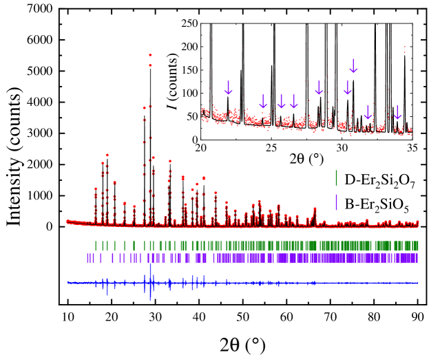
D-type Er2Si2O7 was first prepared in polycrystalline form by the conventional solid state synthesis method. Powders of Er2O3 and SiO2 were weighed in stoichiometric amounts, mixed together and heat treated in air for several days (in 3 or 4 steps) at temperatures in the range 1400-1500 ∘C. The annealed mixture (sample labelled ESO) was reground between each step of the synthesis to ensure good homogeneity and to facilitate the reaction of the starting materials. Nevertheless, even after 4 steps, each of long duration, a phase composition analysis by powder X-ray diffraction (goodness of fit (GOF) = 1.68) reveals that a small quantity of a SiO2 deficient erbium monosilicate impurity phase is present (see Fig. 2). This impurity, B-type Er2SiO5 (monoclinic structure, 2/) 39, persists even after the powder mixture is annealed at a higher temperature (1550-1600 ∘C) and/or for extended periods of time (several days). The sintered material was isostatically pressed into rods (typically 6-8 mm diameter and 70-80 mm long) and sintered at 1500-1550 ∘C in air for several days. The annealed rods were then used for the crystal growth.
3.1.2 Crystal growth
Crystals of erbium disilicate were grown using the FZ method, in static air atmosphere, at ambient pressure. In order to optimise the growth conditions, we have carried out a systematic study, by varying the growth speeds and the rotation rates of the two rods (feed and seed). The crystal growths were carried out at growth rates in the range 5-12 mm/h, and the feed and seed rods were counter-rotated, each at a rate of 10-25 rpm. Initially, a polycrystalline rod was used as a seed and once a good quality crystal boule was obtained, a crystal seed was used for subsequent growths. Er2Si2O7 appears to melt congruently, and no deposition was observed on the quartz tube surrounding the feed and seed rods. The best quality crystals (assessment based on the analysis of the X-ray Laue diffraction) were obtained when growth rates of 10-12 mm/h were employed. The crystal quality appears to be independent of the rotation rate used for the seed rod.
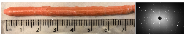
Er2Si2O7 crystal boules prepared were typically 4-6 mm in diameter and 70-85 mm long. The boules tended to have thermally generated cracks in most cases, regardless of the growth rate employed. The crystals developed facets as they grew and two very strong facets were present on more than half the length of the grown crystals. All the erbium disilicate boules were a cloudy pink colour. The crystal boules of erbium disilicate are very fragile and all the crystals broke along the crystal growth axis into two long halves. Moreover, these two pieces cleaved a second time, perpendicular to the growth axis, thus forming crystal fragments of 553 mm3. Figure 3 shows a photograph of a crystal of Er2Si2O7, which was grown using the optimal growth conditions, in air atmosphere, and at a growth speed of 12 mm/h.
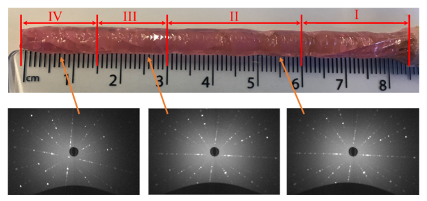
X-ray Laue photographs taken of boules of Er2Si2O7 confirm the good quality of the crystals. Typically, 2-3 grains are present and they extend along the length of each of the boules. A selection of Laue photographs taken along the length of an Er2Si2O7 crystal boule is shown in Fig. 3. When a polycrystalline rod was used as a seed, the Laue patterns of the first 25 mm of the growth (region I) show that this region is either polycrystalline or poor quality crystal. We then note that identical Laue patterns were observed in region II for 30 mm, whereas our Laue examination of the boule at around 55 mm of growth, reveals the presence of 2 overlapping grains extending over a short length of 15 mm (region III) in the boule. For the remaining 15 mm of the boule (region IV), the Laue photographs show the existence of a single grain. The Laue patterns indicate that the axis direction is nearly orthogonal to the growth direction.
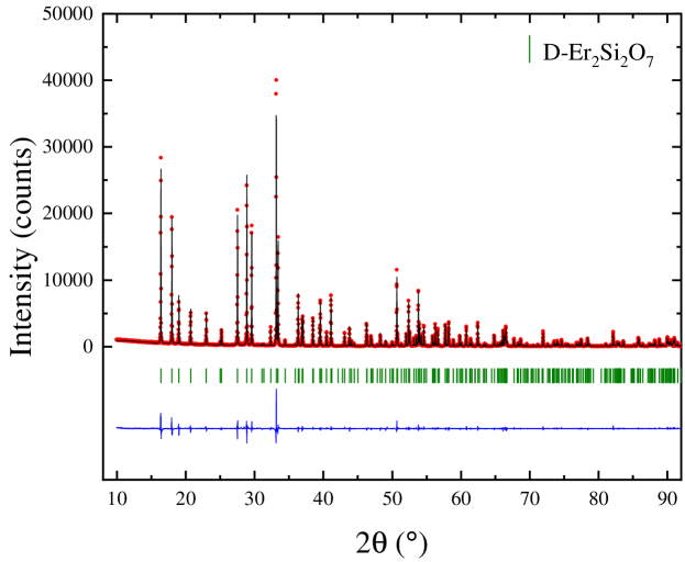
Phase purity analysis was carried out on a ground crystal piece of Er2Si2O7 and the powder X-ray diffraction pattern is shown in Fig. 4. Profile matching (GOF = 2.57) using the Fullprof software suite 38 indicates that the main phase is the monoclinic (21/) D-type Er2Si2O7. There is no evidence that the monoclinic 2/ B-type Er2SiO5 phase is present in the crystal, although it was observed as an impurity in the starting polycrystalline powders. The lattice parameters calculated from the profile matching were determined to be = 4.6908(2) Å, = 5.5615(2) Å and = 10.7991(2) Å, with the angle = 96.040(2)∘. These are in reasonable agreement with previously published results 2.
Composition analysis by EDAX was performed on a cleaved piece from the Er2Si2O7 crystal boule. This showed that the cationic ratio averages of 1:1 for Er:Si for the bulk of the crystal. The average atomic percentages of Er, Si and O were 14.4(1), 17.6(3) and 68.0(3) respectively. Given the limitations of this technique, the results are in reasonable agreement with the expected theoretical values of 18.2 for Er and Si, and 63.6 for O respectively.
3.1.3 Heat capacity
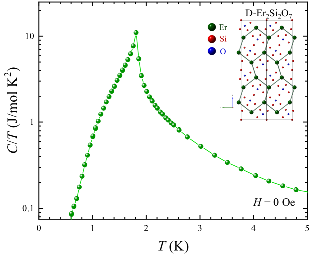
D-type Er2Si2O7 orders magnetically below 1.9(1) K, with a presumed four-sublattice antiferromagnetic arrangement of Ising-like moments 36, 37. Heat capacity measurements performed in zero applied magnetic field on an unaligned fragment of an erbium disilicate crystal show a sharp peak in at 1.80(2) K (see Fig. 5), confirming the ordering of the magnetic ions Er3+. The temperature of the sharp feature observed in the heat capacity measurements further demonstrates that the FZ-grown Er2Si2O7 crystal belongs to the structural type D, because C-type Er2Si2O7 orders below = 2.50(5) K 22, 24. The results are in agreement with the previous results published on a Er2Si2O7 crystal grown using the FZ method 16. Extensive studies of the magnetic ground state of D-type Er2Si2O7 are described elsewhere in a more detailed paper 40.
3.2 Ho2Si2O7
3.2.1 Polycrystalline synthesis
The two structural types of holmium disilicate that are promising from a magnetic point of view are the C and D polymorphs. The magnetic ions are arranged in these crystallographic structures, in such a way that they form a distorted honeycomb-like lattice (see Figs. 1(b-c)).
Ho2Si2O7 was first prepared in polycrystalline form by the conventional solid state synthesis method. Due to the fact that the temperatures of the structural phase transitions are close to one another 2, 33, we have carried out several synthesis attempts in order to determine the optimal conditions for preparing C or D-type holmium disilicate compounds.
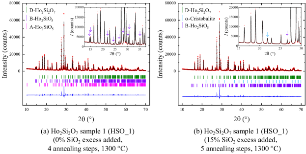
In a first attempt (sample labelled HSO_1), stoichiometric amounts of Ho2O3 and SiO2 powders were ground together and reacted in air for several days (in 4 steps) at 1300 ∘C, with intermediate grindings. Analysis (GOF = 3.81) of the X-ray diffraction pattern collected at room temperature on this powder (see Fig. 6(a)) indicates that the main phase is the D-type Ho2Si2O7. Nevertheless, there are several Bragg peaks which could not be indexed with the monoclinic (21/) space group, and arise due to the presence of a holmium monosilicate impurity, Ho2SiO5. This impurity phase is present in two structural types, a monoclinic (21/) structure (A-type), and a monoclinic (2/) arrangement (B-type) 41.
The difficulty in stabilising the Si-rich phase, Ho2Si2O7, is probably due to the reactivity of the starting silica powder. Felsche previously reported the synthesis of rare-earth disilicates starting with the highly reactive form of SiO2, cristobalite, a very high temperature polymorph of quartz 2. In all our synthesis experiments, we have used a less reactive form of silica. To overcome this drawback, one option is to pre-anneal the starting reagent, silica, in air at high temperatures (1500-1650 ∘C), however, this process does not produce a direct conversion to cristobalite 42. We have thus opted for a second solution, i.e. the synthesis using an excess of SiO2.
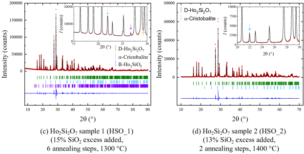
An excess of 15% silica was added to the reacted stoichiometric powder obtained previously. The powder mixture was then heated in air for several days (in 1 step) at 1300 ∘C. Phase purity analysis was carried out and the diffractogram is shown in Fig. 6(b). Profile matching (GOF = 3.71) indicates that the main phase is D-type Ho2Si2O7 and that there are two impurity phases present, unreacted SiO2 crystallising in a tetragonal (41212) structure (-cristobalite 43) and B-type Ho2SiO5. To ensure good homogeneity and to facilitate the reaction, another annealing was carried out for several days at 1300 ∘C. Room temperature powder X-ray diffraction measurements were again performed to determine the phase purity of the polycrystalline material. The X-ray diffraction pattern (see Fig. 6(c)) (GOF = 6.46) reveals the presence of D-type Ho2Si2O7, -cristobalite and B-type Ho2SiO5. In addition, a fourth chemical phase is present (indicated by the existence of one unindexed Bragg peak at 29.1∘ in 2), however, this impurity could not be identified, due to the reduced intensity of the peak. Two additional shoulders can be observed at 20.5 and 21.7∘ in 2, however, due to their extremely reduced intensities, their presence could not be correlated with any known chemical phase. Additional trials were therefore performed in order to optimise the synthesis conditions and obtain pure phase D-type Ho2Si2O7 polycrystalline material.
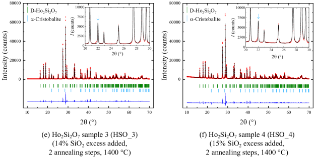
In a second attempt, we have prepared three samples (labelled HSO_2, HSO_2 and HSO_3) starting with stoichiometric amounts of Ho2O3 and different amounts of excess SiO2 (13, 14 and 15%). The starting oxides were mixed together and heat treated in air for several days (in 2 steps) at 1400 ∘C, with an intermediate grinding. The powder X-ray diffraction patterns obtained for the holmium disilicate powders prepared using excess silica are shown in Figs. 6(d)-(f). An analysis of the patterns provided a good fit (GOF = 3.71, 3.70 and 3.75 for 13, 14 and 15% excess SiO2 respectively) to the D-type Ho2Si2O7. Additionally, in all three patterns, there is one Bragg peak which could not be indexed with the monoclinic (21/) space group. This Bragg peak is attributed to the presence of a small amount of unreacted SiO2, in the form of -cristobalite. In addition, the existence of an unindexed shoulder at 21.7∘ in 2, suggests the presence of a third chemical phase, however this impurity could not be identified, due to the reduced intensity of the peak. The best results in the synthesis of D-type Ho2Si2O7 powders were obtained when an excess of 13% silica was used (see Fig. 6(d)).
Throughout the synthesis procedure, extra care has been taken to minimise the amount of unreacted SiO2 in the polycrystalline material used to prepare the feed and seed rods. To ensure a higher reactivity of the starting silica reagent, silica was pre-annealed in air at 1400 ∘C for 24 h. To ensure that the chemical reaction is complete, we have started with a slightly larger amount of excess (14%) than determined previously to be optimal and we have reacted the powder mixtures in air for several days (in 3 steps). The sintered material was isostatically pressed into rods (typically 6-8 mm diameter and 70-80 mm long) and sintered at 1400-1450 ∘C in air for several days. The annealed rods were then used for the crystal growth.
3.2.2 Crystal growth
We have first grown crystal boules of Ho2Si2O7 using the FZ method, in static air atmosphere, at ambient pressure. The crystal growths were carried out at growth rates in the range 5-15 mm/h, and the feed and seed rods were counter-rotated, each at a rate of 15-25 rpm. Polycrystalline rods were used as seed rods. Ho2Si2O7 appears to melt congruently, and no deposition was observed on the quartz tube surrounding the feed and seed rods. The crystals did not develop any facets as they grew and the crystal boules of holmium disilicate were very fragile. X-ray Laue photographs taken of these boules of Ho2Si2O7 reveal a poor crystalline quality of the crystal boules.
The crystal growth of Ho2Si2O7 was also carried out in air at pressures in the range 1-2 bars, a flow of air of 0.1-0.2 L/min. A growth rate of 8 mm/h was used, with the feed and seed rods counter-rotating at a rate of 10 and 25 rpm, respectively. A boule prepared in static air atmosphere, at a growth rate of 15 mm/h was used as a seed for this crystal growth. The Ho2Si2O7 crystal boule obtained was 5 mm in diameter and 75 mm long. The boule tended to have thermally generated cracks, and it developed facets as it grew and two very strong facets were present on more than half the length of the grown boule. All the holmium disilicate crystal boules obtained were a pale orange colour. Figure 7 shows a photograph of a crystal boule of Ho2Si2O7, grown in air atmosphere, at pressures in the range 1-2 bars, in a flow of air of 0.1-0.2 L/min, using a growth speed of 8 mm/h. X-ray Laue photographs taken of this boule of Ho2Si2O7 show that the crystal boule consists of a collection of several grains. Nevertheless, long needle-like single crystals, 2021-2 mm3, could be isolated from the crystal boule. The size of these crystals makes them suitable for the study of the magnetic behaviour of this system.
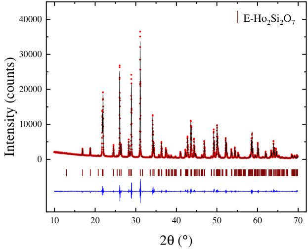
Holmium disilicate does not melt below C, and the D E-type structural phase transition occurs at a lower temperature than the melting point 2. One thus expects a crystal of Ho2Si2O7 grown using the FZ method to crystallise in the structural type stable at the highest temperature ( 1500 ∘C) i.e., E-type. Phase purity analysis of a ground piece of the holmium disilicate crystal boule grown in air atmosphere, at pressures in the range 1-2 bars, a flow of air of 0.1-0.2 L/min, using a growth rate of 8 mm/h (see Fig. 8), shows that the main phase is E-type Ho2Si2O7 (orthorhombic structure), with no significant impurity phases present. Profile matching (GOF = 2.98) was carried out and the lattice parameters were determined to be = 13.6770(3) Å, = 5.0235(3) Å and = 8.1598(3) Å. These are slightly smaller than the previously published results on flux grown crystals of E-type Ho2Si2O7 2, 33.
Composition analysis by EDAX was performed on a cleaved piece from the Ho2Si2O7 crystal boule. Given the limitations of this analysis method, the average atomic percentages of 16.0(6), 16.6(2) and 67.4(6) for Ho, Si and O, respectively, are in reasonable agreement with the expected theoretical values (18.2 for Ho and Si, and 63.6 for O) for the Ho2Si2O7 phase.
3.2.3 Magnetisation
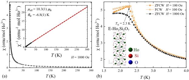
Zero-field-cooled-warming (ZFCW) and field-cooled-warming (FCW) magnetisation versus temperature curves were measured on a ground piece of the E-type Ho2Si2O7. The temperature dependence of the dc magnetic susceptibility, , and reciprocal dc magnetic susceptibility are shown in Fig. 9. The magnetic susceptibility in an applied magnetic field of 1000 Oe exhibits a monotonic increase when cooling from 300 to 1.8 K, and an anomaly is observed at low temperature. A fit of the data to a Curie-Weiss law over an extended temperature range (35-300 K) (see Fig. 9(a) inset) shows that for 35 300 K, E-type Ho2Si2O7 has an effective moment of and a Weiss temperature of K. The effective moment of Ho3+ in holmium disilicate is in agreement with the magnetic moment of a free Ho3+ in the ground state . The value indicates an antiferromagnetic coupling between the Ho spins.
A measurement of the temperature dependence of the susceptibility was also performed in an applied magnetic field of 100 Oe. The examination of the magnetisation at low temperatures (1.8 4 K) reveals a bifurcation of the ZFCW and FCW susceptibility curves below 2.55(5) K, shown in Fig. 9(b). The feature centred around 2.30(5) K suggests the antiferromagnetic ordering between the Ho3+ ions. In order to establish the magnetic structure of E-type Ho2Si2O7 and its evolution with the applied magnetic field, detailed studies of the magnetic behaviour of this system are in progress.
3.3 Tm2Si2O7
3.3.1 Polycrystalline synthesis
The Tm2Si2O7 polymorph stable at high temperature is C-type (see Fig. 1(b)). The arrangement of the magnetic ions in the crystallographic structure is similar to what is observed in Yb2Si2O7, and this motivated us to embark on the study of C-type thulium disilicate.
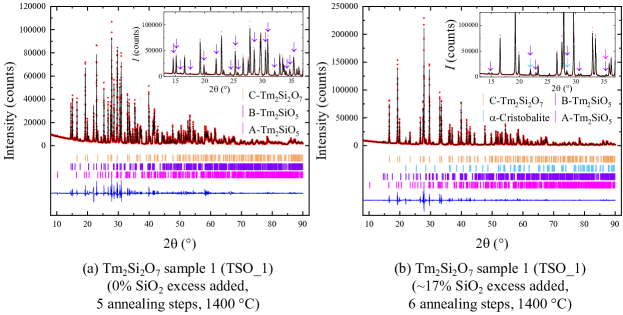
To prepare C-type Tm2Si2O7 powders, stoichiometric amounts of Tm2O3 and SiO2 were thoroughly ground, pressed into pellets and then heated several times in air, for several days, at 1400 ∘C, above the B C-type structural phase transition temperature 2, in 5 steps, with intermediate grinding. A powder X-ray diffraction measurement was carried out at room temperature in order to check the composition of the thulium disilicate polycrystalline sample prepared (labelled TSO_1). Analysis of the pattern (GOF = 7.26) using the FullProf software suite indicates that although the main phase is the monoclinic (2/) C-type structure of Tm2Si2O7, there are several peaks belonging to a thulium monosilicate impurity, Tm2SiO5 (see Fig. 10(a)). This impurity is present in two structural phases, a monoclinic (21/) structure (A-type), and a monoclinic (2/) arrangement (B-type) 44, 45.
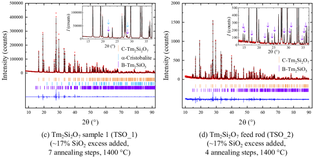
To obtain single phase polycrystalline material of C-type Tm2Si2O7, we have adopted a similar approach to the one used for the synthesis of Ho2Si2O7. An excess ( 17%) of SiO2 was added to the pre-reacted stoichiometric powder obtained. The powder mixture was then pressed into pellets and heated in air for several days (in 1 step) at 1400 ∘C. The X-ray diffraction pattern (GOF = 7.45) collected on this powder shows that the polycrystalline material is a mixture of phases (see Fig. 10(b)). The main phase is C-type Tm2Si2O7, but there are several impurity peaks belonging to A and B-type Tm2SiO5, as well as unreacted SiO2 crystallised in the -cristobalite tetragonal (41212) structure. The powder mixture was pelletised again and reacted in air for several days at 1400 ∘C. Phase purity analysis of the resulting powder by X-ray diffraction (GOF = 7.73) shows the existence of three phases, C-type Tm2Si2O7, B-type Tm2SiO5 and -cristobalite. Figures 10(a-c) show that the amount of the Tm2SiO5 impurity is significantly reduced due to the addition of SiO2.
A larger amount of polycrystalline sample of C-type Tm2Si2O7 was prepared using a stoichiometric amount of Tm2O3 and 17% excess SiO2. The starting materials were mixed together and heat treated in air for several days (in 3 steps) at 1400 ∘C, with an intermediate grinding. The sintered material (sample labelled TSO_2) was then isostatically pressed into a rod (7 mm diameter and 60 mm long) and sintered at 1400 ∘C in air for several days. The annealed rod was then used for the crystal growth. An analysis (GOF = 1.51) of the powder X-ray diffraction pattern collected on a ground piece of the feed rod reveals the presence of two phases, C-type Tm2Si2O7 and B-type Tm2SiO5 (see Fig. 10(d)).
The results of our synthesis experiments suggest that the stabilisation of the disilicate phase in the case of thulium is more difficult than for the two other rare-earth disilicate compounds investigated in this study. This is most likely due to the very similar temperature stability ranges of Tm2SiO5 and Tm2Si2O7 phases 2, 3.
3.3.2 Crystal growth
We have first grown a Tm2Si2O7 crystal boule, in static air atmosphere, at ambient pressure, using growth rates in the range 5-7 mm/h. The feed and seed rods were counter-rotated, each at a rate of 15 rpm. A polycrystalline rod was used as a seed rod. No deposition was observed on the quartz tube surrounding the feed and seed rods, suggesting that Tm2Si2O7 melts congruently. The crystal did not develop any facets and the crystal boule was very fragile. To minimise the thermal stress on the crystal boule and reduce the cracks, the rotation of the seed rod was reduced to 5 rpm. Analysis by X-ray Laue diffraction of the crystalline quality of the Tm2Si2O7 crystal boule obtained revealed the presence of multiple grains.
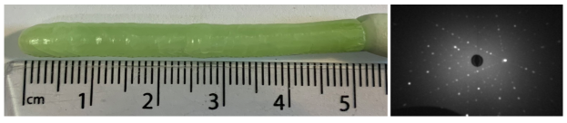
A second crystal growth was performed, using as feed the crystal boule obtained previously. The growth was carried out in air atmosphere, at ambient pressure, in a flow of air of 1-2 L/min, using a growth rate of 10 mm/h (see Fig. 11). The feed and seed rods were counter-rotated, each at a rate of 15-25 rpm. The Tm2Si2O7 crystal boule obtained was 5 mm in diameter and 50 mm long. Thermally generated cracks were observed on the surface of the crystal boule. One facet developed midway during the growth and extended over the remaining length of the grown crystal boule. The thulium disilicate crystal boule obtained was a pale green colour. A visual inspection of the cross-section of the crystal revealed that the boule consists of a semi-transparent shell and an opaque core. Analysis of the X-ray Laue diffraction patterns collected along the sides of this boule of Tm2Si2O7 shows the presence of several grains. Nonetheless, long needle-like single crystals, 10-1521-2 mm3, could be isolated from the outermost layer of the crystal boule. These crystals are suitable for magnetic properties measurements of this material.
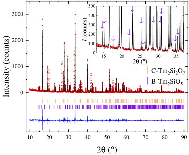
Phase composition analysis by powder X-ray diffraction (GOF = 1.50) of a ground cross-section of Tm2Si2O7 boule (made up of both the outer layer and the inner core) reveals that, although the main phase is C-type Tm2Si2O7, there is a small amount of B-type Tm2SiO5 impurity present in the crystal (see Fig. 12). The lattice parameters of monoclinic (2/) thulium disilicate were determined to be = 6.8276(2) Å, = 8.9105(3) Å and = 4.7067(2) Å, with the angle = 101.834(2)∘. These values are close to the previously published results on flux grown crystals of C-type Tm2Si2O7 2, 25.
Composition analysis by EDAX was performed on a cleaved piece from the Tm2Si2O7 crystal boule. The average atomic percentages of Tm, Si and O were 17.1(2), 16.7(2) and 66.2(3) respectively. Given the limitations of this technique, the results are in reasonable agreement with the expected cationic ratio average of 1:1 for Tm:Si for the Tm2Si2O7 phase.
The problems encountered in the preparation of Tm2Si2O7 samples, both in powder and crystal form, are most likely due to the similar ranges of thermal stability of the thulium monoslicate and disilicate, as well as, to the similar melting temperatures of these compounds (see, for example, the Y-Si-O system 46). Polycrystalline and single crystal synthesis experiments are in progress in order to optimise the conditions and obtain phase C-type Tm2Si2O7 samples.
3.3.3 Heat capacity
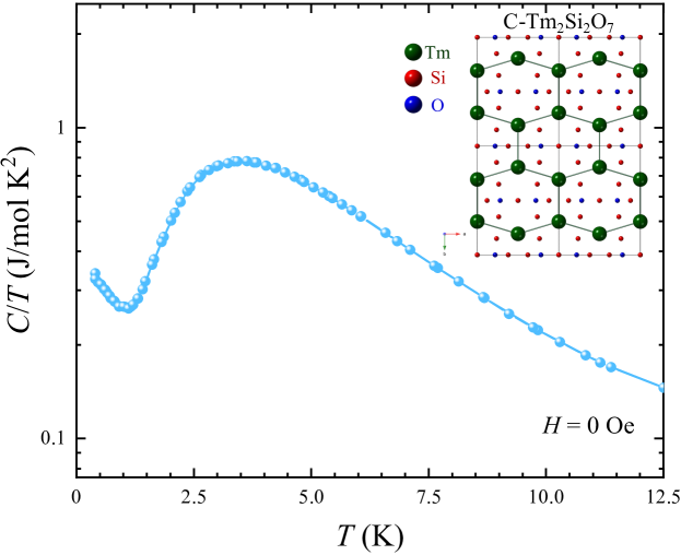
To date, the magnetic properties of C-type Tm2Si2O7 have not been studied. Figure 13 shows the heat capacity measurement performed in zero applied magnetic field on an unaligned fragment of a thulium disilicate crystal. Similar to what is observed for C-type Yb2Si2O7, there is no sign of magnetic ordering of Tm2Si2O7 down to 0.4 K. The broad peak centred around 3.5 K corresponds, in all likelihood, to a Schottky anomaly. The increase of below 1 K suggests the development of short-range magnetic correlations. Detailed investigations at low temperature are being carried out in order to establish the magnetic structure of C-type Tm2Si2O7.
4 Summary and Conclusions
Samples of Si2O7 (with R = Er, Ho, and Tm) compounds were first prepared in polycrystalline form by the conventional solid state synthesis method. The analysis by powder X-ray diffraction of the polycrystalline samples prepared revealed the presence of impurities (in the case of holmium and thulium), in the form of SiO5. We have shown that to reduce the formation of the impurity monosilicate phases in the synthesis, it is necessary to start with an excess of SiO2. Our synthesis efforts with excess SiO2 succeeded in producing samples with considerably reduced levels of the impurity phases and we were able to obtain mainly the desired Si2O7 phase. Detailed studies are now being carried out to further optimise the synthesis conditions in order to obtain phase pure powder samples of the rare-earth disilicate compounds, minimising the amount of unreacted SiO2. The conditions used for the synthesis of the polycrystalline materials, as well as the results of the phase composition analysis of each sample, are summarised in Table 2.
We have been successful in growing crystals of Si2O7 (with R = Er, Ho, and Tm) using the FZ method. All the rare-earth disilicate compounds appear to melt congruently and no evaporation was observed for the growths. The crystal growths were performed using various growth rates, in air atmosphere. All the rare-earth disilicate crystals are very fragile and tend to have thermally generated cracks. In general, crystal boules of better crystalline quality were obtained using average or high speed of growth. A summary of the crystal growth conditions used for the growth is given in Table 3.
The quality and composition of the as-grown Si2O7 boules were investigated using X-ray diffraction techniques. The lattice parameters determined by powder X-ray diffraction are collected in Table 4. Erbium and holmium disilicate crystals were single phase, whereas the thulium crystal boule consists of two chemical phases (monosilicate and disilicate), due to the overlapping ranges of thermal stability of the two thulium silicate compounds, Tm2SiO5 and Tm2Si2O7. The lattice parameters of the Si2O7 crystal boules are in agreement with the previously published results. Despite the difficulties encountered in the preparation of polycrystalline and crystal samples, good size grains could be isolated from the crystal boules to be used for further physical properties characterisation measurements. Further investigations to better understand the stabilisation of Si2O7 phases in their crystal form are in progress.
Magnetic properties measurements confirm an antiferromagnetic ordering of Er3+ ions at = 1.80(2) K in D-type Er2Si2O7. We report, for the first time, that E-type Ho2Si2O7 orders antiferromagnetically below = 2.30(5) K. We also show that Tm2Si2O7 exhibits no signs of long-range magnetic ordering down to 0.4 K, however, short-range magnetic correlations develop below 1 K. Detailed magnetic properties measurements are now being carried out on the Si2O7 (with R = Er, Ho, and Tm) crystals to determine the magnetic ground state of these materials.
| Chemical | Sample | Sintering | Synthesis | SiO2 | Phase composition analysis |
|---|---|---|---|---|---|
| composition | label | temperature | steps | excess | |
| (∘C) | number | (%) | |||
| Er2Si2O7 | ESO | 1400-1500 | 4 | 0 | mainly monoclinic D-type Er2Si2O7 and a few peaks of monoclinic B-type Er2SiO5 |
| Ho2Si2O7 | HSO_1 | 1300 | 4 | 0 | mixture of monoclinic D-type Ho2Si2O7, monoclinic A-type Ho2SiO5 and monoclinic B-type Ho2SiO5 |
| HSO_1 | 1300 | 5 | 15 | mainly monoclinic D-type Ho2Si2O7, a few peaks of monoclinic B-type Ho2SiO5 and -cristobalite SiO2 | |
| HSO_1 | 1300 | 6 | 15 | mainly monoclinic D-type Ho2Si2O7, one strong peak of -cristobalite SiO2, one peak of monoclinic B-type Ho2SiO5 and one peak belonging to an unidentified impurity phase | |
| HSO_2 | 1400 | 2 | 13 | mainly monoclinic D-type Ho2Si2O7 and one small peak of -cristobalite SiO2 | |
| HSO_3 | 1400 | 2 | 14 | mainly monoclinic D-type Ho2Si2O7 and one peak of -cristobalite SiO2 | |
| HSO_4 | 1400 | 2 | 15 | mainly monoclinic D-type Ho2Si2O7 and one peak of -cristobalite SiO2 | |
| Tm2Si2O7 | TSO_1 | 1400 | 5 | 0 | mixture of monoclinic C-type Tm2Si2O7, monoclinic A-type Tm2SiO5 and monoclinic B-type Tm2SiO5 |
| TSO_1 | 1400 | 6 | 17 | mainly monoclinic C-type Tm2Si2O7, a few strong peaks of monoclinic monoclinic B-type Tm2SiO5, and a few peaks of A-type Tm2SiO5 and -cristobalite SiO2 | |
| TSO_1 | 1400 | 7 | 17 | mainly monoclinic C-type Tm2Si2O7 and a few peaks of monoclinic monoclinic B-type Tm2SiO5 and -cristobalite SiO2 | |
| TSO_2 | 1400 | 4 | 17 | mainly monoclinic C-type Tm2Si2O7 and a few peaks of monoclinic monoclinic B-type Tm2SiO5 |
| Si2O7 | Growth | Gas atmosphere/ | Feed & seed | Remarks |
| rate | pressure/flow | rotation rate | ||
| (mm/h) | (rpm) | |||
| Er2Si2O7 | 5-12 | air, ambient | 10-25 | cloudy pink boules |
| 10-12 | air, ambient | 20-25 | grains 553 mm3 ★ | |
| Ho2Si2O7 | 5-15 | air, ambient | 15-25 | pale orange boules |
| 8 | air, 1-2 bars, 0.1-0.2 L/min | 10-25 | grains 2021-2 mm3 ★ | |
| Tm2Si2O7 | 5-7 | air, ambient | 15-5 | pale green boules |
| 10 | air, ambient, 1-2 L/min | 15-25 | grains 10-1521-2 mm3 ★ |
| Chemical | Structure | Space | Lattice parameters | |||
|---|---|---|---|---|---|---|
| composition | (type) | group | Angle | |||
| (Å) | (Å) | (Å) | (∘) | |||
| Er2Si2O7 | monoclinic (D) | 21/ | 4.6908(2) | 5.5615(2) | 10.7991(2) | = 96.040(2) |
| Ho2Si2O7 | orthorhombic (E) | 13.6770(3) | 5.0235(3) | 8.1598(3) | - | |
| Tm2Si2O7 | monoclinic (C) | 2/ | 6.8276(2) | 8.9105(3) | 4.7067(2) | = 101.834(2) |
Financial support was provided by EPSRC, UK through Grant EP/T005963/1. The authors would like to acknowledge the contributions of S. Donner, J. Mileson, M. Minney, and G. Palmer to the preparation of rare-earth silicate compounds through their involvement with undergraduate projects. The authors would also like to thank S. J. York for the EDAX compositional analysis.
References
- Ito and Johnson 1968 Ito, J.; Johnson, H. Synthesis and study of yttrialite. Am. Mineral. 1968, 53, 1940–1952
- Felsche 1970 Felsche, J. Polymorphism and crystal data of the rare-earth disilicates of type R.E.2Si2O7. J. Less-Common Met. 1970, 21, 1–14
- Felsche 1973 Felsche, J. The crystal chemistry of the rare-earth silicates. Struct. Bonding (Berlin, Ger.) 1973, 13, 99–197
- Liu and Fleet 2002 Liu, X.; Fleet, M. E. High-pressure synthesis of a La orthosilicate and Nd, Gd, and Dy disilicates. J. Phys.: Condens. Matter 2002, 14, 11223–11226
- Bretheau-Raynal et al. 1980 Bretheau-Raynal, F.; Tercier, N.; Blanzat, B.; Drifford, M. Synthesis and spectroscopic study of lutetium pyrosilicate single crystals doped with trivalent europium. Mater. Res. Bull. 1980, 15, 639–646
- Pauwels et al. 2000 Pauwels, D.; Le Masson, N.; Viana, B.; Kahn-Harari, A.; van Loef, E.; Dorenbos, P.; van Eijk, C. A novel inorganic scintillator: Lu2Si2O7:Ce3+ (LPS). IEEE Trans. Nucl. Sci. 2000, 47, 1787–1790
- Feng et al. 2010 Feng, H.; Ding, D.; Li, H.; Lu, S.; Pan, S.; Chen, X.; Ren, G. Growth and luminescence characteristics of cerium-doped yttrium pyrosilicate single crystal. J. Alloys Compd. 2010, 489, 645–649
- He et al. 2012 He, F.; Guohao, R.; Yuntao, W.; Jun, X.; Qiuhong, Y.; Jianjun, X.; Mitch, C.; Chenlong, C. Optical and thermoluminescence properties of Lu2Si2O7:Pr single crystal. J. Rare Earths 2012, 30, 775–779
- Fernańdez-Carrión et al. 2013 Fernańdez-Carrión, A. J.; Allix, M.; Ocaña, M.; García-Sevillano, J.; Cusso, F.; Fitch, A. N.; Suard, E.; Becerro, A. I. Crystal Structures and Photoluminescence across the La2Si2O7-Ho2Si2O7 System. Inorg. Chem. 2013, 52, 13469–13479
- Lee et al. 2005 Lee, K. N.; Fox, D. S.; Bansal, N. P. Rare earth silicate environmental barrier coatings for SiC/SiC composites and Si3N4 ceramics. J. Eur. Ceram. Soc. 2005, 25, 1705–1715
- Sun et al. 2008 Sun, Z.; Zhou, Y.; Wang, J.; Li, M. Thermal Properties and Thermal Shock Resistance of - Y2Si2O7. J. Am. Ceram. Soc. 2008, 91, 2623–2629
- Sun et al. 2009 Sun, Z.; Li, M.; Zhou, Y. Thermal properties of single-phase Y2SiO5. J. Eur. Ceram. Soc. 2009, 29, 551–557
- Zhou et al. 2013 Zhou, Y.; Zhao, C.; Wang, F.; Sun, Y.; Zheng, L.; Wang, X. Theoretical Prediction and Experimental Investigation on the Thermal and Mechanical Properties of Bulk - Yb2Si2O7. J. Am. Ceram. Soc. 2013, 96, 3891–3900
- Tian et al. 2016 Tian, Z.; Zheng, L.; Li, Z.; Li, J.; Wang, J. Exploration of the low thermal conductivities of - Y2Si2O7, - Y2Si2O7, - Yb2Si2O7, and - Lu2Si2O7 as novel environmental barrier coating candidates. J. Eur. Ceram. Soc. 2016, 36, 2813–2823
- Luo et al. 2018 Luo, Y.; Sun, L.; Wang, W.; Wu, Z.; Lv, X.; Wang, J. Material-genome perspective towards tunable thermal expansion of rare-earth di-silicates. J. Eur. Ceram. Soc. 2018, 38, 3547–3554
- Nair et al. 2019 Nair, H. S.; DeLazzer, T.; Reeder, T.; Sikorski, A.; Hester, G.; Ross, K. A. Crystal Growth of Quantum Magnets in the Rare-Earth Pyrosilicate Family Si2O7 ( = Yb, Er) Using the Optical Floating Zone Method. Crystals 2019, 9, 196
- Hester et al. 2019 Hester, G.; Nair, H.; Reeder, T.; Yahne, D.; DeLazzer, T.; Berges, L.; Ziat, D.; Neilson, J.; Aczel, A.; Sala, G.; Quilliam, J.; Ross, K. Novel Strongly Spin-Orbit Coupled Quantum Dimer Magnet: Yb2Si2O7. Phys. Rev. Lett. 2019, 123, 027201
- Flynn et al. 2020 Flynn, M. O.; Baker, T. E.; Jindal, S.; Singh, R. R. P. On two phases inside the Bose condensation dome of Yb2Si2O7. arXiv.org, e-Print Arch., Condens. Matter 2020, arXiv:2001.08219
- Smolin and Shepelev 1970 Smolin, Y. I.; Shepelev, Y. F. The crystal structures of the rare earth pyrosilicates. Acta Crystallogr., Sect. B: Struct. Sci., Cryst. Eng. Mater. 1970, 26, 484–492
- Wanklyn et al. 1974 Wanklyn, B. M.; Wondre, F. R.; Ansell, G. B.; Davison, W. Flux growth of rare earth silicates and aluminosilicates. J. Mater. Sci. 1974, 9, 2007–2014
- Wanklyn 1978 Wanklyn, B. M. Effects of modifying starting compositions for flux growth. J. Cryst. Growth 1978, 43, 336–344
- Maqsood et al. 1979 Maqsood, A.; Wanklyn, B. M.; Garton, G. Flux growth of polymorphic rare-earth disilicates, Si2O7 ( = Tm, Er, Ho, Dy). J. Cryst. Growth 1979, 46, 671–680
- Nørlund Christensen et al. 1997 Nørlund Christensen, A.; Hazell, R. G.; Hewat, A. W. Synthesis, Crystal Growth and Structure Investigations of Rare-Earth Disilicates and Rare-Earth Oxyapatites. Acta Chem. Scand. 1997, 51, 37–43
- Maqsood 2000 Maqsood, A. Single crystal growth of polymorphic Er2Si2O7 ceramics. J. Mater. Sci. Lett. 2000, 19, 711–712
- Kahlenberg and Aichholzer 2014 Kahlenberg, V.; Aichholzer, P. Thortveitite-type Tm2Si2O7. Acta Crystallogr., Sect. E: Struct. Rep. Online 2014, 70, i34–i35
- Chi et al. 1998 Chi, L.-S.; Chen, H.-Y.; Zhuang, H.-H.; Huang, J.-S. Synthesis and Crystal Structure of Er2Si2O7. Chin. J. Struct. Chem. 1998, 17, 24–26
- Horiai et al. 2016 Horiai, T.; Kurosawa, S.; Murakami, R.; Pejchal, J.; Yamaji, A.; Shoji, Y.; Chani, V.; Ohashi, Y.; Kamada, K.; Yokota, Y.; Yoshikawa, A. Crystal growth and luminescence properties of Yb2Si2O7 infra-red emission scintillator. Opt. Mater. (Amsterdam, Neth.) 2016, 58, 14–17
- Balakrishnan et al. 1998 Balakrishnan, G.; Petrenko, O. A.; Lees, M. R.; Paul, D. M. Single crystal growth of rare earth titanate pyrochlores. J. Phys.: Condens. Matter 1998, 10, L723–L725
- Koohpayeh et al. 2008 Koohpayeh, S. M.; Fort, D.; Abell, J. S. The optical floating zone technique: A review of experimental procedures with special reference to oxides. Prog. Cryst. Growth Charact. Mater. 2008, 54, 121–137
- Dabkowska and Dabkowski 2015 Dabkowska, H.; Dabkowski, A. Handbook of Crystal Growth (Second Edition); Elsevier, 2015; pp 281–329
- Maqsood and ul Haq 1987 Maqsood, A.; ul Haq, I. Preparation of rare-earth disilicates and their X-ray diffraction studies. J. Mater. Sci. Lett. 1987, 6, 1095–1097
- Maqsood 1997 Maqsood, A. Phase transformations in Er2Si2O7 ceramics. J. Mater. Sci. Lett. 1997, 16, 837–840
- Maqsood 2009 Maqsood, A. Phase transformations in Ho2Si2O7 ceramics. J. Alloys Compd. 2009, 471, 432–434
- Ciomaga Hatnean et al. 2016 Ciomaga Hatnean, M.; Decorse, C.; Lees, M. R.; Petrenko, O. A.; Balakrishnan, G. Zirconate pyrochlore frustrated magnets: crystal growth by the floating zone technique. Crystals 2016, 6, 79
- Sibille et al. 2017 Sibille, R.; Lhotel, E.; Ciomaga Hatnean, M.; Nilsen, G. J.; Ehlers, G.; Cervellino, A.; Ressouche, E.; Frontzek, M.; Zaharko, O.; Pomjakushin, V.; Stuhr, U.; Walker, H. C.; Adroja, D. T.; Luetkens, H.; Baines, C.; Amato, A.; Balakrishnan, G.; Fennell, T.; Kenzelmann, M. Coulomb spin liquid in anion-disordered pyrochlore Tb2Hf2O7. Nat. Commun. 2017, 8, 892
- Maqsood 1981 Maqsood, A. Magnetic properties of D-Er2Si2O7 at low temperatures. J. Mater. Sci. 1981, 16, 2198–2204
- Leask et al. 1986 Leask, M. J. M.; Tapster, P. R.; Wells, M. R. Magnetic properties of D-Er2Si2O7. J. Phys. C: Solid State Phys. 1986, 19, 1173–1187
- Rodríguez-Carvajal 1993 Rodríguez-Carvajal, J. Recent advances in magnetic structure determination by neutron powder diffraction. Phys. B (Amsterdam, Neth.) 1993, 192, 55–69
- Phanon and Čerńy 2008 Phanon, D.; Čerńy, R. Crystal structure of the B-type dierbium oxide ortho-oxosilicate Er2O[SiO4]. Z. Anorg. Allg. Chem. 2008, 634, 1833–1835
- Petrenko et al. 2020 Petrenko, O. A.; Ciomaga Hatnean, M.; Manuel, P.; Orlandi, F.; Khalyavin, D. D.; Balakrishnan, G. in preparation. 2020,
- Maqsood 1984 Maqsood, A. Single crystal preparation of the rare earth oxyorthosilicates SiO5 (=Er, Ho, Dy) by a flux method. J. Mater. Sci. Lett. 1984, 3, 65–67
- Chaklader and Roberts 1961 Chaklader, A. C. D.; Roberts, A. L. Transformation of Quartz to Cristobalite. J. Am. Ceram. Soc. 1961, 44, 35–41
- Downs and Palmer 1994 Downs, R. T.; Palmer, D. C. The pressure behavior of cristobalite. Am. Mineral. 1994, 79, 9–14
- Wang et al. 2001 Wang, J.; Tian, S.; Li, G.; Liao, F.; Jing, X. Preparation and X-ray characterization of low-temperature phases of R2SiO5 (R = rare earth elements). Mater. Res. Bull. 2001, 36, 1855–1861
- Tian et al. 2016 Tian, Z.; Zheng, L.; Wang, J.; Wan, P.; Li, J.; Wang, J. Theoretical and experimental determination of the major thermo-mechanical properties of RE2SiO5 (RE = Tb, Dy, Ho, Er, Tm, Yb, Lu, and Y) for environmental and thermal barrier coating applications. J. Eur. Ceram. Soc. 2016, 36, 189–202
- Abdul-Jabbar et al. 2018 Abdul-Jabbar, N. M.; Poerschke, D. L.; Gabbett, C.; Levi, C. G. Phase equilibria in the zirconia–yttria/gadolinia–silica systems. J. Eur. Ceram. Soc. 2018, 38, 3286–3296