Why epithelial cells collectively move against a traveling signal wave
Abstract
The response of cell populations to external stimuli plays a central role in biological phenomena such as epithelial wound healing and developmental morphogenesis. Wave propagation of a protein signal has been shown to direct collective migration in one direction, but this mechanism based on active matter under a traveling wave is not fully understood. To elucidate how the traveling wave of the protein signal directs collective migration, we study the mechanics of the epithelial cell monolayer, taking into account the signal-dependent coordination of contractile stress and cellular orientation. By constructing an optogenetically controlled experimental cell model, we found that local signal activation induces changes in cell density and orientation with the direction of propagation, increasing the net motion relative to the wave. In addition, the migration exhibits an optimal speed with the propagation speed of the signal. This occurs because the mechanical deformation is not fully relaxed for rapid signal activation, resulting in an optimal signal propagation speed. The presented mechanical model can also be extended to wound healing, providing a versatile model for understanding the interplay between mechanics and signaling in active matter.
I Introduction
The ordered surface structures in living tissue is organized by epithelial cells. However, these cells do not stay in the same place; a group of epithelial cells moves in an aligned overall direction, and such collective migration is an important process that creates complex morphological organization from embryogenesis, morphogenesis, and wound healing [1, 2, 3, 4, 5]. The control of collective migration is thus a major challenge in the fields of physics, biology, and tissue engineering. Collective migration in living epithelial tissues is regulated by a chemical signal of protein [2, 6, 7]. Cells transiently increase the activity of protein signaling that controls contractile force, and the propagation of protein signals occurs between neighboring cells. As the wave of protein signal passes through the cell monolayer, the direction of collective migration is rectified in the direction opposite to that of the wave by accurately determining their migration direction [5, 8]. However, a spatially symmetric wave does not result in a net movement of cells because the forces generated by the wave are equal in magnitude but opposite in sign. How do protein signaling waves affect the collective migration of cells in the opposite direction? How an epithelial cell monolayer can move against a wave of chemical signals is little understood.
The broken time-reversal symmetry resulting from the multiplication of two physical processes provides a fundamental concept capable of addressing this question [9, 10]. If the forces driving cell migration involve more than two processes, time-reversal symmetry can be broken, resulting in net motion even during force equilibration [11, 12, 13, 14, 15]. However, the existence of such a multiplicative effect in collective migration remains elusive. Finding a relationship between directed motion and a traveling signal wave is a significant challenge with implications not only for epithelial tissues but also for colloidal particles [16].
The physics of active matter, known as autonomously moving groups of particles, has advanced for cell biophysics, and mechanical models are revealing the mechanisms of turbulence-like dynamics that occur in a cell monolayer [17, 18, 19, 20]. Although individual cells move randomly, when their density becomes large, a polarity axis of cells is oriented by cell adhesion with neighboring cells, resulting in aligned directions of movement and emergent collective migration. Numerical models of epithelial collective migration have been developed [21, 22, 23, 24], and it is becoming clear that orientation dynamics that align polarity and local cell density changes are coupled to produce spatiotemporally ordered structures. Thus, there is a growing interest in understanding the physical principles of complex tissue formation by understanding the collective dynamics of cell populations.
In this study, we investigate the mechanics of collective migration along a protein signal wave. We show that by considering such a protein signal, which regulates both local cell contraction and the cell orientation field in a cell monolayer, directed collective migration can occur in the direction opposite to the signal wave. Our results indicate that the traveling signal wave rectifies the direction of collective migration using only mechanical factors. Through experimental investigation, we explain key aspects of directed collective migration, such as the nonlinear increase in migration speed with signal intensity and the optimal response to the signal wave. We conclude that wave-directed migration results from the interplay of signal-dependent contraction and friction in cellular mechanics.
II Theoretical model of collective migration under signaling wave
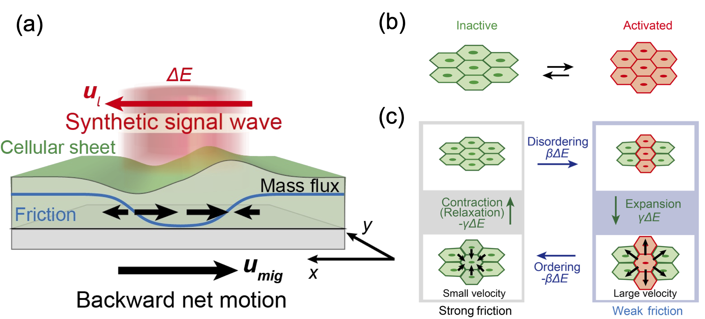
To study how signaling waves control collective migration, we use continuum mechanics. The cell population regulated by intracellular protein signaling can be described as a continuum in the two-dimensional coordinate (Fig. 1(a)). This theoretical model aims to show the mechanism that controls collective migration (backward net motion) along a wave of protein signaling as it travels through a cell monolayer. Although collective migration is regulated by complex signaling pathways in real cells, it is simplified here to assume that the cell population continuously switches between active and inactive states as it receives the protein signal (Fig. 1(b)). The basal level of the protein signal is given by and takes the same constant value at infinity regardless of the direction. The signal propagates with intensity = and velocity . The collective migration is driven by internal forces while maintaining a force-free condition [25, 26, 27, 28, 29, 30]. Cell population adheres to a substrate and balances both contractile stresses generated by the cells and frictional stress. We consider that both the contractile force and the frictional stresses depend on the magnitude of signal activation. The force balance is described by
| (1) |
where is local cell density, is the velocity field of the cell population, and is the frictional coefficient matrix against cell migration per unit area. Cells adhere to each other via cell-cell adhesion junction, and is the internal stress [31, 32]. For epithelial cell monolayer typically moving at a slow velocity, the inertia term is negligible and Eq. (1) is rewritten as
| (2) |
In addition, we define the internal stress by
| (3) |
where is the identity matrix, the first term is density-dependent passive stress by pressure , and the second term is signal-dependent stress generated by molecular motor protein (protein concentration ) with the constant [31]. However, there is no net motion under changes in the density when the friction coefficient does not change with signal activation [29, 30]. This is because the net internal stress (the integral of over the cell population) must be zero for force-free conditions. The active stress by molecular motors is not sufficient to rectify directionality in motion relative to the traveling signal wave.
The key concept that unravels directed net motion is the multiplication of anisotropic frictional force and active contractile force regulated by protein signal (Fig. 1(c)). Inside a cell population, cells have an orientation, and the ease of movement is determined along that orientation [33, 34, 35]. The orientation of the cell monolayer is expressed by the director with orientation angle . This orientation also characterizes the direction of cell migration. We assume that the cell population is oriented in the -axis direction along which the signal wave propagates. If this orientation is changed by signaling activity, such anisotropic mobility can be altered depending on the orientation field and spatially biased by .
We define the tensor of orientation field as where the scalar order parameter is determined by the average of within coarse-grained region. can change in an signal-dependent manner (i.e., ). The anisotropic friction is given by
| (4) |
where is the scalar friction parameter representing orientational anisotropy [34]. If the velocity is perpendicular to the orientation angle, a negative sign (or positive sign) of means a larger (or smaller) friction coefficient. Eq. (4) then gives the viscous resistance force as
| (5) | |||||
Consider the situation where the cell population can move in the direction of the -axis along which the ERK wave propagates, but the motion in the -axis direction perpendicular to the wave propagation is small, and . Substituting Eqs. (3) and (5) into Eq. (2), the force-balance relation for the aforementioned situation becomes
| (6) |
and
| (7) |
| (8) |
We consider that momentum transport according to the velocity gradient occurs mostly in -direction (along the wave) and little velocity change occurs in -direction (perpendicular to the wave). In this condition, the 2nd term in Eq. (8) is negligibly small. Then, the following equation holds
| (9) |
where denoting the ERK dependence of the friction coefficient. Eq. (9) can be written using (the ERK dependence of ) as follows
| (10) |
These results indicate that the ERK activity dependence of , which depends on the orientation of the cell population, can determine the velocity gradient.
Next, given that the signal continues to activate the cell body until the wave passes, the contractile stress is induced and gets stronger at the rear end of the wave [36, 37]. The resultant gradient of the contractile stresses changes the local cell density . The change in local cell density follows the continuity equation:
| (11) |
when the growth and division of cells are negligible for short time scales ( min). Since the protein signal travels at a constant velocity , we rewrite Eq. (11) in the coordinates of the moving frame (, see Appendix),
| (12) |
The dependence of the cell density on the signal activity can then be regarded as if the rate of density change is determined by the first order of the signal change with coefficient . By using , Eq. (12) leads to Finally, by converting the coordinates from the moving frame to the experimental frame ( and ), we obtain the velocity gradient as
| (13) |
By integrating Eq. (13), is
| (14) |
where is derived from the integration of . Eq. (13) denotes that the signal-dependent change of cell density induces the mass flux. By inserting Eq. (14) into Eq. (9),
| (15) |
Then, the migration velocity is
| (16) |
allowing non-zero net motion (Fig. 1(a) and (c)). Notably, the positive sign of means that the cell population migrates opposite to the direction of the approaching signal wave and drives the net motion in one direction persistently.
III ERK signaling in MDCK cell monolayer as an experimental model
Our next focus is to experimentally verify the theoretical model using epithelial cells that form monolayer structures on the substrate. We focused on the fact that a cell type known as Madin-Darby canine kidney (MDCK) cells exhibit collective migration coupled to intracellular signaling activation. It is known that in the monolayer of MDCK cells, the activity of ERK MAP kinase signaling (ERK signaling) propagates like a traveling wave and drives collective migration in the opposite direction of the signal wave [2, 5, 38]. During collective migration, ERK signaling regulates contractility through the myosin molecular motor. Such ERK activity propagates through the cell monolayer like a traveling wave, exhibiting rectified migration. The presence of cells with high ERK activity in a cell population can also induce spatial changes in the contractile force gradient and orientation order between cells. Based on the above considerations, the ERK-regulated MDCK cell monolayer is one of the appropriate experimental systems for our theoretical model.
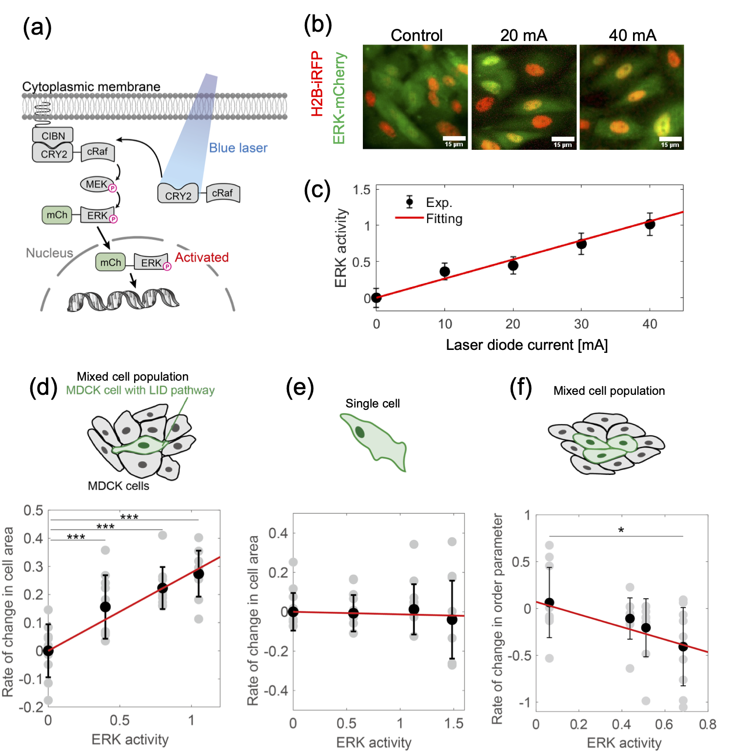
The MDCK cells used in the experiment can be controlled by an optogenetic tool of the ERK pathway through light-induced dimerization (LID) of CRY2-CIBN [3, 5] (Fig. 2(a)). This tool allows the manipulation of ERK activity at the single cell level in a light-responsive manner (Fig. 2(b)). The level of activity was assessed quantitatively by measuring the translocation of ERK-mCherry protein to the nucleus (Fig. 2(c) and Fig. S1 [39]). We decided to use these MDCK cells to test whether light can regulate ERK signaling activity and thereby induce changes in cell density and orientation within the monolayer.
In addition to manipulating ERK signaling with light, we included the MDCK cell population in a light-unresponsive cell population to create differences in signal intensity between cells. We then generated a mosaic cell population by mixing these optogenetic MDCK cells with normal cells in a 1:9 ratio. This heterogeneous cell population was then exposed to blue light, which induced ERK signaling only in the light-responsive cells. By applying such optogenetic activation for 30 minutes, we tested whether the activated cells exhibited an increase in cell spreading area. The activated cell experienced stretching forces at the interface between the activated and non-activated cells (Fig. 2(d)). However, no area change was observed in a single isolated cell without surrounding cells (Fig. 2(e)). Traction force microscopy analysis revealed that intracellular contractility decreased in MDCK cells with elevated ERK activity (Fig. S2) [40, 39]. This suggests that cells with increased ERK activity have decreased contractility upon light stimulation, while surrounding cells maintain their contractility, resulting in an enlarged area due to pulling from the surrounding cells. Thus, because the occupied area is increased by ERK activity, local activation of ERK signaling results in reduced cell density if the volume of cell monolayer is conserved, implying .
Next, we examined the change in cell orientation that may be involved in anisotropic friction. To investigate whether such changes are caused by ERK activation, we also optogenetically activated ERK activity and analyzed changes in the degree of orientation order in cell populations. The value of the orientation order parameter before light exposure was set to , and after optogenetically upregulating ERK activity for 30 minutes, the difference in the orientation order parameter was analyzed at different levels of ERK activity. As the level of ERK activity was increased, the rate of change of the order parameter decreased (Fig. 2(f)), indicating that the orientation of the cell population became more random due to ERK activation. We then need to clarify how friction changes with orientation order. Previous studies have reported that in MDCK cell populations, velocity tends to increase when cells move parallel to the angle of orientation [33, 41]. From Eq. (4), the decrease in friction in the direction of alignment corresponds to a negative friction parameter, (this trend is opposite to neural progenitor cells, which are considered to have [34]). According to , the change of the friction coefficient in our experiment (, ) is .
IV Collective migration under a synthetic ERK wave
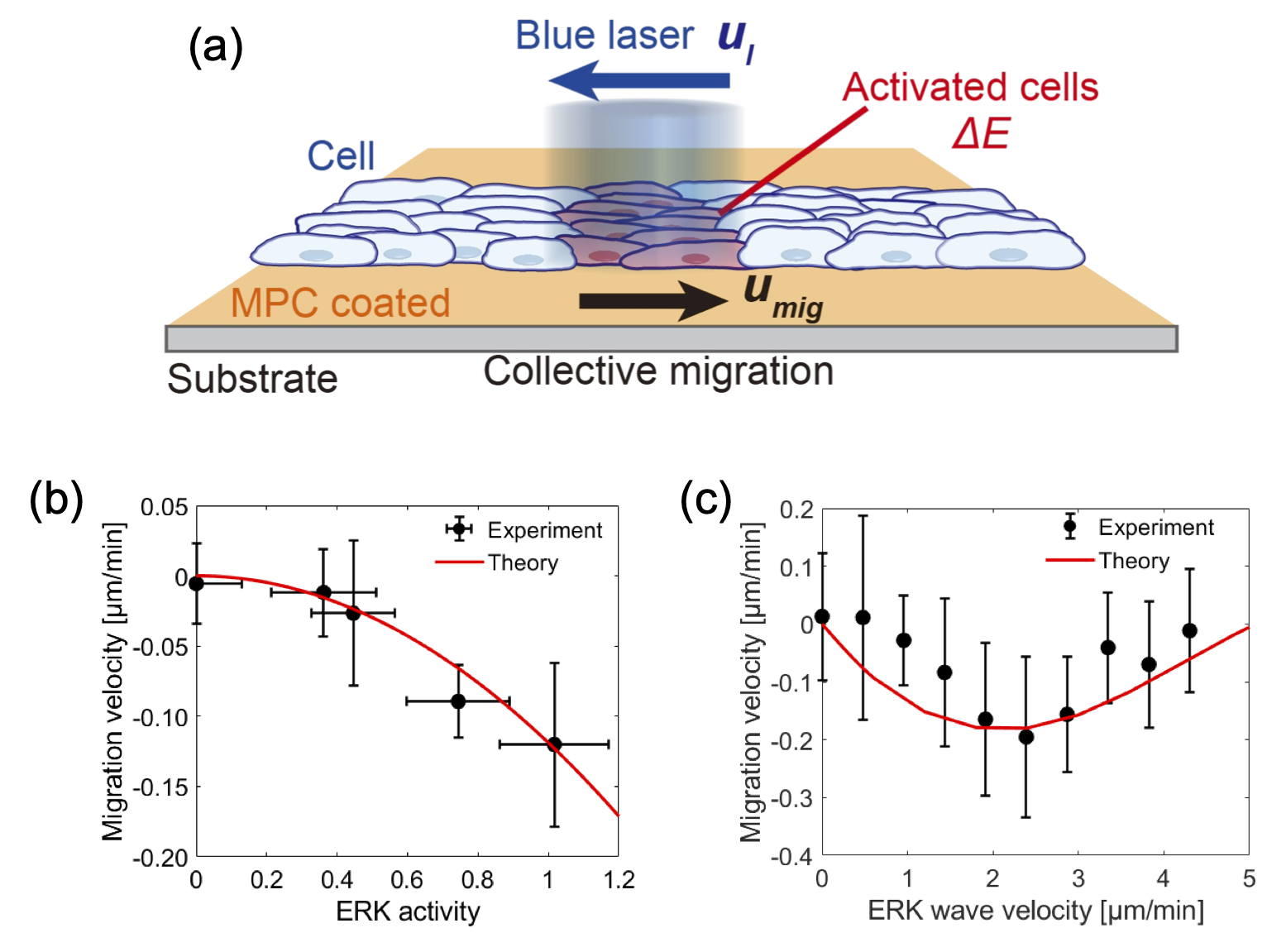
Experiments on the optogenetic control of ERK suggest that the MDCK cell population responds to an increase in ERK signaling with friction change of and the density change of . Eq. (16) states , which suggests that the motion is in the opposite direction to the wave of the ERK signal. To test this, we designed a synthetic ERK wave that mimics the natural signal wave by unidirectional sweeping of focused laser light (Fig. 3(a)). This synthetic ERK wave travels at a constant speed while is regulated by adjusting the laser intensity. We found that the velocity of cell migration was always negative value, indicating that the cell population with increased ERK activity moves in the opposite direction of the ERK wave (Fig. 3(b)). Moreover, as seen in Eq. (16), the speed of collective migration is proportional to the square of ERK activity, . In the experiment, the migration velocity showed a quadratic increase for (Fig. 3(b)). Our theoretical model is consistent with the experimental investigations, and in particular, the quadratic increase of migration speed implies that the two ERK-dependent processes in density and friction is involved in rectifying collective migration under the traveling signal wave.
We also measured the dependence on ERK wave velocity and found that the maximum migration velocity was reached at a certain wave velocity of . (Fig. 3(c)). Such optimal wave velocity in directed collective migration has also been reported in earlier studies [5, 16, 38] but its mechanism was little described from mechanical views. To resolve this point, we consider viscoelastic deformation and restoration proceeding with cell migration. The extended deformation coefficient is , where is the characteristic time of mechanical restoration ( [42]), is the wavelength of the ERK wave ( = ). By considering such relaxation time in density change in [43], the migration velocity is extended to a general form:
| (17) |
affects migration speed according to the relationship between the relaxation time of density () and the propagation time of the wave (). For slowly propagating ERK wave (), the migration velocity and thus migration speed linearly increases with ERK wave speed. In contrast, for rapidly propagating ERK wave (), the migration velocity plateaus as . Hence, the relaxation process can explain the gradual increase of at slow wave speeds.
To further investigate the mechanism that optimizes ERK wave velocity, we consider another relaxation process, the decay of signaling activity. The relaxation dynamics of ERK signaling is given by
| (18) |
where is the point source of the signal wave and is the relaxation time of activated signal [5]. In MDCK cells, the duration of signal activation can be defined as the time scale with a typical cell size of . If this activation time is sufficiently longer than the ERK signal relaxation time (), is fully activated by the saturation level and no change appears in for the slowly propagating ERK wave. On the other hand, if the activation time is shorter than the relaxation time (), as when the wave moves faster, becomes small, and in turn the associated changes in contractile stress and reduction of friction are damped over time. This leads to a slowing of for a rapidly propagating ERK wave.
Therefore, the interplay between density relaxation and signal activation may lead to the optimal wave speed in ERK-mediated collective migration. Our model predicts the optimal wave velocity to be , which is comparable to the experimental value (Fig. 3(c)).
V Wound healing of epithelial cell monolayer
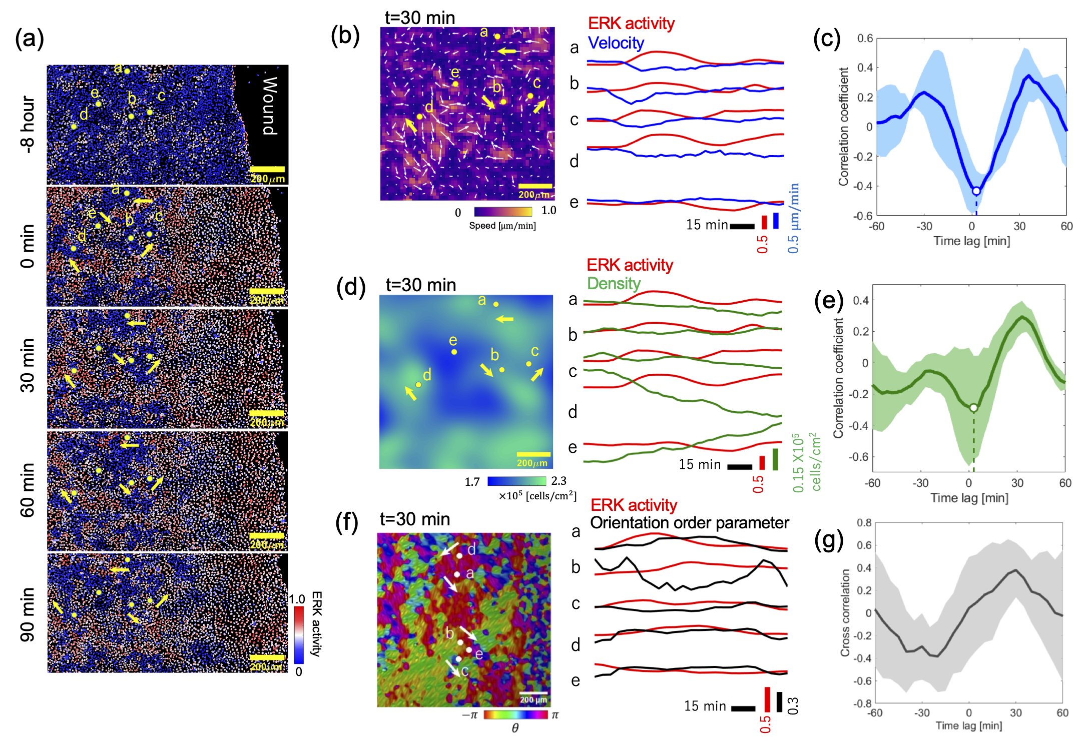
The theoretical model of wave-directed collective migration motivates us to experimentally investigate the interplay between signaling waves and the mechanics of the epithelial cell monolayer during wound healing. Upon injury to the cell monolayer, directed collective migration of cells is initiated, leading to wound closure. The initiation of collective migration applies mechanical stimuli to the leading cell layer, which activates ERK signaling. Visualization of ERK activity using a protein-based FRET sensor shows that the highly active region (red cells) is localized near the wound site, indicating that ERK signaling propagates from the leading edge of wounded cell populations (Fig. 4(a)). It has been shown that cells collectively migrate in the direction opposite to the propagation of the ERK wave, and then the leading edge of the migrating cells can fill the existing wound [5, 38, 4]. By measuring the intensity of ERK signal and cell migration relative to the direction of ERK wave propagation, it was observed that the speed of cell motility increased as the intensity of the signal decreased (Fig. 4(b)), indicating a negative correlation between motility speed and changes in ERK signal (Fig. 4(c)). We also examined the change in cell density associated with ERK activity during wound healing. Local cell density decreased with increasing signaling activity (Fig. 4(d) and (e)). This negative correlation between cell density and ERK signaling activity is consistent with previous studies [5, 36].
We then analyzed the orientational dynamics of the MDCK cells nduced by ERK signaling activity. The orientation angle in the cell monolayer was calculated from brightfield images (Fig. 4(f), left)[39]. The orientation field was then used to calculate the scalar order parameter by averaging over a region of interest. We analyzed the signal-dependent change of and found that this order parameter decreased as ERK activity increased (Fig. 4(f), right). This result implies that the increased ERK activity at the wound causes the domain size of the well-oriented cell population to be smaller in the vicinity of the wound region. We also calculated the correlation between ERK signaling activity and . The cross-correlation function shows a negative value with a time delay (Fig. 4(g)), indicating local orientation becomes less organized as ERK activity increases. By assuming that local orientation order is related to anisotropic friction, the ERK signal wave may facilitate the reorientation of cells along the direction of wave propagation, making it easier for the cell population to move in that direction.
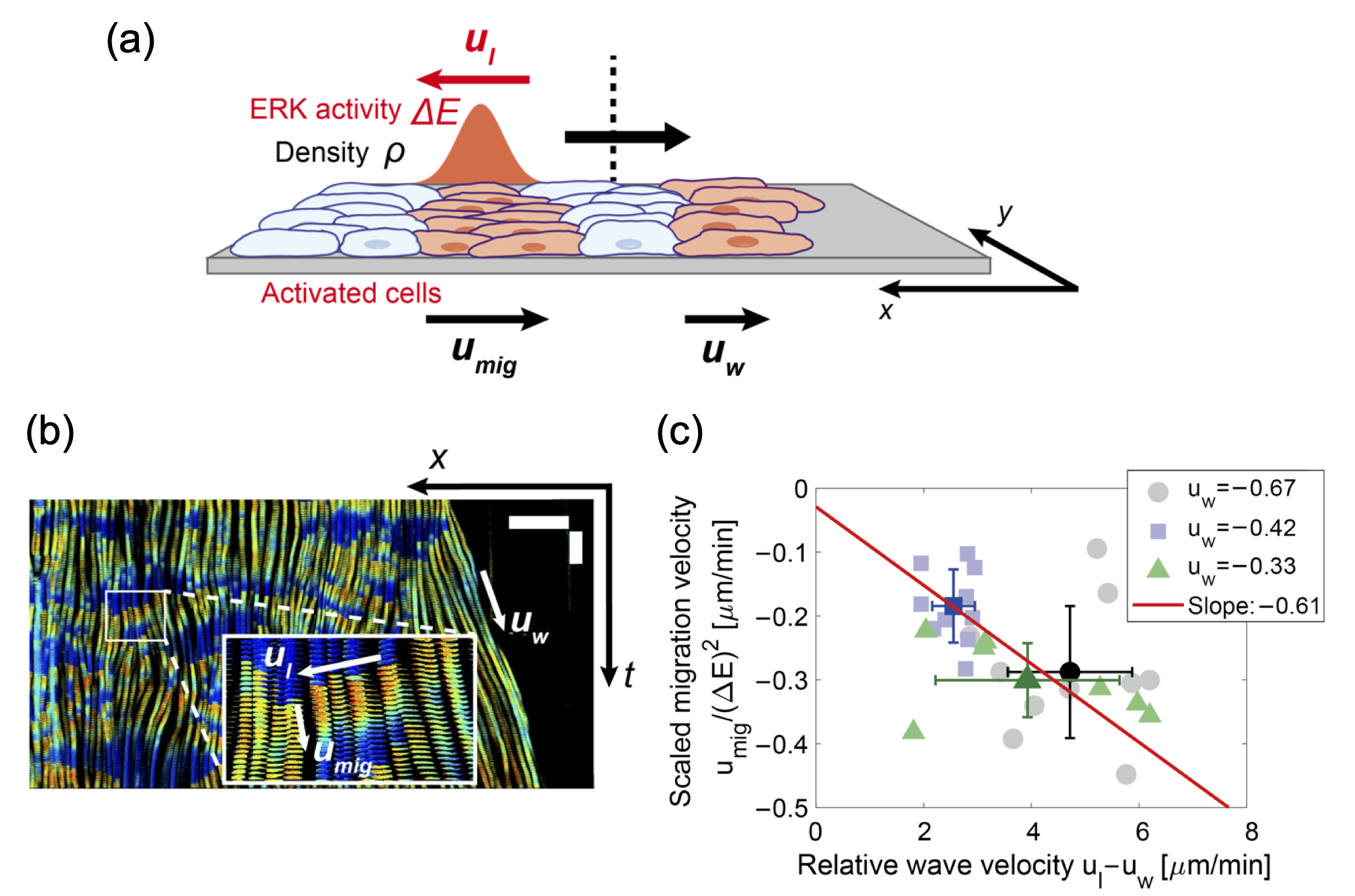
To extend our theoretical model to wound healing [44, 45, 46, 47], we consider a cell monolayer whose leading edge can move in one direction (Fig. 5(a)). We assume that the ERK signal propagates from the wound site at a speed within the cell monolayer, while the leading cells move toward the wound region at a constant speed . This motion of the leading cells induces the outward density flux of cells (Fig. 5(b)). The cell population then moves at velocity against the protein signal wave generated from the wound. Given that the supply of cell population maintains the continuous cell monolayer, Eq. (11) is rewritten as . At the moving frame of a traveling signal wave, the density flux is , and this expression indicates that the effective velocity of the signal wave is as a system standing at the tip of the signal wave. The migration velocity Eq. (17) can be given by
| (19) |
Eq. (19) indicates that even if the leading edge of the cell population moves, the migration speed appears similar expression, and the only difference in the form that the wave velocity is stretched by , meaning that our theoretical model can be extended to the process of wound healing. To compare theoretical models with wound healing experiments, we simultaneously measured signal activity and ERK wave velocity using a FRET-based ERK sensor, in addition to analyzing the rate of leading edge spreading in wound healing. We plotted the migration velocity scaled by the ERK signal level, , for the stretched velocity of the ERK wave, (Fig. 5(c)), and found the linear relationship as proposed by Eq. (19). The slope of this curve is then expressed as the product of the coefficients of the ERK-dependent density change and the friction change . The value of in the wound healing experiment is comparable to that obtained by optogenetic manipulation ( in Fig. 3(b)).
VI Discussion
In this study, the coordination of changes in cell density and friction as a function of signal activation can induce a net collective migration rectified against a traveling signal wave. Around the trailing end of the signal wave, the friction coefficient decreases and the contractile stress increases. As a result, the pulling force from the rear end drives the net backward movement of the cells. On the other hand, at the front end of the wave, the cells are stretched, which accentuates the velocity of motion in the opposite direction of the wave. To summarize the mechanism of this collective migration, the driving force results from the multiplicative effect of active stress and friction change generated by the ERK signal. The signal-dependent active stress causes local density change, and the density flow is generated by the tug-of-war between neighboring cells. Such density flow is biased by the ERK-dependent cell orientation, which generates an anisotropic friction gradient. Thus, the two ERK-dependent effects work together to drive directed collective migration, and its speed exhibits quadratic increase () to ERK signal intensity.
Previous theoretical models have suggested that ERK activation may affect the frictional force of cell motility by assuming the change in magnitude [5, 48]. In Eq. (4), we consider that the signal-dependent mobility is rather controlled by the orientation dependence, not the magnitude of the friction coefficient. This is based on the fact that we measured adhesion strength in single cells with increased ERK activity and found no ERK dependence in adhesion magnitude (Fig. S3 in [39] and [49, 50]). This finding suggests that the critical change in friction is likely to be orientation among cell-cell interactions. However, our results do not rule out the possibility that the magnitude of the friction coefficient may depend on ERK activation at the level of cell populations. Recent reports have shown that the interplay between contractile stress and polarity of epithelial cells [38, 51] are involved in directed collective migration. Thus, elucidating the mechanism of signal- and force-dependent orientation changes remains a challenge for the future [52].
To further study multi-cellular systems from physical modeling, quantitative methods such as optogenetics will become increasingly essential. We have shown that there is an optimal wave speed for driving collective migration by optogenetic control of synthetic signal waves. This is because the relaxation time of density change and signal transduction can determine the most efficient wave speed. The cell monolayer can control collective migration by regulating the phase difference between the two cyclical changes in density and friction while maintaining force balance. Further understanding of the mechanical coordination of contraction and friction, including force-signaling feedback [53], may provide physical insights into the broad spectrum of active dynamics organized by a traveling signal wave. Understanding how signal transduction systems regulate directed collective migration and ordered pattern formation not only in epithelial cells but also in other cell types, such as stem cells [34, 54], will help us understand the principles by which cell populations build functional forms while adapting to complex geometric constraints [55, 56, 57]. It has been shown that such interplay of mechanical force and protein signaling is involved in apoptosis [58] and tissue regeneration [59]. Therefore, elucidating the physical mechanisms underlying these mechanical-chemical interactions is a major challenge for future research.
Acknowledgements
This work was supported by Grant-in-Aid for Scientific Research on Innovative Areas (JP16H00805, JP17H05234, and 18H05427 to YTM, JP19H05798 to KA), Grant-in-Aid for Transformative Research Areas (A) (JP23H04711 and JP23H04599 to YTM), Grant-in-Aid for Scientific Research (B) (JP20H01872 and JP23H01144 to YTM, JP18H02444 and JP22H02625 to KA), Grant-in-Aid for Young Scientists (JP22K14014 to TF), JST FOREST Grant (JPMJFR2239 to YTM), CREST JST Grant (JPMJCR1654 to KA), AMED-CREST Grant (JP20GM0810002 to SK), NIBB Collaborative Research Program (18-355 to YTM) and Joint Research of ExCELLS (23EXC205 to YTM).
References
- Friedl and Gilmour [2009] P. Friedl and D. Gilmour, Collective cell migration in morphogenesis, regeneration and cancer, Nature Reviews Molecular Cell Biology 10, 445 (2009).
- Matsubayashi et al. [2004] Y. Matsubayashi, M. Ebisuya, S. Honjoh, and E. Nishida, Erk activation propagates in epithelial cell sheets and regulates their migration during wound healing, Current Biology 14, 731 (2004).
- Aoki et al. [2013] K. Aoki, Y. Kumagai, A. Sakurai, N. Komatsu, Y. Fujita, C. Shionyu, and M. Matsuda, Stochastic erk activation induced by noise and cell-to-cell propagation regulates cell density-dependent proliferation, Molecular Cell 52, 529 (2013).
- Hiratsuka et al. [2015] T. Hiratsuka, Y. Fujita, H. Naoki, K. Aoki, Y. Kamioka, and M. Matsuda, Intercellular propagation of extracellular signal-regulated kinase activation revealed by in vivo imaging of mouse skin, eLife 4, e05178 (2015).
- Aoki et al. [2017] K. Aoki, Y. Kondo, H. Naoki, T. Hiratsuka, R. E. Itoh, and M. Matsuda, Propagating wave of erk activation orients collective cell migration, Developmental Cell 43, 305—317.e5 (2017).
- O’Brien et al. [2004] L. E. O’Brien, K. Tang, E. S. Kats, A. Schutz-Geschwender, J. H. Lipschutz, and K. E. Mostov, Erk and mmps sequentially regulate distinct stages of epithelial tubule development, Developmental Cell 7, 21 (2004).
- Weijer [2009] C. J. Weijer, Collective cell migration in development, Journal of Cell Science 122, 3215 (2009).
- Londono et al. [2014] C. Londono, M. J. Loureiro, B. Slater, P. B. Lücker, J. Soleas, S. Sathananthan, J. S. Aitchison, A. J. Kabla, and A. P. McGuigan, Nonautonomous contact guidance signaling during collective cell migration, Proceedings of the National Academy of Sciences USA 111, 1807 (2014).
- Purcell [1977] E. M. Purcell, Life at low reynolds number, American Journal of Physics 45, 3 (1977).
- Najafi and Golestanian [2004] A. Najafi and R. Golestanian, Simple swimmer at low reynolds number: Three linked spheres, Physical Review E 69, 062901 (2004).
- Kumar et al. [2008] K. V. Kumar, S. Ramaswamy, and M. Rao, Active elastic dimers: Self-propulsion and current reversal on a featureless track, Physical Review E 77, 020102 (2008).
- Qiu et al. [2014] T. Qiu, T.-C. Lee, A. G. Mark, K. I. Morozov, R. Münster, O. Mierka, S. Turek, A. M. Leshansky, and P. Fischer, Swimming by reciprocal motion at low reynolds number, Nature Communications 5, 5119 (2014).
- Lauga [2011] E. Lauga, Life around the scallop theorem, Soft Matter 7, 3060 (2011).
- Leoni and Sens [2017] M. Leoni and P. Sens, Model of cell crawling controlled by mechanosensitive adhesion, Physical Review Letters 118, 228101 (2017).
- Mai and Camley [2020] M. H. Mai and B. A. Camley, Hydrodynamic effects on the motility of crawling eukaryotic cells, Soft Matter 16, 1349 (2020).
- Lozano and Bechinger [2019] C. Lozano and C. Bechinger, Diffusing wave paradox of phototactic particles in traveling light pulses, Nature Communications 10, 2495 (2019).
- Garcia et al. [2015] S. Garcia, E. Hannezo, J. Elgeti, J.-F. Joanny, P. Silberzan, and N. S. Gov, Physics of active jamming during collective cellular motion in a monolayer, Proceedings of the National Academy of Sciences 112, 15314 (2015).
- Blanch-Mercader et al. [2018] C. Blanch-Mercader, V. Yashunsky, S. Garcia, G. Duclos, L. Giomi, and P. Silberzan, Turbulent dynamics of epithelial cell cultures, Physical Review Letters 120, 208101 (2018).
- Mueller et al. [2019] R. Mueller, J. M. Yeomans, and A. Doostmohammadi, Emergence of active nematic behavior in monolayers of isotropic cells, Physical Review Letters 122, 048004 (2019).
- Lin et al. [2021] S.-Z. Lin, W.-Y. Zhang, D. Bi, B. Li, and X.-Q. Feng, Energetics of mesoscale cell turbulence in two-dimensional monolayers, Communications Physics 4, 21 (2021).
- Nagai and Honda [2001] T. Nagai and H. Honda, A dynamic cell model for the formation of epithelial tissues, Philosophical Magazine B 81, 699 (2001).
- Fletcher et al. [2014] A. G. Fletcher, M. Osterfield, R. E. Baker, and S. Y. Shvartsman, Vertex models of epithelial morphogenesis, Biophysical Journal 106, 2291 (2014).
- Barton et al. [2017] D. L. Barton, S. Henkes, C. J. Weijer, and R. Sknepnek, Active vertex model for cell-resolution description of epithelial tissue mechanics, PLoS Computational Biology 13, e1005569 (2017).
- Lin et al. [2018] S.-Z. Lin, S. Ye, G.-K. Xu, B. Li, and X.-Q. Feng, Dynamic migration modes of collective cells, Biophysical Journal 115, 1826 (2018).
- Wang and Wolynes [2011] S. Wang and P. G. Wolynes, On the spontaneous collective motion of active matter, Proceedings of the National Academy of Sciences USA 108, 15184 (2011).
- Tanimoto and Sano [2014] H. Tanimoto and M. Sano, A simple force-motion relation for migrating cells revealed by multipole analysis of traction stress, Biophysical Journal 106, 16 (2014).
- Ebata et al. [2020] H. Ebata, K. Moriyama, T. Kuboki, and S. Kidoaki, General cellular durotaxis induced with cell-scale heterogeneity of matrix-elasticity, Biomaterials 230, 119647 (2020).
- Tarama and Yamamoto [2018] M. Tarama and R. Yamamoto, Mechanics of cell crawling by means of force-free cyclic motion, Journal of the Physical Society of Japan 87, 044803 (2018).
- Weinert et al. [2008] F. M. Weinert, J. A. Kraus, T. Franosch, and D. Braun, Microscale fluid flow induced by thermoviscous expansion along a traveling wave, Physical Review Letters 100, 164501 (2008).
- Fukuyama et al. [2018] T. Fukuyama, S. Nakama, and Y. T. Maeda, Thermal molecular focusing: tunable cross effect of phoresis and light-driven hydrodynamic focusing, Soft Matter 14, 5519 (2018).
- Banerjee et al. [2015] S. Banerjee, K. J. Utuje, and M. C. Marchetti, Propagating stress waves during epithelial expansion, Physical Review Letters 114, 228101 (2015).
- Yabunaka and Marcq [2017] S. Yabunaka and P. Marcq, Emergence of epithelial cell density waves, Soft Matter 13, 7046 (2017).
- Saw et al. [2017] T. B. Saw, A. Doostmohammadi, V. Nier, L. Kocgozlu, S. Thampi, Y. Toyama, P. Marcq, C. T. Lim, J. M. Yeomans, and B. Ladoux, Topological defects in epithelia govern cell death and extrusion, Nature 544, 212 (2017).
- Kawaguchi et al. [2017] K. Kawaguchi, R. Kageyama, and M. Sano, Topological defects control collective dynamics in neural progenitor cell cultures, Nature 545, 327 (2017).
- Doostmohammadi et al. [2018] A. Doostmohammadi, J. Ignés-Mullol, J. M. Yeomans, and F. Sagués, Active nematics, Nature Communications 9, 3246 (2018).
- Saraswathibhatla et al. [2022] A. Saraswathibhatla, J. Zhang, and J. Notbohm, Coordination of contractile tension and cell area changes in an epithelial cell monolayer, Physical Review E 105, 024404 (2022).
- Yang et al. [2018] J.-M. Yang, S. Bhattacharya, H. West-Foyle, C.-F. Hung, T.-C. Wu, P. A. Iglesias, and C.-H. Huang, Integrating chemical and mechanical signals through dynamic coupling between cellular protrusions and pulsed erk activation, Nature Communications 9, 4673 (2018).
- Hino et al. [2020] N. Hino, L. Rossetti, A. Marín-Llauradó, K. Aoki, X. Trepat, M. Matsuda, and T. Hirashima, Erk-mediated mechanochemical waves direct collective cell polarization, Developmental Cell 53, 646 (2020).
- [39] See Supplemental Material at http://link.aps.org/supplemental/ for experimental materials and methods, for a detailed description of theoretical caluclation .
- Ueki and Kidoaki [2015] A. Ueki and S. Kidoaki, Manipulation of cell mechanotaxis by designing curvature of the elasticity boundary on hydrogel matrix, Biomaterials 41, 45 (2015).
- Balasubramaniam et al. [2021] L. Balasubramaniam, A. Doostmohammadi, T. B. Saw, G. H. N. S. Narayana, R. Mueller, T. Dang, M. Thomas, S. Gupta, S. Sonam, A. S. Yap, et al., Investigating the nature of active forces in tissues reveals how contractile cells can form extensile monolayers, Nature Materials 20, 1156 (2021).
- Iyer et al. [2019] K. V. Iyer, R. Piscitello-Gómez, J. Paijmans, F. Jülicher, and S. Eaton, Epithelial viscoelasticity is regulated by mechanosensitive e-cadherin turnover, Current Biology 29, 578 (2019).
- Hamadi et al. [2005] A. Hamadi, M. Bouali, M. Dontenwill, H. Stoeckel, K. Takeda, and P. Ronde, Regulation of focal adhesion dynamics and disassembly by phosphorylation of fak at tyrosine 397, Journal of Cell Science 118, 4415 (2005).
- Zaritsky et al. [2014] A. Zaritsky, D. Kaplan, I. Hecht, S. Natan, L. Wolf, N. S. Gov, E. Ben-Jacob, and I. Tsarfaty, Propagating waves of directionality and coordination orchestrate collective cell migration, PLOS Computational Biology 10, 1 (2014).
- Basan et al. [2013] M. Basan, J. Elgeti, E. Hannezo, W.-J. Rappel, and H. Levine, Alignment of cellular motility forces with tissue flow as a mechanism for efficient wound healing, Proceedings of the National Academy of Sciences USA 110, 2452 (2013).
- Brugués et al. [2014] A. Brugués, E. Anon, V. Conte, J. H. Veldhuis, M. Gupta, J. Colombelli, J. J. Muñoz, G. W. Brodland, B. Ladoux, and X. Trepat, Forces driving epithelial wound healing, Nature Physics 10, 683 (2014).
- Tetley et al. [2019] R. J. Tetley, M. F. Staddon, D. Heller, A. Hoppe, S. Banerjee, and Y. Mao, Tissue fluidity promotes epithelial wound healing, Nature Physics 15, 1195 (2019).
- Asakura et al. [2021] Y. Asakura, Y. Kondo, K. Aoki, and H. Naoki, Hierarchical modeling of mechano-chemical dynamics of epithelial sheets across cells and tissue, Scientific Reports 11, 4069 (2021).
- Yoshikawa et al. [2011] H. Y. Yoshikawa, F. F. Rossetti, S. Kaufmann, T. Kaindl, J. Madsen, U. Engel, A. L. Lewis, S. P. Armes, and M. Tanaka, Quantitative evaluation of mechanosensing of cells on dynamically tunable hydrogels, Journal of the American Chemical Society 133, 1367 (2011).
- Yu et al. [2018] L. Yu, J. Li, J. Hong, Y. Takashima, N. Fujimoto, M. Nakajima, A. Yamamoto, X. Dong, Y. Dang, Y. Hou, et al., Low cell-matrix adhesion reveals two subtypes of human pluripotent stem cells, Stem Cell Reports 11, 142 (2018).
- Jain et al. [2020] S. Jain, V. M. Cachoux, G. H. Narayana, S. de Beco, J. D’alessandro, V. Cellerin, T. Chen, M. L. Heuzé, P. Marcq, R.-M. Mège, et al., The role of single-cell mechanical behaviour and polarity in driving collective cell migration, Nature Physics 16, 802 (2020).
- Cavanaugh et al. [2022] K. E. Cavanaugh, M. F. Staddon, T. A. Chmiel, R. Harmon, S. Budnar, A. S. Yap, S. Banerjee, and M. L. Gardel, Force-dependent intercellular adhesion strengthening underlies asymmetric adherens junction contraction, Current Biology 32, 1986 (2022).
- Boocock et al. [2021] D. Boocock, N. Hino, N. Ruzickova, T. Hirashima, and E. Hannezo, Theory of mechanochemical patterning and optimal migration in cell monolayers, Nature Physics 17, 267 (2021).
- Guillamat et al. [2022] P. Guillamat, C. Blanch-Mercader, G. Pernollet, K. Kruse, and A. Roux, Integer topological defects organize stresses driving tissue morphogenesis, Nature Materials 21, 588 (2022).
- Deforet et al. [2014] M. Deforet, V. Hakim, H. G. Yevick, G. Duclos, and P. Silberzan, Emergence of collective modes and tri-dimensional structures from epithelial confinement, Nature Communications 5, 3747 (2014).
- Shigeta et al. [2022] K. Shigeta, T. Fukuyama, R. Takahashi, K. Beppu, A. Tanaka, and Y. T. Maeda, Collective motion of epithelial cells along a wrinkled 3d-buckled hydrogel, RSC Advances 12, 20174 (2022).
- Ienaga et al. [2023] R. Ienaga, K. Beppu, and Y. T. Maeda, Geometric confinement guides topological defect pairings and emergent flow in nematic cell populations, Soft Matter 19, 5016 (2023).
- Gagliardi et al. [2021] P. A. Gagliardi, M. Dobrzyński, M.-A. Jacques, C. Dessauges, P. Ender, Y. Blum, R. M. Hughes, A. R. Cohen, and O. Pertz, Collective erk/akt activity waves orchestrate epithelial homeostasis by driving apoptosis-induced survival, Developmental Cell 56, 1712 (2021).
- De Simone et al. [2021] A. De Simone, M. N. Evanitsky, L. Hayden, B. D. Cox, J. Wang, V. A. Tornini, J. Ou, A. Chao, K. D. Poss, and S. Di Talia, Control of osteoblast regeneration by a train of erk activity waves, Nature 590, 129 (2021).