Fluorescence-Detected Circular Dichroism of a Chiral Molecular Monolayer with Dielectric Metasurfaces
Abstract
Strong enhancement of molecular circular dichroism has the potential to enable efficient asymmetric photolysis, a method of chiral separation that has conventionally been impeded by insufficient yield and low enantiomeric excess. Here, we study experimentally how predicted enhancements in optical chirality density near resonant silicon nanodisks boost circular dichroism. We use fluorescence-detected circular dichroism spectroscopy to measure indirectly the differential absorption of circularly polarized light by a monolayer of optically active molecules functionalized to silicon nanodisk arrays. Importantly, the molecules and nanodisk antennas have spectrally-coincident resonances, and our fluorescence technique allows us to deconvolute absorption in the nanodisks from the molecules. We find that enhanced fluorescence-detected circular dichroism signals depend on nanophotonic resonances in good agreement with simulated differential absorption and optical chirality density, while no signal is detected from molecules adsorbed on featureless silicon surfaces. These results verify the potential of nanophotonic platforms to be used for asymmetric photolysis with lower energy requirements.
keywords:
dielectric nanoparticles, optical chirality, enantiomer separation, Mie resonances, circular dichroismMechanical and Aerospace Engineering, University of California San Diego, 9500 Gilman Drive, La Jolla, California 92093, USA
{tocentry}
![[Uncaptioned image]](/html/2008.11270/assets/20200609_TOC.png)
Chirality, or handedness, is a fundamental property of all living organisms, from biological building blocks such as DNA and amino acids to macroscopic structures. Chirality also features prominently in many synthetic molecules, with over 50% of pharmaceuticals and 40% of agrochemicals existing as enantiopure forms or racemic mixtures.1, 2 These enantiomers can have distinct efficacy in biological systems, making the ability to distinguish mirror-image molecules with high sensitivity and maximize enantiomeric excess (e.e.) in asymmetric synthesis crucial tasks. Circular dichroism (CD), defined as the selective absorption of circularly polarized light (CPL), is commonly used to differentiate enantiomers via CD spectroscopy, though it typically requires relatively high sample concentrations or long optical path lengths.3, 4 This differential absorption between left- and right-CPL, (), has also inspired efforts to use CPL as a reagent in enantioselective synthesis or for photolysis as early as 1929.5, 6 However, due to the low differential absorption cross section of molecules, the yield & e.e. achieved by decomposition of optical isomers with light alone falls below industrially relevant state-of-the-art techniques to maximize enantiopurity.7, 8, 9
A variety of methods have sought to improve sensitivity of chiral differentiation in CD spectroscopy. For example, nonlinear spectroscopies utilizing second harmonic generation that are sensitive to surfaces and interfaces10, 11, 12 and single molecule spectroscopy have enabled enantiomeric detection at the monolayer to few-to-single molecule regime.13, 14 To expand upon these spectroscopic techniques, superchiral electromagnetic fields arising from the interference of chiral plane waves have been shown to increase dissymmetry in chiral excitation, but the position of the superchiral fields within the resulting standing wave nodes limits utility.15, 16 Recently, nanophotonic architectures have received considerable attention due to their potential to manipulate chiral evanescent near fields while maintaining high field strength.17, 18, 19, 20, 21, 22, 23, 24, 25, 26 These near fields have been predicted to enhance the enantioselective rates of molecular absorption and can be achieved using plasmonic 27, 28, 29, 30, 31, 32, 33, 34and high-refractive-index nanostructures. 35, 36, 37, 38, 39, 40, 41 In experiments, many of these approaches enable highly effective enantiomeric sensors, but often exhibit negligible spectral overlap between chiral molecular absorption and nanoantenna resonances, which is necessary to acheive enantioselective photolysis with high yield and e.e.42, 36, 37 Therefore, rather than enhancing differential absorption rates by the molecules themselves, resulting CD signals in the visible and near-IR are mainly due to intrinsic (extrinsic) chirality of 3D (2D) nanostructures, or induced CD in lossy platforms.43 Furthermore, these techniques struggle to distinguish molecular absorption from total absorption making it difficult to unveil and optimize the near-field mechanism behind chiral-optical enhancements in molecules.44, 45
Here, we demonstrate enhanced enantioselective absorption in chiral molecular monolayers using nanostructures with optical resonances spectrally matched to the molecular CD. Sub-wavelength, periodic arrays of silicon disks (hereafter, “metasurfaces”) are functionalized with self-assembled monolayers of fluorescently-labeled oligonucleotide strands with visible-frequency CD (see Figure 1a), attributed to dye binding within the helical DNA environment. We use fluorescence-detected circular dichroism (FDCD) to perform a background-free measurement, as the dye fluorescence is distinguishable from that in the metasurface or substrate. While negligible FDCD is observed on unpatterned films, we observe strong, red-shifting FDCD on disks with increasing radius, in agreement with calculations. We show that our method can distinguish conformation in molecular monolayers, which we validate with in-situ measurements of FDCD sign reversal during DNA dehybridization. Our results exhibit enhancement of intrinsic molecular CD, en-route to enantioselective photolysis.
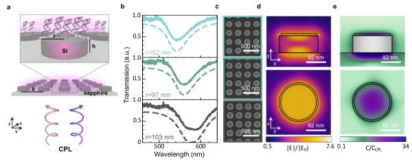
Metasurfaces were engineered to have concurrent electric and magnetic dipolar Mie resonances coinciding with the molecular monolayer absorption, a condition known as the first Kerker condition at which optical chirality density was found to be highly enhanced in our previous work.37 These overlapping modes enhance optical chirality density due to strong electric and magnetic near fields that maintain the phase properties of incident CPL, and disk nanoantennas enable facile tuning of such resonances (see SI and Figure S1 for further detail).37, 38, 45, 46, 47, 48, 49, 50, 51 Nanodisk arrays were fabricated in single-crystalline silicon layers grown on sapphire substrates. The fabricated arrays are , with nominal disk height =80nm, disk radii 90-105nm, and pitch =300nm. Figure 1b includes experimental transmission spectra of three example bare silicon metasurfaces with nominal disk radii of =92, 97, and 103nm, with representative SEMs in Figure 1c. We simulate these metasurfaces with a layer of chiral medium on top, where layer height =10nm, and layer radius, -10nm (see SI), finding that the simulated transmission (dotted line, Figure 1b) is in good agreement with experiments. Dips in the transmission spectra correspond to concurrent electric and magnetic Mie resonances; the simulated electric field plots of Figure 1d are indicative of these overlapping resonances and show strong electric fields extending into the chiral layer.37 Near these resonances, and just blue-shifted (=565nm vs. =570nm), optical chirality density in the chiral layer reaches a maximum enhancement (Figure 1e). Here, we define the optical chirality density as ) and the enhancement factor as C/, where , the optical chirality density of CPL alone.16, 52, 18 The small shift between peak C/C and resonance center wavelength occurs due to the balance between the relative phases and intensities of the electric & magnetic fields.36
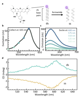
The fluorophore, 590, was covalently attached to the 5’-end of a 25 base pair DNA sequence with a thiol modification on the 3’-end (Figure 2a, see SI for details). The complex absorbs strongly from 500-650nm, with a peak in excitation at 600nm and an emission maximum at 615nm that depends on the excitation wavelength (Figure 2b,c). When hybridized with its fully complementary strand, the dye-dsDNA complex exhibits a positive CD signal in its visible absorption band.53, 54, 55, 56 Interestingly, the sign of the CD signal is reversed when the DNA molecules are denatured as the dye molecules experience the different local environment of the single strand’s secondary structure (Figures 2d, S2).57 This enantiomer-reversal-like behavior is particularly useful to probe changes in the sign of the CD signal on the same substrates without varying surface density.
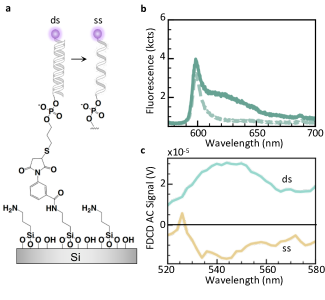
The metasurfaces were functionalized with self-assembled monolayers of the dye-DNA complexes with widely-used silane chemistry (Figure 3a).58, 59 Terminal amine groups on vapor-deposited films of (3-aminopropyl)trimethoxysilane were crosslinked to pre-hybridized thiol-modified DNA strands using m-maleimidobenzoyl-N-hydroxysuccinimide ester (See SI). Monolayers of DNA prepared by silanization on Si/SiO2 typically feature surface densities of 1012 molecules/cm2, which represents an approximate upper bound to the limit of detection of our technique.60, 61 To validate surface functionalization, metasurface fluorescence spectra were collected both before and after DNA assembly. Strong fluorescence near the long-pass edge at 600nm can be attributed to background fluorescence from the sapphire substrate. However, a significant increase in emission intensity from 610-650nm following functionalization confirms that fluorescence from the monolayer is distinguishable from that of the substrate (Figure 3b).
.
To detect the CD of the monolayers indirectly using fluorescence, we built a table-top polarization-sensitive spectrometer that performs a lock-in measurement to collect excitation spectra (see SI). For this measurement, we use a 635nm bandpass filter that transmits the fluorescence of the dye alone. First, we studied the FDCD signal dependence on the hybridization state of the DNA monolayers which, from solution/ensemble-CD measurements, reverses sign between double- and single-stranded forms. Monolayer-functionalized metasurfaces were mounted within a cuvette of 1x phosphate buffered saline (PBS) to maintain the helical tertiary structure of dsDNA when tethered to surfaces during measurements (Figure S3). Using a metasurface of =92nm functionalized with dye-dsDNA complexes, a positive FDCD signal is measured from 520-580nm (Figure 3c). Then, PBS was removed and replaced with formamide to lower the DNA melting temperature below room temperature. Upon denaturing, the FDCD signal reverses sign or is destroyed completely (Figures 3c, S4). Importantly, because the dye-functionalized ssDNA remains tethered to the surfaces, the change in FDCD signal can be attributed to partial or complete dehybridization of adsorbed DNA rather than removal of fluorescent probes from the surfaces.
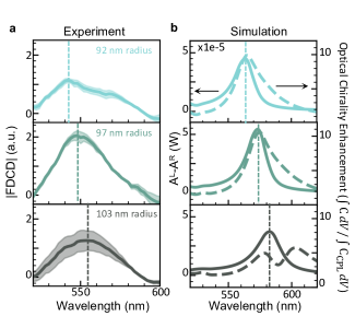
We repeat the double-stranded DNA measurements on the three metasurfaces characterized in Figure 1 to study the spectral influence of the metasurface resonance on the FDCD signal. These results (Figure 4a) show a red-shifting FDCD signal with increasing disk size. Consistent with in Figure 1, the peak is blue-shifted from the resonance center wavelength. In contrast, upon repeating the measurement on an identically-sized square of unpatterned silicon on the same sapphire substrate, no significant FDCD signal is observed (Figure S5), illustrating that the detected signal can primarily be attributed to the electromagnetic near-field enhancements. To confirm that the measured FDCD signal arises primarily from molecular circular dichroism, we perform full-field simulations, calculating A and within a 10-nm-thick chiral layer above the silicon disks. To account for coupling effects between induced electric and magnetic dipoles in the chiral medium, these simulations use a wavelength-independent Pasteur parameter with strength typical of an on-resonant chiral molecule (see SI). 32, 45, 34 Figure 4b shows these results, indicating that the peak in molecular A occurs just blue-shifted from the dip in transmission, and red shifts with increasing disk size, corresponding well with experiments. Further, the observed lineshape and spectral position of the simulated A are in good agreement with those of the optical chirality enhancement in the molecular layer, seen in the solid and dotted lines of Figure 4b, respectively. We note that the optical chirality calculated in the chiral layer on the 103-nm-metasurface exhibits two peaks, in contrast to the single peak in A, likely due to higher enhancements in electric field associated with the blue-shifted mode as the resonances separate. Importantly, A within the silicon nanodisks in simulation (Figure S6) is given by a bisignate lineshape not seen in our measurement. This result further indicates that the measured FDCD signal arises primarily from enhanced molecular CD due to high optical chirality density in the near field, as opposed to induced CD in the disks themselves.44 We attribute the non-negligible ( 20nm) red shift between simulation and experiment to a combination of an oxide layer on the silicon surface, tapering of disk side-walls, and wavelength-dependent chiral medium properties which were not captured in our model.
In summary, we demonstrate enhancement of intrinsic molecular CD in a monolayer via overlap of molecular CD with nanophotonic resonances using FDCD. We study the circular dichroism of 590-functionalized DNA strands, with a solution-phase CD signal from 500-650nm. Interestingly, the sign of this CD signal is dependent on DNA conformation, with dye-dsDNA exhibiting a positive signal and dye-ssDNA exhibiting a negative signal. The complexes are functionalized to silicon metasurfaces fabricated with optical resonances in the CD band of interest and characterized using a table-top FDCD spectrometer. We exploit the enantiomer-reversal-like behavior of the different conformations to probe surface chirality by de-hybridizing the dsDNA, observing a sign-reversal when metasurfaces are present but negligible signal in their absence. Finally, we show that the FDCD signal red shifts with increasing disk size, and thus that the metasurface’s resonant features are central to molecular CD enhancement. Full-field simulations confirm that both differential absorption and optical chirality density are enhanced within the chiral medium near the disk resonances. This indicates that the experimental FDCD enhancement is due to enhanced optical chirality density, as predicted in prior works, thus providing important insights toward efficient enantioselective photolysis.
1 Acknowledgements
The authors thank Dr. Mark Lawrence, Dr. Aitzol García-Etxarri, Dr. Michal Vadai, Dr. Claire McClellan, and David Barton for insightful discussions. The authors gratefully acknowledge the Gordon and Betty Moore Foundation for funding through a Moore Inventors Fellowship under grant number 6881, and the National Science Foundation under grant number 1905209. M.L.S. acknowledges a National Defense Science and Engineering graduate fellowship. L.V.P. acknowledges support from a Swiss National Science Foundation Early Postdoc Mobility fellowship under project number P2EZP2_181595. Work was performed in part in the nanoStanford labs (Stanford Nano Shared Facilities and the Stanford Nanoafabrication Facility), which are supported by the National Science Foundation as part of the National Nanotechnology Coordinated Infrastructure under award ECCS-1542152.
1.1 Supporting Information
Supporting Information contains materials and methods, a summary of findings of dye-DNA functionalized to unpatterned silicon, simulation details, and raw data and data analysis procedure, as well as additional data.
References
- Nguyen et al. 2006 Nguyen, L. A.; He, H.; Pham-Huy, C. Chiral drugs: an overview. International journal of biomedical science: IJBS 2006, 2, 85
- Jeschke 2018 Jeschke, P. Current status of chirality in agrochemicals. Pest management science 2018, 74, 2389–2404
- Kelly et al. 2005 Kelly, S. M.; Jess, T. J.; Price, N. C. How to study proteins by circular dichroism. Biochimica et Biophysica Acta (BBA)-Proteins and Proteomics 2005, 1751, 119–139
- Zhao et al. 2017 Zhao, Y.; Askarpour, A. N.; Sun, L.; Shi, J.; Li, X.; Alù, A. Chirality detection of enantiomers using twisted optical metamaterials. Nature communications 2017, 8, 1–8
- Kuhn and Braun 1929 Kuhn, W.; Braun, E. Photochemische erzeugung optisch aktiver stoffe. Naturwissenschaften 1929, 17, 227–228
- Feringa and Wynberg 1978 Feringa, B.; Wynberg, H. Biomimetic asymmetric oxidative coupling of phenols. Bioorganic Chemistry 1978, 7, 397–408
- Inoue 1992 Inoue, Y. Asymmetric photochemical reactions in solution. Chemical reviews 1992, 92, 741–770
- Flores et al. 1977 Flores, J. J.; Bonner, W. A.; Massey, G. A. Asymmetric photolysis of (RS)-leucine with circularly polarized ultraviolet light. Journal of the American Chemical Society 1977, 99, 3622–3625
- Balavoine et al. 1974 Balavoine, G.; Moradpour, A.; Kagan, H. Preparation of chiral compounds with high optical purity by irradiation with circularly polarized light, a model reaction for the prebiotic generation of optical activity. Journal of the American Chemical Society 1974, 96, 5152–5158
- Lin et al. 2014 Lin, L.; Wang, T.; Lu, Z.; Liu, M.; Guo, Y. In situ measurement of the supramolecular chirality in the langmuir monolayers of achiral porphyrins at the air/aqueous interface by second harmonic generation linear dichroism. The Journal of Physical Chemistry C 2014, 118, 6726–6733
- Burke et al. 2003 Burke, B. J.; Moad, A. J.; Polizzi, M. A.; Simpson, G. J. Experimental confirmation of the importance of orientation in the anomalous chiral sensitivity of second harmonic generation. Journal of the American Chemical Society 2003, 125, 9111–9115
- Petralli-Mallow et al. 1993 Petralli-Mallow, T.; Wong, T.; Byers, J.; Yee, H.; Hicks, J. Circular dichroism spectroscopy at interfaces: a surface second harmonic generation study. The Journal of Physical Chemistry 1993, 97, 1383–1388
- Hassey-Paradise et al. 2009 Hassey-Paradise, R.; Cyphersmith, A.; Tilley, A. M.; Mortsolf, T.; Basak, D.; Venkataraman, D.; Barnes, M. D. Dissymmetries in fluorescence excitation and emission from single chiral molecules. Chirality: The Pharmacological, Biological, and Chemical Consequences of Molecular Asymmetry 2009, 21, E265–E276
- Cyphersmith et al. 2012 Cyphersmith, A.; Surampudi, S.; Casey, M. J.; Jankowski, K.; Venkataraman, D.; Barnes, M. D. Chiroptical dissymmetries in fluorescence excitation from single molecules of (M-2) helicene dimers. The Journal of Physical Chemistry A 2012, 116, 5349–5352
- Tang and Cohen 2011 Tang, Y.; Cohen, A. E. Enhanced enantioselectivity in excitation of chiral molecules by superchiral light. Science 2011, 332, 333–336
- Tang and Cohen 2010 Tang, Y.; Cohen, A. E. Optical chirality and its interaction with matter. Physical Review Letters 2010, 104, 163901
- Gansel et al. 2009 Gansel, J. K.; Thiel, M.; Rill, M. S.; Decker, M.; Bade, K.; Saile, V.; von Freymann, G.; Linden, S.; Wegener, M. Gold helix photonic metamaterial as broadband circular polarizer. Science 2009, 325, 1513–1515
- Poulikakos et al. 2018 Poulikakos, L. V.; Thureja, P.; Stollman, A.; Leo, E. D.; Norris, D. J. Chiral Light Design and Detection Inspired by Optical Antenna Theory. Nano Letters 2018, 18, 4633–4640
- Esposito et al. 2015 Esposito, M.; Tasco, V.; Cuscuná, M.; Todisco, F.; Benedetti, A.; Tarantini, I.; de Giorgi, M.; Sanvitto, D.; Passaseo, A. Nanoscale 3D chiral plasmonic helices with circular dichroism at visible frequencies. ACS Photonics 2015, 2, 105–114
- Ferry et al. 2015 Ferry, V. E.; Hentschel, M.; Alivisatos, A. P. Circular dichroism in off-resonantly couple plasmonic nanosystems. Nano Letters 2015, 15, 8336–8341
- Duan et al. 2015 Duan, X.; Yue, S.; Liu, N. Understanding complex chiral plasmonics. Nanoscale 2015, 7, 17237–17243
- Yao and Liu 2018 Yao, K.; Liu, Y. Enhancing circular dichroism by chiral hotspots in silicon nanocube dimers. Nanoscale 2018, 10, 8879–8786
- Kuzyk et al. 2012 Kuzyk, A.; Schreiber, R.; Fan, Z.; Pardatscher, G.; Roller, E.-M.; Högele, A.; Simmel, F. C.; Govorov, A. O.; Liedl, T. DNA-based self-assembly of chiral plasmonic nanostructures with tailored optical response. Nature 2012, 483, 311–314
- Govorov et al. 2010 Govorov, A. O.; Fan, Z.; Hernandez, P.; Slocik, J. M.; Rajesh R, N. Theory of circular dichroism of nanomaterials comprising chiral molecules and nanocrystals: plasmon enhancement, dipole interactions, and dielectric effects. Nano Letters 2010, 10, 1374–1382
- Wang et al. 2015 Wang, L.-Y.; Smith, K. W.; Dominguez-Medina, S.; Moody, N.; Olson, J. M.; Zhang, H.; Chang, W.-S.; Kotov, N.; Link, S. Circular differential scattering of single chiral self-assembled gold nanorod dimers. ACS Photonics 2015, 2, 1602–1610
- Zhao et al. 2012 Zhao, Y.; Belkin, M. A.; Alù, A. Twisted optical metamaterials for planarized ultrathin broadband circular polarizers. Nature communications 2012, 3, 1–7
- Schäferling et al. 2014 Schäferling, M.; Yin, X.; Engheta, N.; Giessen, H. Helical plasmonic nanostructures as prototypical chiral near-field sources. ACS Photonics 2014, 1, 530–537
- Schäferling et al. 2012 Schäferling, M.; Dregely, D.; Hentschel, M.; Giessen, H. Tailoring enhanced optical chirality: Design principles for chiral plasmonic nanostructures. Physical Review X 2012, 2, 031010
- Hendry et al. 2010 Hendry, E.; Carpy, T.; Johnston, J.; Popland, M.; Mikhaylovskiy, R.; Lapthorn, A.; Kelly, S.; Barron, L.; Gadegaard, N.; Kadodwala, M. Ultrasensitive detection and characterization of biomolecules using superchiral fields. Nature Nanotechnology 2010, 5, 783–787
- Hendry et al. 2012 Hendry, E.; Mikhaylovskiy, R.; Barron, L.; Kadodwala, M.; Davis, T. Chiral electromagnetic fields generated by arrays of nanoslits. Nano Letters 2012, 12, 3640–3644
- Kneer et al. 2018 Kneer, L. M.; Roller, E.-M.; Besteiro, L. V.; Schreiber, R.; Govorov, A. O.; Liedl, T. Circular dichroism of chiral molecules in DNA-assembled plasmonic hotspots. ACS nano 2018, 12, 9110–9115
- García-Guirado et al. 2018 García-Guirado, J.; Svedendahl, M.; Puigdollers, J.; Quidant, R. Enantiomer-selective molecular sensing using racemic nanoplasmonic arrays. Nano letters 2018, 18, 6279–6285
- Tullius et al. 2015 Tullius, R.; Karimullah, A. S.; Rodier, M.; Fitzpatrick, B.; Gadegaard, N.; Barron, L. D.; Rotello, V. M.; Cooke, G.; Lapthorn, A.; Kadodwala, M. “Superchiral” spectroscopy: detection of protein higher order hierarchical structure with chiral plasmonic nanostructures. Journal of the American Chemical Society 2015, 137, 8380–8383
- Nesterov et al. 2016 Nesterov, M. L.; Yin, X.; Schäferling, M.; Giessen, H.; Weiss, T. The role of plasmon-generated near fields for enhanced circular dichroism spectroscopy. Acs Photonics 2016, 3, 578–583
- García-Extarri and Dionne 2013 García-Extarri, A.; Dionne, J. Surface-enhanced circular dichroism spectroscopy mediated by nonchiral nanoantennas. Physical Review B 2013, 87, 235409
- Ho et al. 2017 Ho, C.-S.; García-Extarri, A.; Zhao, Y.; Dionne, J. Enhancing enantioselective absorption using dielectric nanospheres. ACS Photonics 2017, 4, 197–203
- Solomon et al. 2019 Solomon, M. L.; Hu, J.; Lawrence, M.; García-Etxarri, A.; Dionne, J. A. Enantiospecific optical enhancement of chiral sensing and separation with dielectric metasurfaces. ACS Photonics 2019, 6, 43–49
- Hu et al. 2020 Hu, J.; Lawrence, M.; Dionne, J. A. High Quality Factor Dielectric Metasurfaces for Ultraviolet Circular Dichroism Spectroscopy. ACS Photonics 2020, 7, 36–42
- Mohammadi et al. 2018 Mohammadi, E.; Tsakmakidis, K. L.; Askarpour, A. N.; Dehkhoda, P.; Tavakoli, A.; Altug, H. Nanophotonic platforms for enhanced chiral sensing. ACS Photonics 2018, 5, 2669–2675
- Ben-Moshe et al. 2013 Ben-Moshe, A.; Maoz, B. M.; Govorov, A. O.; Markovich, G. Chirality and chiroptical effects in inorganic nanocrystal systems with plasmon and exciton resonances. Chemical Society Reviews 2013, 42, 7028–7041
- Zhang et al. 2017 Zhang, W.; Wu, T.; Wang, R.; Zhang, X. Amplification of the molecular chiroptical effect by low-loss dielectric nanoantennas. Nanoscale 2017, 9, 5701–5707
- Balavoine et al. 1974 Balavoine, G.; Moradpour, A.; Kagan, H. Preparation of Chiral Compounds with High Optical Purity by Irradiation with Circularly Polarized Light, a Model Reaction for the Prebiotic Generation of Optical Activity. Journal of the American Chemical Society 1974, 96, 5152–5158
- Lee et al. 2017 Lee, S.; Yoo, S.; Park, Q.-H. Microscopic origin of surface-enhanced circular dichroism. ACS Photonics 2017, 4, 2047–2052
- Garcia-Guirado et al. 2019 Garcia-Guirado, J.; Svedendahl, M.; Puigdollers, J.; Quidant, R. Enhanced chiral sensing with dielectric nano-resonators. Nano Letters 2019,
- Mohammadi et al. 2019 Mohammadi, E.; Tavakoli, A.; Dehkhoda, P.; Jahani, Y.; Tsakmakidis, K. L.; Tittl, A.; Altug, H. Accessible superchiral near-fields driven by tailored electric and magnetic resonances in all-dielectric nanostructures. ACS Photonics 2019, 6, 1939–1946
- Graf et al. 2019 Graf, F.; Feis, J.; Garcia-Santiago, X.; Wegener, M.; Rockstuhl, C.; Fernandez-Corbaton, I. Achiral, helicity preserving, and resonant structures for enhanced sensing of chiral molecules. ACS Photonics 2019, 6, 482–491
- Staude et al. 2013 Staude, I.; Miroshnichenko, A. E.; Decker, M.; Fofang, N. T.; Liu, S.; Gonzales, E.; Dominguez, J.; Luk, T. S.; Neshev, D. N.; Brener, I.; Kivshar, Y. Tailoring directional scattering through magnetic and electrical resonances in subwavelength silicon nanodisks. ACS Nano 2013, 7, 7824–7832
- Siday et al. 2019 Siday, T.; Vabishchevich, P. P.; Hale, L.; Harris, C. T.; Luk, T. S.; Reno, J. L.; Brener, I.; Mitrofanov, O. Terahertz detection with perfectly-absorbing photoconductive metasurface. Nano Letters 2019, 19, 2888–2896
- Yang et al. 2018 Yang, C.-Y.; Yang, J.-H.; Yang, Z.-Y.; Zhou, Z.-X.; Sun, M.-G.; Babicheva, V. E.; Chen, K.-P. Nonradiating silicon nanoantenna metasurfaces as narrowband absorbers. Acs Photonics 2018, 5, 2596–2601
- Decker et al. 2015 Decker, M.; Staude, I.; Falkner, M.; Dominguez, J.; Neshev, D. N.; Brener, I.; Pertsch, T.; Kivshar, Y. S. High-efficiency dielectric Huygens’ surfaces. Advanced Optical Materials 2015, 3, 813–820
- Decker and Staude 2016 Decker, M.; Staude, I. Resonant dielectric nanostructures: a low-loss platform for functional nanophotonics. Journal of Optics 2016, 18, 103001
- Barron 2009 Barron, L. D. Molecular Light Scattering and Optical Activity; Cambridge University Press, 2009
- Kringle et al. 2018 Kringle, L.; Sawaya, N. P.; Widom, J.; Adams, C.; Raymer, M. G.; Aspuru-Guzik, A.; Marcus, A. H. Temperature-dependent conformations of exciton-coupled Cy3 dimers in double-stranded DNA. The Journal of chemical physics 2018, 148, 085101
- Nordén and Tjerneld 1982 Nordén, B.; Tjerneld, F. Structure of methylene blue–DNA complexes studied by linear and circular dichroism spectroscopy. Biopolymers: Original Research on Biomolecules 1982, 21, 1713–1734
- Neelakandan et al. 2014 Neelakandan, P. P.; Zeidan, T. A.; McCullagh, M.; Schatz, G. C.; Vura-Weis, J.; Kim, C. H.; Wasielewski, M. R.; Lewis, F. D. Ground and excited state electronic spectra of perylenediimide dimers with flexible and rigid geometries in DNA conjugates. Chemical Science 2014, 5, 973–981
- Tuite and Nordén 2018 Tuite, E. M.; Nordén, B. Linear and circular dichroism characterization of thionine binding mode with DNA polynucleotides. Spectrochimica Acta Part A: Molecular and Biomolecular Spectroscopy 2018, 189, 86–92
- Stokes et al. 2007 Stokes, G. Y.; Gibbs-Davis, J. M.; Boman, F. C.; Stepp, B. R.; Condie, A. G.; Nguyen, S. T.; Geiger, F. M. Making “sense” of DNA. Journal of the American Chemical Society 2007, 129, 7492–7493
- Nakatsuka et al. 2018 Nakatsuka, N.; Yang, K.-A.; Abendroth, J. M.; Cheung, K. M.; Xu, X.; Yang, H.; Zhao, C.; Zhu, B.; Rim, Y. S.; Yang, Y.; Weiss, P. S.; Stojanović, M. N.; Andrews, A. M. Aptamer–field-effect transistors overcome Debye length limitations for small-molecule sensing. Science 2018, 362, 319–324
- Cheung et al. 2020 Cheung, K. M.; Abendroth, J. M.; Nakatsuka, N.; Zhu, B.; Yang, Y.; Andrews, A. M.; Weiss, P. S. Detecting DNA and RNA and Differentiating Single-Nucleotide Variations via Field-Effect Transistors. Nano letters 2020,
- Strother et al. 2000 Strother, T.; Cai, W.; Zhao, X.; Hamers, R. J.; Smith, L. M. Synthesis and characterization of DNA-modified silicon (111) surfaces. Journal of the American Chemical Society 2000, 122, 1205–1209
- Pirrung 2002 Pirrung, M. C. How to make a DNA chip. Angewandte Chemie International Edition 2002, 41, 1276–1289