Molecular beam epitaxy growth and surface structure of Sr1-xNdxCuO2 cuprate films
Abstract
We report epitaxial growth and surface structure of infinite-layer cuprate Sr1-xNdxCuO2 films on SrTiO3(001) substrates by combining ozone-assisted molecular beam epitaxy and in-situ scanning tunneling microscopy. Careful substrate temperature and flux control has been used to achieve single phase, stoichiometric and -axis oriented films. The surface of the films is usually characterized by mixed CuO2 surface and grid-like superstructure. The superstructure exhibits a periodicity of 3.47 nm that corresponds to a coincidence lattice between overlayer peroxide SrO2 and underlying CuO2 plane, and gives rise to conductance spectrum that is distinct from the Mott-Hubbard band structure of CuO2. At higher Nd composition 0.1, a surface characteristic of the hole-doped CuO2 emerges, which we ascribe to the intake of apical oxygens in the intervening Sr planes.
Infinite-layer (IL) CuO2 ( = Ca, Sr, Ba) compounds exhibit the simplest crystal structure among cuprates, in which the major superconducting CuO2 is alternatively separated by alkaline earth cations along the crystallographic -axis Siegrist et al. (1988). Partial substitution of divalent ions by trivalent ions such as La3+ and Nd3+ leads to electron-doped superconductivity with a record transition temperature of 43 K Smith et al. (1991); Chen et al. (2002); Armitage et al. (2010). More remarkably, IL compounds represent a rare category of cuprate superconductors with surface termination of the superconducting CuO2 planes Koguchi et al. (1995); Harter et al. (2015); Zhong et al. (2018). Given that most cuprates are terminated with non-CuO2 charge reservoir layers upon cleaving, e.g. BiO for bismuth-based cuprates, this peculiar feature provides an unprecedented opportunity to directly characterize the superconducting CuO2 planes by surface-sensitive experiments Zhong et al. (2020), compared to previous studies Damascelli et al. (2003); Fischer et al. (2007); Ye et al. (2013). A systematic direct measurement of the major CuO2 planes may help understand eventually the microscopic mechanism of high- superconductivityChen et al. (2002); Zhong et al. (2020); Harter et al. (2012); Misra et al. (2002); Lv et al. (2015, 2016); Zhong et al. (2016). However, IL cuprates with tetragonal structure are thermodynamically unstable. It is nearly impossible to synthesize single crystals by conventional solid state methods, and only some powder form of IL samples was obtained using high pressure techniquesTakano et al. (1989); Er et al. (1994).
Epitaxial films of IL cuprates can be stabilized and prepared on appropriate substrates by using pulsed laser deposition (PLD)Gupta et al. (1994); Leca et al. (2006); Tomaschko et al. (2012) or reactive molecular beam epitaxy (MBE) techniqueKarimoto et al. (2001); Krockenberger and Yamamoto (2011); Krockenberger et al. (2012); Ikeda et al. (2019). However, the as-grown thin films are often characterized with several competing phases, such as Sr2CuO3, Sr14Cu24O41 and orthorhombic SrCuO2 Krockenberger et al. (2018), as summarized in Table I. Furthermore, due to the limited solubility of trivalent ions in IL compounds, oxygen-deficient or -redundant superstructures with a relatively larger out-of-plane lattice parameter, referred as a long-c phase, occur at elevated doping Zhong et al. (2020); Leca et al. (2006); Karimoto et al. (2001); Gupta et al. (1994). In this study, we combine ozone-assisted MBE and in-situ scanning tunneling microscopy (STM) to solve these problems, aiming to establish growth procedures for single phase crystalline Sr1-xNdxCuO2 (SNCO, 0.08 0.12) thin films. We emphasize that, compared to alternative shutter-controlled deposition, our method for composition/phase control is self-regulated, without the complicated calibration of the composition by shutter time.
The experiments were performed on a commercial ultrahigh vacuum (UHV) STM apparatus (Unisoku), connected to an ozone-assisted MBE chamber for in-situ film growth. Nb-doped SrTiO3(001) substrates were firstly degassed at 600C, and subsequently annealed at 1250C under UHV for 20 minutes to get the clean surface. Prior to film epitaxy, fluxes of all metal sources (Sr, Nd and Cu) were precisely calibrated in sequence by using a standard crystal microbalance (QCM, Inficon SQM160H). Epitaxial thin films were then prepared by co-deposition of high-purity metal sources from standard Knudsen cells under an ozone flux beam of 1.1 10-5 Torr. The growth rate is 0.4 unit cell per minute, and the flux ratio between Nd and Cu sources is used to calculate the nominal composition . Polycrystalline PtIr tips were cleaned by electron-beam heating and calibrated on MBE-grown Ag/Si(111) films. Tunneling spectra were measured using a standard lock-in technique with a small bias modulation of 10 mV at 937 Hz. After in-situ STM measurements at 78 K, the samples were taken out from the UHV chamber for X-ray diffraction (XRD) measurements using the monochromatic Cu Kα1 radiation with a wavelength of 1.5406 Å.
| Space group | a(Å) | b(Å) | c(Å) | Ref | |
|---|---|---|---|---|---|
| IL tetragonal SrCuO2 | P4/mmm | 3.9269 | = a | 3.4346 | Siegrist et al. (1988) |
| Orthorhombic SrCuO2 | Cmcm | 3.5770 | 16.342 | 3.9182 | Matsushita et al. (1995) |
| Orthorhombic Sr2CuO3 | Immm | 12.702 | 3.911 | 3.4990 | Hyatt et al. (2004) |
| Orthorhombic Sr14Cu24O41 | Amma | 11.488 | 13.414 | 27.428 | Abbamonte et al. (2004) |
| Tetragonal SrO2 | I4/mmm | 3.55 | = a | 6.55 | Middleburgh et al. (2013) |
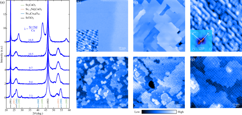
Growth of IL SNCO epitaxial films demands for precise control of the substrate temperature and cation stoichiometry. Similar to previous reports Mihailescu et al. (2014), we found that tetragonal IL SNCO films start to crystallize at 500 and change to orthorhombic phase above 610. Thus, = 550 was chosen for both good crystallinity and avoiding high temperature orthorhombic phase. Figure 1(a) shows the XRD patterns of as-grown films as a function of the nominal flux ratio = (Sr+Nd)/Cu, with a smaller Nd/Cu flux ratio of 0.10. Apparently, IL SNCO phase coexists with Sr-deficient spin ladder Sr14Cu24O41 at lower of 7.3. This is understandable because Sr has a higher vapor pressure of 1.8 10-2 Torr and is very volatile at = 550. Meanwhile, Sr is easily oxidized in ozone atmosphere, which reduces its effective flux during the growth. The two factors explain why a larger 9.4 is required to prepare single phase IL films, as demonstrated by the XRD spectra in Fig. 1(a). Evidently, the cation stoichiometry of SNCO is quasi-self-regulating, resembling, to some extent, the growth of GaAs and metal chalcogenides Li et al. (2010); Song et al. (2011). We note that the self-regulation of stoichiometry is somewhat limited and the IL SNCO phase forms only in a narrow window of . A larger of 16.8 converts the epitaxial films to a more thermodynamically stable Sr2CuO3 phase [see Fig. 1(a)].
Our STM characterization corroborates the flux-ratio-dependent phase evolution. At = 7.3, the chain-like surface characteristic of spin ladder Sr14Cu24O41(010) occurs [Fig. 1(b)], whereas single phase Sr2CuO3 overwhelms the others under Sr-rich condition [Figs. 1(c) and 1(d)]. Fast Fourier transform (FFT) analysis inserted in Fig. 1(d) indicates that the in-plane lattice constants are b = 3.9 0.1 Å and c = 3.5 0.1 Å, consistent with the expected value for orthorhombic Sr2CuO3(100) surface in Table I. The single phase IL SNCO films are prepared at an intermediate and display atomically flat surface [Figs. 1(e)-1(g)], which are separated by gird-like superstructure. The grid-like feature gradually becomes prominent with increasing and covers the whole surface at 10.5.
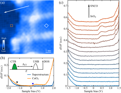
To identify the two apparently distinct surfaces of IL SNCO films, we acquire atomically-resolved STM images, as illustrated in Fig. 2(a). The flat surface has a square lattice with a periodicity of 3.9 Å, matching well CuO2-terminated IL SNCO Smith et al. (1991); Zhong et al. (2020). This is indeed supported by the site-dependent differential conductance dI/dV spectra in Fig. 2(b). On the flat surface, the tunneling dI/dV spectrum features a fundamental Mott-Hubbard band structure of the cuprate CuO2 planes, accompanied by metallic-like states within the charge-transfer gap Zhong et al. (2020). It is worth noting that the Fermi level is closer to the upper Hubband (UHB) than the charge-transfer band (CTB), in line with the electron doping by the Nd3+ substitution for Sr2+ ions.
In contrast, the grid-like superstructure is characterized by a larger in-plane unit cell of 5.0 Å (marked by the white square), rotated by 45 relative to the CuO2 unit cell in Fig. 2(a). A possible surface reconstruction of SNCO(001) R45 could be safely excluded since the measured periodicity of 5.0 Å deviates substantially from the times ( 5.6 Å) of in-plane lattice constant of SNCO. Moreover, tunneling dI/dV spectrum of gird-like superstructure shows an extremely large band gap ( 2.8 eV) and is significantly different from that of CuO2 plane [Fig. 2(b)]. This is confirmed by the linecut dI/dV spectra across one step edge between the grid-like superstructure and the CuO2 surface in Fig. 2(c). These observations, together with the populated gird-like superstructure at elevated [Figs. 1(e)-1(g)], strongly suggest that the superstructure originates from a totally different compound, most probably linking with strontium. Tetragonal strontium peroxide SrO2 has a lattice constant of 3.55 Å in the a-b plane (Table I) Middleburgh et al. (2013), coinciding with 1/ of the measured unit cell periodicity of 5.0 Å in Fig. 2(a). In other words, the grid-like surface might correspond to SrO2 in nature, which exhibits an enlarged surface structure, i.e. SrO2(001) R45. Considering that no excess phase other than IL SNCO is found in the bulk-sensitive XRD spectra at intermediate [Fig. 1(a)], the SrO2 ought to occur only at the topmost CuO2 surface of epitaxial SNCO films.
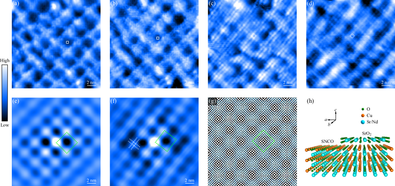
By acquiring bias-dependent STM images with atomic-scale resolution in Figs. 3(a)-3(d), we further confirm this conclusion for the grid-like superstructure. Intriguingly, the SrO2(001) R45 surface switches to SrO2(001) structure as the bias polarity is reversed from negative to positive. This hints that the emergent surface structures, irrespective of and , may most likely stem from charger ordering in SrO2 Renner et al. (2002); Iwaya et al. (2011). The surface structure switching should be due to a bias-dependent lateral variation of local density of states in SrO2 Maroutian et al. (2003), which requires further theoretical investigations. Notwithstanding, the grid-like superstructure remains unchanged in both dimension and orientation. The measured periodicity is 34.7 1.4 Å on average, which is approximately 10 times the Sr-Sr atom spacing ( 3.55 Å) in SrO2 according to the autocorrelation analysis in Figs. 3(e) and 3(f). Additionally, the possible charge ordering of SrO2 is apparently visible (see the white dashes) in Fig. 3(f) that enables to deduce the zero angle of intersection between the respective lattices of SrO2 and grid-like superstructure. Note that the latter periodicity of 34.7 1.4 Å coincides nicely with 9 times of the lattice constant of SNCO films Smith et al. (1991); Bobrovskii et al. (1997), a coincidence lattice between the SrO2 overlayer and CuO2-terminated SNCO films is proposed to be responsible for the grid-like superstructure [Figs. 3(g) and 3(h)]. Figure 3(g) illustrates a simulated Moiré pattern by reasonably assuming = 3.55 Å and = 3.94 Å, which matches well our results [Figs. 3(e) and 3(f)].
The coincidence lattice for the superstructure, rather than a simple topographic Moiré pattern between the SrO2 overlayer and underlying CuO2, is based on two experimental findings. One is the significant dependence of the apparent corrugation of grid-like superstructure on the applied sample voltage in Figs. 3(a)-3(d). For example, the corrugation of superstructure is more apparent at negative biases. The other finding relates to the local distortion in the grid-like superstructure and the accompanying charge ordering, which is unexpected for Moiré pattern. Instead, it can be the local structural distortion in coincidence lattice to yield the bias-dependent corrugation, distorted superstructure and charge ordering.
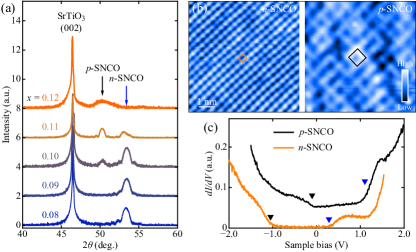
Next we explore the dependence of SNCO films on the nominal composition of Nd. As shown in Fig. 4(a) are five XRD spectra of IL SNCO films at varied . Analogous to La-doped Sr1-xLaxCuO2 (SLCO) IL epitaxial films Zhong et al. (2020), a second phase with a larger -axis lattice constant emerges at 0.1, coexists and becomes dominant with increasing . The emergent new phase is characteristic of CuO2(001) surface structure [Fig. 4(b)] and exhibits a hole-doped behavior with the closer to CTB [see the black curve in Fig. 4(c)], which we dub as -SNCO. In contrast, the electron doped -SNCO films always display a bare CuO2(001) surface, even in the two-phase coexisting SNCO films for = 0.12 [Figs. 4(b) and 4(c)]. Without loss of generality, we attribute the CuO2(001) surface reconstruction and emergent -type behavior in SNCO films as the considerable incorporation of apical oxygens in the intervening Sr planesZhong et al. (2020), which overwhelm the electron doping by Nd3+ donors. In any case, the observed tunneling dI/dV spectra are of striking resemblance, except for an energy shift in . This echoes the self-modulation doping schemeZhong et al. (2020), namely doping the intervening Sr layers changes the fundamental Mott-Hubbard band structure of CuO2(001) little.
Finally we comment on implication from the observed SrO2 overlayers. Based on the step height in Fig. 2(a), we readily estimate the thickness of SrO2 overlayer, to wit, only half of unit cell ( 3.3 Å). Evidently, the top SrO2 layer is insulating and exhibits a large semiconducting gap of 2.8 eV [Fig. 2(b)]. Notably, the surface stacking of one SrO2 layer on CuO2 is structurally similar to the BiO-terminated Bi2Sr2CaCu2O8+δ Damascelli et al. (2003); Fischer et al. (2007); Misra et al. (2002), i.e. insulating Sr(Bi) oxides on CuO2. Here, the measured dI/dV spectra appear sharply different between SrO2 and CuO2, and thus how the cuprate database from the vacuum-cleaved BiO planes represents the spectral properties of buried CuO2 merits further investigations.
Acknowledgements.
The work is financially supported by the Ministry of Science and Technology of China (Grants No. 2017YFA0304600, No. 2018YFA0305603), the National Natural Science Foundation of China (Grants No. 11774192, No. 11634007), and in part by Beijing Innovation Center for Future Chips, Tsinghua University.References
- Siegrist et al. (1988) T. Siegrist, S. M. Zahurak, D. W. Murphy, and R. S. Roth, Nature 334, 231 (1988).
- Smith et al. (1991) M. G. Smith, A. Manthiram, J. S. Zhou, J. B. Goodenough, and J. T. Markert, Nature 351, 549 (1991).
- Chen et al. (2002) C. T. Chen, P. Seneor, N. C. Yeh, R. P. Vasquez, L. D. Bell, C. U. Jung, J. Y. Kim, M.-S. Park, H.-J. Kim, and S. I. Lee, Phys. Rev. Lett. 88, 227002 (2002).
- Armitage et al. (2010) N. P. Armitage, P. Fournier, and R. L. Greene, Rev. Mod. Phys. 82, 2421 (2010).
- Koguchi et al. (1995) K. Koguchi, T. Matsumoto, and T. Kawai, Science 267, 71 (1995).
- Harter et al. (2015) J. W. Harter, L. Maritato, D. E. Shai, E. J. Monkman, Y. Nie, D. G. Schlom, and K. M. Shen, Phys. Rev. B 92, 035149 (2015).
- Zhong et al. (2018) Y. Zhong, S. Han, Y. Wang, Z. Luo, D. Zhang, L. Wang, W. Li, K. He, C. L. Song, X. C. Ma, and Q. K. Xue, Phys. Rev. B 97, 245420 (2018).
- Zhong et al. (2020) Y. Zhong, J.-Q. Fan, R.-F. Wang, S. Wang, X. Zhang, Y. Zhu, Z. Dou, X.-Q. Yu, Y. Wang, D. Zhang, J. Zhu, C.-L. Song, X.-C. Ma, and Q.-K. Xue, Phys. Rev. Lett. 125, 077002 (2020).
- Damascelli et al. (2003) A. Damascelli, Z. Hussain, and Z. X. Shen, Rev. Mod. Phys. 75, 473 (2003).
- Fischer et al. (2007) Ø. Fischer, M. Kugler, I. Maggio-Aprile, C. Berthod, and C. Renner, Rev. Mod. Phys. 79, 353 (2007).
- Ye et al. (2013) C. Ye, P. Cai, R. Yu, X. Zhou, W. Ruan, Q. Liu, C. Jin, and Y. Wang, Nat. Commun. 4, 1365 (2013).
- Harter et al. (2012) J. W. Harter, L. Maritato, D. E. Shai, E. J. Monkman, Y. Nie, D. G. Schlom, and K. M. Shen, Phys. Rev. Lett. 109, 267001 (2012).
- Misra et al. (2002) S. Misra, S. Oh, D. J. Hornbaker, T. DiLuccio, J. N. Eckstein, and A. Yazdani, Phys. Rev. Lett. 89, 087002 (2002).
- Lv et al. (2015) Y. F. Lv, W. L. Wang, J. P. Peng, H. Ding, Y. Wang, L. Wang, K. He, S. H. Ji, R. Zhong, J. Schneeloch, G. D. Gu, C. L. Song, X. C. Ma, and Q. K. Xue, Phys. Rev. Lett. 115, 237002 (2015).
- Lv et al. (2016) Y. F. Lv, W. L. Wang, H. Ding, Y. Wang, Y. Ding, R. Zhong, J. Schneeloch, G. D. Gu, L. Wang, K. He, S. H. Ji, L. Zhao, X. J. Zhou, C. L. Song, X. C. Ma, and Q. K. Xue, Phys. Rev. B 93, 140504 (2016).
- Zhong et al. (2016) Y. Zhong, Y. Wang, S. Han, Y. F. Lv, W. L. Wang, D. Zhang, H. Ding, Y. M. Zhang, L. Wang, K. He, R. D. Zhong, J. A. Schneeloch, G. D. Gu, C. L. Song, X. C. Ma, and Q. K. Xue, Sci. Bull. 61, 1239 (2016).
- Takano et al. (1989) M. Takano, Y. Takeda, H. Okada, M. Miyamoto, and T. Kusaka, Physica C 159, 375 (1989).
- Er et al. (1994) G. Er, S. Kikkawa, and F. Kanamaru, Physica C 235, 983 (1994).
- Gupta et al. (1994) A. Gupta, B. Mercey, H. Hervieu, and B. Raveau, Chem. Mater. 6, 1011 (1994).
- Leca et al. (2006) V. Leca, D. H. A. Blank, G. Rijnders, S. Bals, and G. Van Tendeloo, App. Phys. Lett. 89, 092504 (2006).
- Tomaschko et al. (2012) J. Tomaschko, V. Leca, T. Selistrovski, S. Diebold, J. Jochum, R. Kleiner, and D. Koelle, Phys. Rev. B 85, 024519 (2012).
- Karimoto et al. (2001) S.-i. Karimoto, K. Ueda, M. Naito, and T. Imai, App. Phys. Lett. 79, 2767 (2001).
- Krockenberger and Yamamoto (2011) Y. Krockenberger and H. Yamamoto, Physica C 471, 185 (2011).
- Krockenberger et al. (2012) Y. Krockenberger, K. Sakuma, and H. Yamamoto, App. Phys. Express 5, 043101 (2012).
- Ikeda et al. (2019) A. Ikeda, Y. Krockenberger, and H. Yamamoto, Phys. Rev. Materials 3, 064803 (2019).
- Krockenberger et al. (2018) Y. Krockenberger, A. Ikeda, K. Kumakura, and H. Yamamoto, J. App. Phys. 124, 073905 (2018).
- Matsushita et al. (1995) Y. Matsushita, Y. Oyama, M. Hasegawa, and H. Takei, Journal of Solid State Chemistry 114, 289 (1995).
- Hyatt et al. (2004) N. C. Hyatt, L. Gray, I. Gameson, P. P. Edwards, and S. Hull, Phys. Rev. B 70, 214101 (2004).
- Abbamonte et al. (2004) P. Abbamonte, G. Blumberg, A. Rusydi, A. Gozar, P. Evans, T. Siegrist, L. Venema, H. Eisaki, E. D. Isaacs, and G. A. Sawatzky, Nature 431, 1078 (2004).
- Middleburgh et al. (2013) S. C. Middleburgh, K. P. D. Lagerlof, and R. W. Grimes, J. Am. Ceram. Soc. 96, 308 (2013).
- Mihailescu et al. (2014) C. N. Mihailescu, I. Pasuk, M. Straticiuc, C. R. Nita, D. Pantelica, and J. Giapintzakis, Appl. Surf. Sci. 320, 852 (2014).
- Li et al. (2010) Y. Y. Li, G. Wang, X. G. Zhu, M. H. Liu, C. Ye, X. Chen, Y. Y. Wang, K. He, L. L. Wang, X. C. Ma, H. J. Zhang, X. Dia, Z. Fang, X. C. Xie, Y. Liu, X. L. Qi, J. F. Jia, S. C. Zhang, and Q. K. Xue, Adv. Mater. 22, 4002 (2010).
- Song et al. (2011) C. L. Song, Y. L. Wang, Y. P. Jiang, Z. Li, L. Wang, K. He, X. Chen, X. C. Ma, and Q. K. Xue, Phys. Rev. B 84, 020503 (2011).
- Renner et al. (2002) C. Renner, G. Aeppli, B.-G. Kim, Y.-A. Soh, and S.-W. Cheong, Nature 416, 518 (2002).
- Iwaya et al. (2011) K. Iwaya, R. Shimizu, T. Ohsawa, T. Hashizume, and T. Hitosugi, Phys. Rev. B 83, 125117 (2011).
- Maroutian et al. (2003) T. Maroutian, S. Degen, C. Becker, K. Wandelt, and R. Berndt, Phys. Rev. B 68, 155414 (2003).
- Bobrovskii et al. (1997) V. Bobrovskii, A. Mirmelstein, A. Podlesnyak, I. Zhdakhin, B. Goshchitskii, E. Mitberg, V. Zubkov, T. D’yachkova, N. Kadyrova, E. Khlybov, F. Fauth, and A. Furrer, Physica B 234, 818 (1997).