Symmetry breaking in single crystal SrTiO3 plates: EPR manifestations
Abstract
Electron paramagnetic resonance studies of the impurity Fe3+ and Mn4+ centers indicate that the symmetry of the crystal structure of millimeter-scale strontium titanate plates above K lowers in the bulk from cubic to tetragonal, different from that of the conventional low-temperature antiferrodistortive phase. It is shown that the effect does not originate from the residual stress; its magnitude (observed tetragonal distortion) and particular manifestations depend on plate orientation, surface quality and the geometry of a sample. Peculiarities of the observed phenomenon are presented and possible scenario of its realization is discussed.
pacs:
73.20.Hb, 76.30.-v, 76.30.KgI Introduction
Strontium titanate SrTiO3 (STO) is a well-studied model representative of highly-polarizable ABO3 oxides Müller and Burkard (1979); Barrett (1952); Müller (1958); Fleury, Scott, and Worlock (1968); Courtens (1972); Cowley (1996); Yamanaka et al. (2000); Kvyatkovskii (2001). Having cubic perovskite-type structure at room temperature, it obeys a model second-order cubic-to-tetragonal antiferrodistorsive (AFD) phase transition at K. Due to its unique properties and ongoing discoveries of new phenomena, STO stays the subject of intense investigations. A variety of beneficial effects arising in STO with doping is especially attractive: it can be endowed with the properties of a ferroelectric Lemanov, Smirnova, and Tarakanov (1997), ferromagnet Taniuchi et al. (2016), conductor Tufte and Chapman (1967) and superconductor Schooley, Hosler, and Cohen (1964); Schooley et al. (1965), exhibit memristor effect Karg et al. (2006); Szot et al. (2006). This leads to the fact that STO is considered nowadays a material of a choice in a wide range of applications. Further improvement of its performance demands a detailed knowledge of the prevailing defects structure and its impact on the macroscopic material properties.
Strontium titanate has been well studied in the macroscopic crystalline form. The modifications of the STO properties in the conditions of a restricted dimensionality are investigated in nanoparticles and thin films Makarova et al. (2010); Tyunina et al. (2009). Surface Alexandrov et al. (2009) and interface Oja et al. (2012) properties and the related phenomena are also intensively studied. Oriented single-crystal plates of SrTiO3 are widely used as the substrates for thin films and heterostructures in oxide-based electronics. Therefore, the effects that either occur at the surface of the oriented STO plates or are induced by a surface become important. However, the problem of a mutual influence of the surface and the bulk of crystalline STO has not been addressed thoroughly enough; the extent to which the properties of the bulk are determined by the surface remains unclear. Here, we show that in small millimeter-scale crystalline STO samples this interrelation is quite pronounced and well defined.
Electron paramagnetic resonance (EPR) of the impurity centers that disturb negligibly doped material (paramagnetic probes) is a powerful experimental method for studying a structure of a compound and phase transformations in it. Precisely, the analysis of the EPR spectra modifications of the impurity Fe3+ and Gd3+ ions Unoki and Sakudo (1967); Rimai and DeMars (1962) allowed for the first time to determine the symmetry of the low-temperature AFD-phase of STO despite the smallness of the distortion magnitude.
In EPR studies, a surface quality and a shape of millimeter-sized samples are usually not considered, as it is generally accepted that the volume of the near-surface region perturbed by the interface is much smaller than the total volume of a sample. Under an assumption that the impurities of the interest are uniformly distributed over a sample volume, EPR spectra should be dominated by the signals from the doped centers located in the unperturbed matrix. However, during our investigations of the high-quality Verneuil grown SrTiO3:Mn and SrTiO3:Fe crystals, it was found that this conventional preposition systematically failed. In particular, the structure of the EPR spectra of well-studied cubic Mn4+ and Fe3+ centers at K was found dependent on sample geometry, orientation of its faces relative to crystallographic directions, and face quality. Impurity Mn4+ and Fe3+ ions are good complementary paramagnetic probes for tracking modifications of STO crystal structure. Both ions form the impurity centers of the cubic symmetry, and even a tiny distortion of the nearest surrounding manifests itself in noticeable change of the EPR spectra.
This paper presents a detailed study of the symmetry breaking that takes place in single-crystal STO samples, predominantly with a shape of a thin though macroscopic ( mm thick) plate, using the EPR spectroscopy of the impurity Mn4+ and Fe3+ ions. It is shown that at , the symmetry of the (001)- and (110)-oriented plates with imperfect faces, in the bulk, is lowered to tetragonal with the structure different from the AFD-phase. We show also that the effect itself does not originate from the residual stress. Occurrence of the effect and its magnitude are determined by sample geometry, size, and quality of its faces. Symmetry breaking occurs at the temperatures well above the and is related to particular compliance of the STO crystal structure along the -directions.
II Samples and experimental details
Samples were cut from the high-quality single crystals of STO:Mn grown at Furuuchi Chemical Corp. (Tokyo, Japan) and STO:Fe were provided by Prof. S. Kapphan (Osnabrueck, Germany). Samples were orientated using the Bruker D8 Advance X-ray diffractometer equipped with an Euler cradle with an accuracy of degree. All the investigated samples had a right square prism shape with the base side a and height h. The two limit cases were thin plates with (majority of the samples) and bars with . The quality of sample faces had two grades: coarsely ground (CG) on a diamond faceplate with a roughness of nm and finely polished (FP) with a roughness of nm. The roughness was determined as the width of the Gaussian distribution of heights about the mean value according to atomic force microscopy (AFM, Bruker Dimension Fastscan) over the area of m2 for the CG-faces and m2 for the FP-ones.
The EPR spectra were measured with a Bruker ESP300 commercial X-band spectrometer. The temperature of the samples in the range of K was varied using a liquid nitrogen-flow Oxford Instruments ER4111VT cryogenic system. High Q-factor double-mode ER4116DM rectangular cavity operating in the TE102 mode was used.
The modeling and the analysis of the EPR spectra were performed with the use of the Easyspin module Stoll and Schweiger (2006) for Matlab. In the case of a small tetragonal distortion with predominant cubic symmetry, the spin Hamiltonian of a center has the form Abraham and Bleaney (1970)
| (1) |
where the first term describes the Zeeman effect, the second corresponds to the hyperfine interaction, the rest describe the “fine” structure of the spectra due to the anisotropy of the crystal field (the third term represents the cubic symmetry term, the fourth and the fifth – the tetragonal one); g is the Lande g-factor, is the Bohr magneton, are equivalent Stevens operators. For the Mn4+ ion (electron spin , nuclear spin ) the terms containing operators are absent, and for the Fe3+ ion (, ), hyperfine interaction term is absent in Eq. 1. In practice, the parameter values obtained from the fits of the orientation dependences of the EPR resonance fields of the Fe3+ ions turned out to be zero within the fit accuracy. In this case, the value is a good measure of the axial component of the crystal field. We note also that the anisotropies of the g-factor and the hyperfine interaction constant A are neglected due to the small deviation of the symmetry from the cubic one.
III Results and discussion
Figure 1, a and b, shows the EPR spectra of the well-known cubic-symmetry centers of the Fe3+ and Mn4+ impurity ions in single-crystal STO samples. The samples had a square plate shape with a size of mm3, with the larger faces orientation of (001) and side faces of (100) and (010), respectively. For the SrTiO3:Fe3+ crystal, EPR spectra of the sample with a shape of a bar with a size of mm3 are presented also. All faces of these samples were of the CG-grade. The spectra were measured at temperatures corresponding to the cubic phase of STO that significantly exceeded the (300 K for STO:Fe and 150 K for STO:Mn). For both the Fe3+ and Mn4+ ions, it is clearly seen that the spectra measured in the crystallographically equivalent orientations [001] and [100] differ from each other; the spectrum in the [010] direction is identical to the [100]-one (not shown in order to avoid the figure complication).

EPR spectra simulations based on the spin Hamiltonian (1) are shown in Fig. 1, a and b, by red lines. In order to describe the observed differences between the spectra measured with B and B, a non-zero -parameter had to be introduced. Its value allowing for satisfactory Fe3+ ion EPR spectra simulations in the plate- and the bar-shaped STO samples differed in sign and had the magnitudes of MHz and MHz, respectively. For the Mn4+ ions in the STO plate, the value of the -parameter was MHz. Orientation dependences of the EPR resonance fields of the Fe3+ centers in the (001)-oriented STO plate with the magnetic field B rotated in the (001) and (100) planes are shown in Fig. 1, c. Clearly, the spectrum structure is almost identical at angle values of 0 and 180 degrees (B) and reveals a notable difference at 90-degree orientations (B and B for (001)-and (100)-rotations, respectively). Small visible difference in the spectrum structures in these planes at 0 and 180 degrees is due to a small -degree misalignment of the rotation plane. The solid lines show the fits of the data using spin Hamiltonian (1). Here, the axial crystal field component MHz had to be taken into account. In all cases, for Fe3+ center, other parameter values were , MHz; for Mn4+, , MHz. These values are in full accordance with the known data Müller (1958); Müller and Burkard (1979) and were used for simulation of the unperturbed cubic Fe3+ center spectrum in Fig. 1, a. Experimental EPR spectra of unperturbed cubic-symmetry Fe3+ centers in the cube-shaped samples cut from the same boule are shown below. Thus, for two transition metal ions, Fe3+ and Mn4+, with different electronic configurations, both having an orbital singlet ground state, a symmetry lowering of the impurity centers to tetragonal is observed in thin plates and bars. This clearly indicates the anisotropy of the crystal field in the Ti-site that originates from the STO matrix and is not related to the properties of the impurity centers themselves. The only source of anisotropy for the studied samples was their shape, namely, the difference in size along [001] direction, on one hand, and [010] and [100], on the other. Impurity centers of Fe3+ ions due to a full and clear splitting of the EPR spectrum into five fine-structure components are evidently more suitable for studies of the crystal field variations than Mn4+; therefore, all further results were obtained on the SrTiO3:Fe crystals. More details on the results obtained on STO:Mn samples can be found in Ref. Gabbasov et al., 2018.
Temperature dependence of the parameter for the STO:Fe plate- and bar-shaped samples is presented in Fig. 1, d. For both samples, tetragonal lattice distortion is found in the range from 300 K down to the K. For a plate, value turns to zero; on further cooling, typical EPR spectra for the AFD-state with a set of symmetry-related centers are observed Unoki and Sakudo (1967). For a bar, value did not reach zero value but suddenly switched nearby the from a simple structure originating from a single center to a number the symmetry-related ones. Note that indicated values are of the same order as the maximum value of 11.2 MHz found for the Fe3+ centers in STO at low temperatures in the AFD-phase. Thus, the magnitude of the distortion in thin plates and bars is far from negligible. Also, the signs of the parameters in combination with the signs of other parameters of the spin-Hamiltonian (1) determined in Ref. Dobrov, Vieth, and Browne, 1959 indicate that the crystal structure of the (001)-plates is compressed and that of the [001]-bar is expanded along the [001]-direction.
Studying the effects like the one we report, one should consider a residual strain due to mechanical treatments as a source of the phenomena. Therefore, we studied the dependence of the effect on the surface quality in a thin (001)-oriented STO:Fe plate. EPR spectra of the same mm3 piece of STO:Fe with varied faces quality are compared in Fig. 2. Initially, both large faces of the plate were of the CG-grade roughness (CG/CG). Then, one of the large faces was carefully polished to achieve the FP-grade (FP/CG). The third spectrum was measured after fine polishing of both large faces of the plate (FP/FP). EPR spectrum was measured at each step. Note that during polishing the plate thickness decreased by less than of its original value (400 m). Now, we compare the spectra of the plates with the same quality of large faces (CG/CG vs FP/FP). First, the widths of the fine structure components for these two cases is approximately identical. This indicates a substantial homogeneity of the bulk of the samples. Second, polishing of both large faces of the plate has led to a noticeable decrease in anisotropy: the fine structure of the EPR spectrum of the Fe3+ ions became closer to the structure of the cubic center (dotted lines in Fig. 2). Axial crystal field -parameter values are MHz and MHz for CG/CG and FP/FP surface grades, respectively. Thus, the observed effect is undoubtedly related to the quality of the faces: the worse the quality, the greater the anisotropy. The second important conclusion is that the effect is not associated with a residual stress accumulated during sample processing. Fine polishing represents a rough impact, accompanied by mechanical destruction of surface irregularities, thus it is hard to expect that residual deformations under this kind of treatment can decrease. However, we observed a significant reduction of the tetragonal distortion.
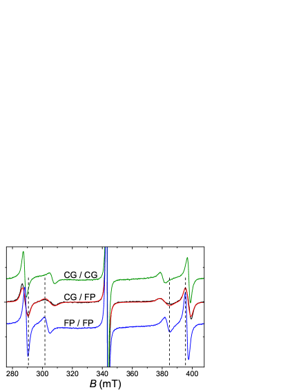
In the Fe3+-ion EPR spectrum of a (001)-plate with different quality of large faces (CG/FP), in the out-of-plane orientation (B) a significant broadening of the fine structure components is observed. Moreover, the resonance fields for the transitions at mT and mT (most sensitive to deformations) are distributed approximately within the values for CG/CG and FP/FP plates. The width of these components in the in-plane orientation (B) did not experience any significant modification with polishing. This observation unambiguously reflects the the distribution of the axial deformation along the plate normal. Figure 2 also presents the result of the EPR spectrum simulation for the Fe3+ centers with a linear distribution of axial deformation between the values characteristic for CG/CG and FP/FP plates. It can be seen that such an approximation makes it possible to fairly well describe the structure of the spectrum and the shape of its components. This fact, in turn, indicates that the surface quality is a factor determining to the high extent the magnitude of the axial deformation in the near-surface area of a plate.
To achieve a deeper insight, we studied the dependence of the axial distortion value and its distribution over the volume on a sample shape. Below we present the results for two STO:Fe samples with the right square prism shape: the first was initially a thin plate with mm and mm, and the second at the beginning had a height larger than a base side ( mm and mm). All faces of both samples were of the CG-grade. In the course of this experiment, the samples’ height was reduced by grinding in a step-by-step manner; EPR spectrum was acquired at each step. The results for the plate are shown in Fig. 3, a. A clear evolution of the spectrum structure is observed. The inset shows the dependence of the axial distortion () on thickness: we find that the magnitude of the distortion grows with the plate thickness decrease and is nicely proportional to . Small linewidths of the fine structure components of the spectra indicate near-uniform distribution of the axial distortion over the sample volume.
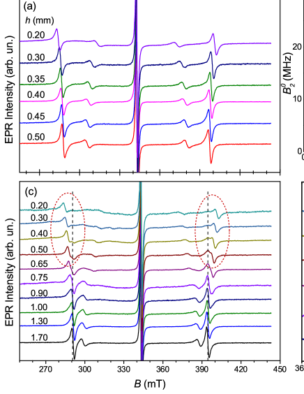
The dependence of the EPR spectrum structure on the height of the second sample () is qualitatively different (Fig. 3, b). Here, at relatively large thicknesses ( mm), the observed EPR spectrum is characteristic for Fe3+ centers free of the axial distortion, identical to that of the cubic Fe3+ impurity in STO Müller (1958); Unoki and Sakudo (1967). At mm, the signals from two paramagnetic Fe3+ centers with different local symmetry can be distinguished in the EPR spectrum. One of them is the cubic-symmetry center, another possesses the tetragonal symmetry. Axial distortion and relative intensity of the signal of the axial center increase with the decrease of the height . At a thickness of mm, the signals of the axial Fe3+ centers dominate in the spectrum. Considering the Fe3+ centers as paramagnetic probes, we conclude that initially the majority of the sample volume had the cubic symmetry. With a decrease of the sample height, the volume fraction with an axial symmetry first became noticeable and later took up the entire volume of the sample. It is important that in the observed spectrum the signals from the two centers with well-defined magnitude of the distortion are clearly revealed, and it is certainly not an average signal over two extreme cases. Such a set of experimental facts convincingly indicates the formation of a low (axial) symmetry phase in macroscopic, with hundreds of microns thickness, near-surface volumes with a small transient area between it and a high-symmetry (cubic) phase.
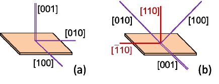
The observed tendency of the STO crystal plates to lower the symmetry reveals a pronounced selectivity. So, for square (001)-oriented plates, the structure of the EPR spectra of Fe3+ ions is different for B and B, while for B and B, the spectra are identical. Thus, in (001)-plates the directions [001] and [100] are inequivalent, and the directions [100] and [010] lying in the plane are equivalent (Fig. 4). That is, the crystal structure of the STO matrix has axial (tetragonal) symmetry with the -axis parallel to the normal to the plane. Comparison of the EPR spectra of the (110)-oriented STO:Fe plate shows that crystallographically equivalent out-of-plane and in-plane directions are equivalent; simultaneously, the in-plane [001] direction, on the one hand, and [100] and [010], which make up an angle of 45 degrees with the plane, on the other hand, turn out to be structurally inequivalent, while [100] and [010] are equivalent (Fig. 4). This is possible for a plate with a tetragonal crystal structure with the -axis along [001] lying in its plane. For the (111)-oriented STO plate, in which all the directions are equivalent relative to the plane of the sample and the directions are inequivalent, all the directions of the - and -types, according to the structure of the Fe3+-ion EPR spectra, are structurally equivalent within each type. Thus, a symmetry lowering manifests itself in the samples (plates) where the four-fold cubic axes are inequivalent with respect to large faces with an imperfect surface; the symmetry is lowered to tetragonal. The SrTiO3 (111)-plate retains cubic symmetry. Equivalency of the out-of-plane [110] and in-plane directions of the (110)-plate clearly indicates that the observed effect is not associated with the residual strain accumulated during surface treatment.
A question on polar or non-polar character of the revealed tetragonal phase will be addressed by us in a separate paper. Briefly, an observation of quadratic electric-field effect for both the STO:Fe and STO:Mn (001)-oriented thin plates clearly shows that the state is non-polar. The described set of experimental observations unambiguously indicates the structural instability in oriented STO single crystal plates with respect to a symmetry lowering from cubic to tetragonal phase, the last possessing a simple compressed (for plates) along the [001] direction structure, different from the antiferrodistortive one. This instability is inherent to the cubic phase, and the transition to a low-symmetry state is induced by the surface.
Existence of the near-surface lattice distortions in the high-symmetry phase of crystals above the phase transition temperature was considered theoretically in Refs. Levanyuk and Minyukov, 1983; Kaganov and Omel’yanchuk, 1971, and for STO it was extensively studied experimentally (see Ref. Dejneka, Trepakov, and Jastrabik, 2010 and references therein). However, STO surface distortions investigated in these works are conjugated to the AFD transition order parameter accompanied by the lattice cell doubling and differ in symmetry from the ones we found. In addition, they are restricted to the near-surface layer that is several lattice constants thick, much smaller than in our case.
The fact that the tetragonal STO phase manifested in our experiments has a structure different from the known cubic and AFD phases leads to the assumption that it can be stabilized, at least at T K, due to the influence of an imperfect, specifically oriented surface. We cannot exclude some influence of crystal structural imperfections, stresses and defects that arise during grinding and polishing, however, they do not determine the effect under discussion. In our opinion, the boundary conditions, including the state of the surface, as well as the geometry and dimensions of the sample, are crucial. The proposed concept requires further investigations.
The effect we report is of fundamental, qualitative character, since in the small, less than a millimeter size samples with the shape of oriented plates or bars with imperfect surfaces, a symmetry lowering of the crystal structure to tetragonal is observed. The value of the distortion in this new tetragonal phase is of the same order as that found in the AFD-phase below 10 K. The effect described in this work can represent a significant source of the variety of phenomena observed in strontium titanate, and should be taken into account when interpreting various experimental observations. It can cause sample dependence of particular effects in STO. At the same time, it can potentially become a technologically significant degree of freedom for fine-tuning of the STO substrate structure for the synthesis of thin-films on it: (i) (001)-plates with imperfect faces are compressed along the [001] direction; (110)-plates are elongated along [001] and (ii) surface roughness and a plate thickness define the magnitude of a distortion. Importantly, -oriented STO plate possesses the cubic symmetry regardless of the surface roughness.
The data that support the findings of this study are available from the corresponding author upon reasonable request.
Acknowledgements.
The work was performed with the support from the subsidy allocated to Kazan Federal University for the state assignment in the sphere of scientific activities No. FZSM-2020-0050 and from MS̀MT, Project No. SOLID21 - CZ.02.1.01/0.0/0.0/16_019/0000760.References
- Müller and Burkard (1979) K. A. Müller and H. Burkard, “SrTiO3: An intrinsic quantum paraelectric below 4 K,” Physical Review B 19, 3593 (1979).
- Barrett (1952) J. H. Barrett, “Dielectric constant in perovskite type crystals,” Physical Review 86, 118 (1952).
- Müller (1958) K. A. Müller, Paramagnetische Resonanz von Fe3+ in SrTiO3 Einkristallen, Ph.D. thesis, ETH Zurich (1958).
- Fleury, Scott, and Worlock (1968) P. Fleury, J. Scott, and J. Worlock, “Soft phonon modes and the 110 K phase transition in SrTiO3,” Phys. Rev. Lett. 21, 16 (1968).
- Courtens (1972) E. Courtens, “Birefringence of SrTiO3 produced by the 105 K Structural Phase Transition,” Phys. Rev. Lett. 29, 1380 (1972).
- Cowley (1996) R. A. Cowley, “The phase transition of strontium titanate,” Philosophical Transactions of the Royal Society of London. Series A: Mathematical, Physical and Engineering Sciences 354, 2799–2814 (1996).
- Yamanaka et al. (2000) A. Yamanaka, M. Kataoka, Y. Inaba, K. Inoue, B. Hehlen, and E. Courtens, “Evidence for competing orderings in strontium titanate from hyper-raman scattering spectroscopy,” EPL (Europhysics Letters) 50, 688 (2000).
- Kvyatkovskii (2001) O. Kvyatkovskii, “Quantum effects in incipient and low-temperature ferroelectrics,” Phys. Solid State 43, 1401 (2001).
- Lemanov, Smirnova, and Tarakanov (1997) V. V. Lemanov, E. P. Smirnova, and E. A. Tarakanov, “Ferroelectricity in SrTiO3:Pb,” Ferroelectrics Lett. Sec. 22, 69 (1997).
- Taniuchi et al. (2016) T. Taniuchi, Y. Motoyui, K. Morozumi, T. Rödel, F. Fortuna, A. Santander-Syro, and S. Shin, “Imaging of room-temperature ferromagnetic nano-domains at the surface of a non-magnetic oxide,” Nature Commun. 7, 1 (2016).
- Tufte and Chapman (1967) O. N. Tufte and P. W. Chapman, “Electron mobility in semiconducting strontium titanate,” Phys. Rev. 155, 796–802 (1967).
- Schooley, Hosler, and Cohen (1964) J. Schooley, W. Hosler, and M. L. Cohen, “Superconductivity in semiconducting SrTiO3,” Phys. Rev. Lett. 12, 474 (1964).
- Schooley et al. (1965) J. Schooley, W. Hosler, E. Ambler, J. Becker, M. L. Cohen, and C. Koonce, “Dependence of the superconducting transition temperature on carrier concentration in semiconducting SrTiO3,” Physical Review Letters 14, 305 (1965).
- Karg et al. (2006) S. Karg, G. Meijer, D. Widmer, and J. Bednorz, “Electrical-stress-induced conductivity increase in SrTiO3 films,” Applied Physics Letters 89, 072106 (2006).
- Szot et al. (2006) K. Szot, W. Speier, G. Bihlmayer, and R. Waser, “Switching the electrical resistance of individual dislocations in single-crystalline SrTiO3,” Nature Materials 5, 312–320 (2006).
- Makarova et al. (2010) M. Makarova, A. Dejneka, J. Franc, J. Drahokoupil, L. Jastrabik, and V. Trepakov, “Soft chemistry preparation methods and properties of strontium titanate nanoparticles,” Optical Materials 32, 803 (2010).
- Tyunina et al. (2009) M. Tyunina, J. Narkilahti, J. Levoska, D. Chvostova, A. Dejneka, V. Trepakov, and V. Zelezny, “Ultrathin SrTiO3 films: epitaxy and optical properties,” J. Phys. Cond. Matt. 21, 232203 (2009).
- Alexandrov et al. (2009) V. Alexandrov, E. Kotomin, J. Maier, and R. Evarestov, “First-principles study of bulk and surface oxygen vacancies in SrTiO3 crystal,” The European Physical Journal B 72, 53–57 (2009).
- Oja et al. (2012) R. Oja, M. Tyunina, L. Yao, T. Pinomaa, T. Kocourek, A. Dejneka, O. Stupakov, M. Jelinek, V. Trepakov, S. van Dijken, and R. M. Nieminen, “ ferromagnetic interface between nonmagnetic perovskites,” Physical Review Letters 109, 127207 (2012).
- Unoki and Sakudo (1967) H. Unoki and T. Sakudo, “Electron spin resonance of Fe3+ in SrTiO3 with special reference to the 110 K phase transition,” Journal of the Physical Society of Japan 23, 546–552 (1967).
- Rimai and DeMars (1962) L. Rimai and G. DeMars, “Electron paramagnetic resonance of trivalent gadolinium ions in strontium and barium titanates,” Physical Review 127, 702 (1962).
- Stoll and Schweiger (2006) S. Stoll and A. Schweiger, “Easyspin, a comprehensive software package for spectral simulation and analysis in EPR,” Journal of magnetic resonance 178, 42–55 (2006).
- Abraham and Bleaney (1970) A. Abraham and B. Bleaney, “Electron paramagnetic resonance of transition ions,” Clarendon, Oxford (1970).
- Gabbasov et al. (2018) B. Gabbasov, I. Gracheva, S. Nikitin, D. Zverev, A. Dejneka, V. Trepakov, and R. Yusupov, “Experimental evidences of the shape-induced structural distortion of SrTiO3 single crystals from impurity Mn4+ ions electron paramagnetic resonance,” Magnetic Resonance in Solids 20 (2018).
- Dobrov, Vieth, and Browne (1959) W. Dobrov, R. Vieth, and M. Browne, “Electron Paramagnetic Resonance in SrTiO3,” Physical Review 115, 79 (1959).
- Levanyuk and Minyukov (1983) A. P. Levanyuk and C. A. Minyukov, “Evolution of crystal structure distortions on the surface in the vicinity of structural phase transitions,” Physics Solid State 25, 2617 (1983).
- Kaganov and Omel’yanchuk (1971) M. I. Kaganov and A. M. Omel’yanchuk, “Phenomenological theory of phase transition in a thin ferromagnetic plate,” Sov. Phys. JETP 34, 895 (1971).
- Dejneka, Trepakov, and Jastrabik (2010) A. Dejneka, V. Trepakov, and L. Jastrabik, “Spectroscopic ellipsometry of SrTiO3 crystals applied to antiferrodistortive surface phase transition,” Physica Status Solidi (b) 247, 1951–1955 (2010).