A Review on the State of the Art in Non Contact Sensing for COVID-19
Abstract
COVID-19 disease, caused by SARS-CoV-2, has resulted in a global pandemic recently. With no approved vaccination or treatment, governments around the world have issued guidance to their citizens to remain at home in efforts to control the spread of the disease. The goal of controlling the spread of the virus is to prevent strain on hospital. In this paper, we have focus on how non-invasive methods are being used to detect the COVID-19 and assist healthcare workers in caring for COVID-19 patients. Early detection of the COVID-19 virus can allow for early isolation to prevent further spread. This study outlines the advantages and disadvantages and a breakdown of the methods applied in the current state-of-the-art approaches. In addition, the paper highlights some future research directions, which are required to be explored further to come up with innovative technologies to control this pandemic.
1 Introduction
Since late 2019, countries around the world have been experiencing a global pandemic through the surfacing and spread of the potentially fatal COVID-19 (COronaVIrusDisease 2019) caused by SARS-CoV-2 (Severe Acute Respiratory syndrome CoronaVirus 2) virus [1]. The COVID-19 causes victims to develop a fever and display respiratory difficulties causing coughing or shortness of breath [2, 3, 4]. Data collected from victims of the virus shows that the majority of deaths occurred in patients with underlying health issues with elderly people being at a higher risk of death [5]. The first confirmed case of the virus is considered to be in Wuhan, China in December 2019 with some of the early cases thought to be traced to seafood markets trading live animal species such as bats and snakes [6, 7, 8, 9]. The virus has been discovered to likely be related to bats. It is suspected that the virus may have transmitted to humans through bats which were being sold as food items [10, 11]. The exact cause of the virus is still unknown and it has also been suggested that the virus could originate from pangolins, which are natural hosts of corona viruses [12]. Pangolins is unlikely linked to the outbreak as the corona viruses found on the animal differ to COVID-19 [13]. Although it is possible the pangolin could have served as a intermediate host. As a result, these markets were shutdown in China [14]. The virus rapidly spread throughout China and eventually spread throughout the world. The virus was officially declared a global pandemic by the world health organisation (WHO) on 30th January 2020 [15, 16]. Although new discoveries are being made at the time of writing this paper, the virus has been found to be highly contagious and this has led to the rapid spread throughout the world [17]. The virus is spread primarily through respiratory droplets from an infected person [18]. These droplets can be dispensed by an infected person when coughing or sneezing. The droplets can then infect others directly via the eyes, mouth or nose when they are within a one meter radius of an infected person. The droplets can also be passed to others indirectly due to their long-term presence on surfaces [19]. Another leading factor in the rapid spread is that those infected with COVID-19, can be contagious during the early stages of infection while they are showing no symptoms [20]. This leads to people believing they are not sick while unknowingly spreading the virus. One of the main challenges of the COVID-19 pandemic is the how the spread of the virus can be controlled. The rapid spread of the COVID-19 virus has highlighted how the world’s population interacts when faced with a pandemic [21]. Governments around the world have outlined guidelines to their citizens to adhere to lock down rules. Currently, the best strategy to control the spread of COVID-19 is to ensure socially distancing people until a vaccine or an effective treatment can be produced [22, 23]. The National Health Service (NHS) of the United Kingdom is expecting an increased demand for their services as more COVID-19 patients are admitted and staff sick leave increases as staff members contract the disease [24]. Technology is being rapidly introduced in healthcare applications to develop systems that can ease the demand of the health service [25, 26, 27]. Any assistance via healthcare technology will free up valuable clinical resources to focus on other areas of care. In this paper we look at the state of the art non-contact sensing techniques and how these technologies can be used to assist in the care and detection of people suffering from COVID-19.
2 Non-contact sensing to detect COVID-19 symptoms
Non contact sensing is the ability to detect information without direct contact with a subject. In terms of healthcare, non contact can be used monitor the human body without devices physically touching the body. Non contact techniques are considered highly valuable in dealing with a highly infectious disease such as COVID-19 as contact may contribute to the spread of disease. Healthcare sensing technologies aim to collect information from a person which can be processed by Artificial Intelligence (AI) to provide decision support or directly analysed by a clinician to diagnose a disease or monitor existing conditions. Non-contact remote sensing technology is able to sense such healthcare markers without introducing anything to the body (e.g. wearable devices). Wearable devices can be uncomfortable for some which will entice users to remove the device and results in misplacement or damage [28]. The non-contact techniques can assist in the detection of COVID-19 and the care of patients suffering from COVID-19. Vital sign monitoring can provide great assistance in the fight against COVID-19 for a number of reasons. Although COVID-19 affects the respiratory system [29, 30], it has also been shown to take affect on the cardiovascular system [16]. Therefore non-contact sensing that monitors these vital signs can be used to aid in the detection and treatment of COVID-19. Examples of Non-contact techniques described in this paper include computed tomography (CT) scans, X-rays, Camera Technology, Ultrasound Technology, Radar Technology and Radio Frequency (RF) signal sensing. Table I details the advantages and disadvantages of each technique. Table II provides a summary table of the current literature contained within this review paper.
| Non-Invasive Techniques | Advantages | Disadvantages |
|---|---|---|
| CT Scans | High Accuracy, | High cost, |
| High image resolution | Exposure to Radiation, | |
| Non-portable, | ||
| Professional required for image analysis | ||
| X-rays | High Accuracy, | High cost, |
| High image resolution | Non-portable, | |
| Exposure to Radiation, | ||
| Professional required for image analysis | ||
| Camera | High Accuracy, | High cost of Camera |
| Technology | AI can be used for data analysis | Camera needs to be operated by professional |
| Ultrasound | High Accuracy, | Patients need to |
| Technology | AI can be used for data analysis | be prepared before |
| Possibility of portability | Ultrasound scan | |
| Radar | High accuracy, | High power consumption, |
| Technology | AI can be used for data analysis | High cost |
| RF signals | Low cost, | Vulnerable to noise |
| High Accuracy | ||
| AI can be used for data analysis |
| Title of Paper | Author(s) and Year | Key themes | Authority |
|---|---|---|---|
| Coronavirus (covid-19) classification using ct images by machine learning methods | Barstugan, M., Ozkaya, U., & Ozturk, S. (2020), | CT scan images are used to experiment with various methods of feature extraction and deep learning algorithms to achieve the best results | Peer reviewed paper. 157 citations on Google scholar. 157 citations on Google scholar. |
| POCOVID-Net: automatic detection of COVID-19 from a new lung ultrasound imaging dataset (POCUS) | Born, J., Brändle, G., Cossio, M., Disdier, M., Goulet, J., Roulin, J., & Wiedemann, N. (2020) | Lung Ultrasound videos of COVID-19, Pneumonia and non-infected patients used deep learning for classification. | Peer reviewed paper. 2 citations on Google scholar. |
| CSAIL device lets doctors monitor COVID-19 patients from a distance | Conner-simons 2020 | Radio Frequencies have been used to monitor the vital signs of patients in a contactless manner to protect healthcare workers | Article found on MIT Computer Science & Artificial Intelligence Laboratory website. |
| Can Radar Remote Life Sensing Technology Help to Combat COVID-19? | Islamcan 2020 | Radar systems have been used to monitor the vital signs of patients in a contact less manner to protect healthcare workers | Paper uploaded on researchgate.net. |
| Combining Visible Light and Infrared Imaging for Efficient Detection of Respiratory Infections such as COVID-19 on Portable Device | Jiang, Z., Hu, M., Fan, L., Pan, Y., Tang, W., Zhai, G., & Lu, Y. (2020) | RGB-Terminal camera footage used in a BiGRU neural network model between healthy and ill. | Peer reviewed paper. |
| Automatic detection of coronavirus disease (covid-19) using x-ray images and deep convolutional neural networks | Narin, A., Kaya, C., & Pamuk, Z. (2020) | X-ray scan images are used in a ResNet-50 Convolutional Neural Network (CNN) to distinguish between COVID-19 and Non-infected scan images. | Peer reviewed paper. 102 citations on Google scholar. |
| Artificial intelligence distinguishes COVID-19 from community acquired pneumonia on chest CT | Li, L., Qin, L., Xu, Z., Yin, Y., Wang, X., Kong, B., & Cao, K. (2020) | CT scan images are used in a COVNet neural network to distinguish between COVID-19, Pneumonia and Non-infected scan images. | Peer reviewed paper. 157 citations on Google scholar. |
| Automated detection of COVID-19 cases using deep neural networks with X-ray images | Ozturk, T., Talo, M., Yildirim, E. A., Baloglu, U. B., Yildirim, O., & Acharya, U. R. (2020) | Xray images are processed using the DarkNet neural network to test binary classification between COVID and Non-infected and multiclass classification between COVID, Pneumonia and Non-infected. | Peer reviewed paper. 22 citations on Google scholar. |
| Lung infection quantification of COVID-19 in ct images with deep learning | Shan, F., Gao, Y., Wang, J., Shi, W., Shi, N., Han, M., … & Shi, Y. (2020) | CT scan images are used in Deep learning to identify COVID-19. Human in the loop technique is used to focus on increasing accuracy | Peer reviewed paper. 52 citations on Google scholar. |
| Abnormal respiratory patterns classifier may contribute to large-scale screening of people infected with COVID-19 in an accurate and unobtrusive manner | Wang, Y., Hu, M., Li, Q., Zhang, X. P., Zhai, G., & Yao, N. (2020) | The paper details that COVID-19 patients display Tachypnea (Rapid breathing). The paper looks at taking Depth images to identify the breathing patterns of volunteers using deep learning | Peer reviewed paper. 24 citations on Google scholar. |
| Covid-19 screening on chest x-ray images using deep learning-based anomaly detection | Zhang, J., Xie, Y., Li, Y., Shen, C., & Xia, Y. (2020) | X-ray images are used with deep learning to identify if samples are COVID-19 or Pneumonia | Peer reviewed paper. 32 citations on Google scholar. |
2.1 CT Scanning
An example of a non invasive technique to detect COVID-19 is using Computed Tomography (CT) scans [31]. This process involves taking several X-ray images of a persons chest to create a 3D image of the lungs. The images can be reviewed by professionals to look for abnormalities in the lungs. The activity in the lungs is more prominent in the later stages of infection, however ultimately the CT scans showed a sensitivity of 86 % - 98 % [32]. This technique is non-contact as nothing is directly introduced into the body of the patient. CT scans are able to achieve high precision with high image resolution however the technology used to perform CT scans is expensive. The equipment is not portable and it requires skilled professional for image analysis. Another disadvantage of CT scanning is that the patient is exposed to radiation [33]. The radiation levels in CT scans have been found to result in an estimated cancer mortality risk of 0.08% within a 45 year old adult [34]. Recently, AI has been used on CT images for diagnosis of COVID-19 [35].
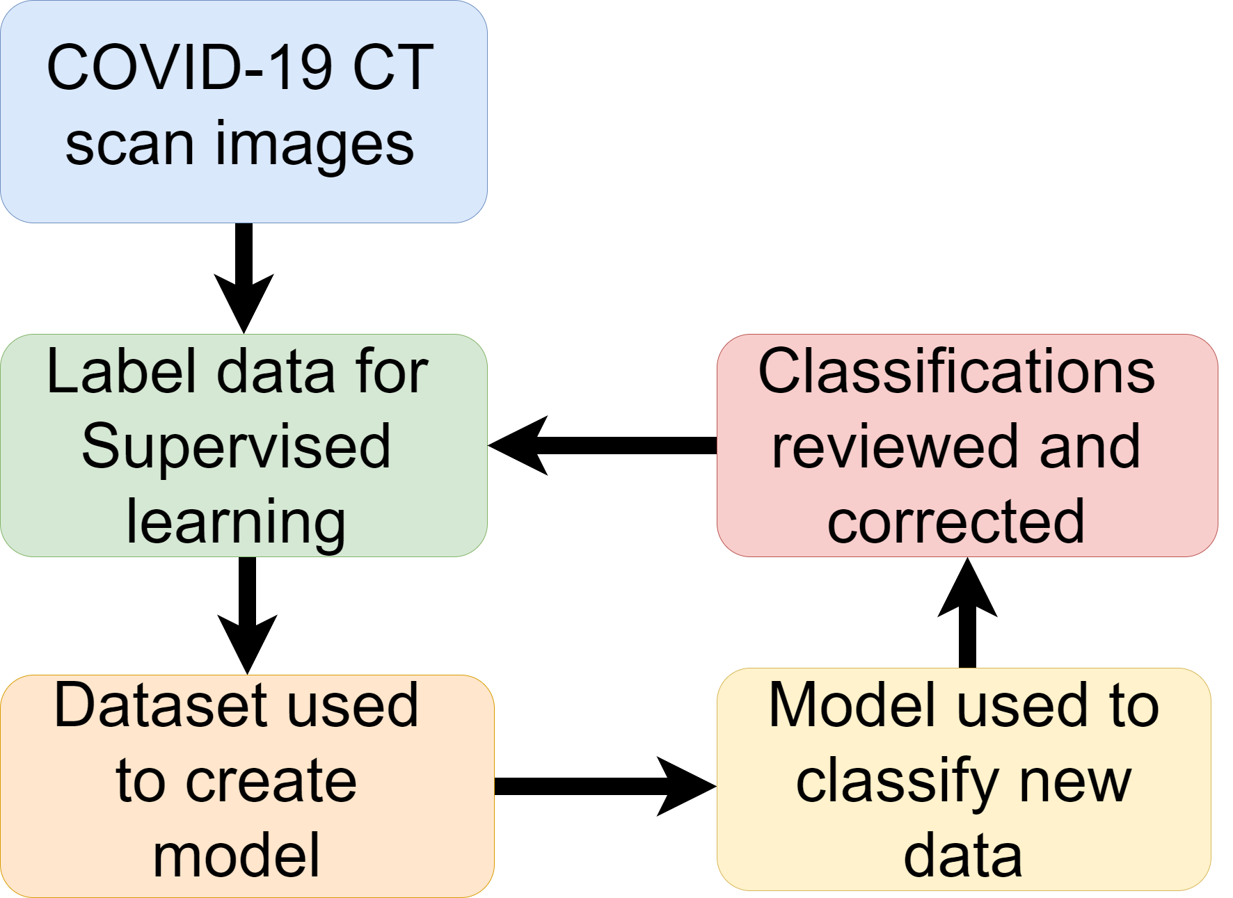
Fei Shan et al. [36] developed a deep learning model which was able to detect COVID-19 and also the level of infection. Their model adopted human-in-the-loop (HITL) strategy. HITL is when specialists are used to label a small amount of training data. Then an initial model is trained. Then this initial model is then used to classify new data. The specialist then correct any incorrect labels and the dataset can be used to train further models. This task can be iterated numerous times to reduce the tedious task of labeling large amounts of data. The experiment used 249 confirmed cases of COVID-19 for training. The experiment achieved a high result of 91.6 % accuracy. The experiment of this paper used 3 iterations. The first iteration made classifications on the validation data using 36 labeled images as a dataset with an an accuracy score of 85.1 %. The labels are then corrected and added to the second iteration. The second iteration used 114 images for training and achieved an accuracy result of 91.0 %. The labels are then corrected and passed to the third iteration The third iteration is used on all 249 training images and achieved an accuracy result of 91.6 %. The improved accuracy greatly reduces the human involvement and time devoted to labeling the full data. Figure 1 displays a flow chart of the process of HITL.
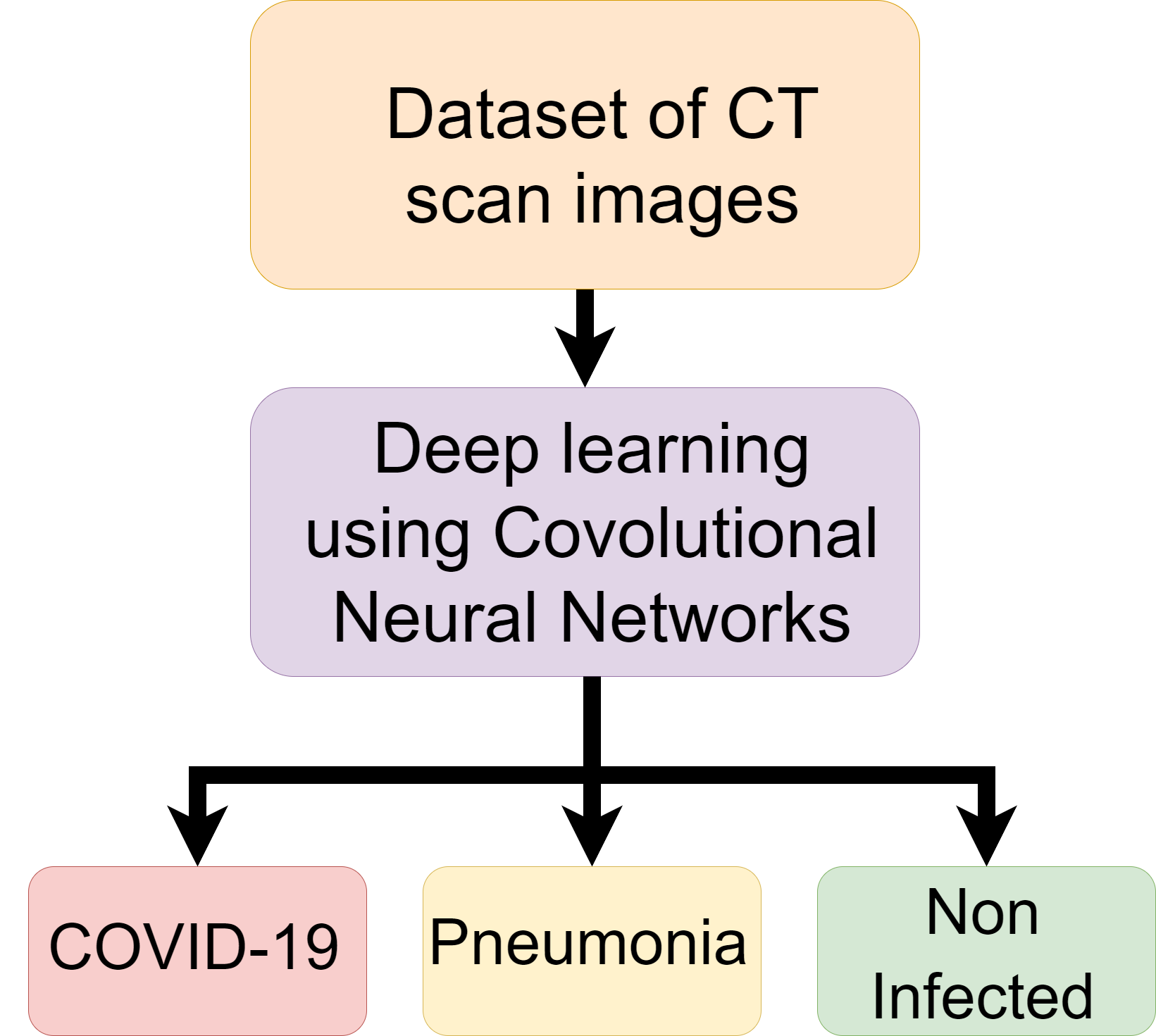
Li, Lin, et al. [37] used a COVNet - a deep learning neural network to predict COVID-19. The data used included 400 COVID-19 CT images, 1396 Pneumonia CT images and 1173 non infected CT images. The model takes CT images as input and extracts Pneumonia features from the images. The features are combined and the neural network is applied to make predictions on COVID-19, Pneumonia or non infected. Results found that the model was able to predict COVID-19 in patients with 90 % sensitivity. The model proved to not only be able to detect infected and non infected lungs but was also able to differentiate between COVID-19 and Pneumonia [37]. Figure 2 shows the process followed in this research.
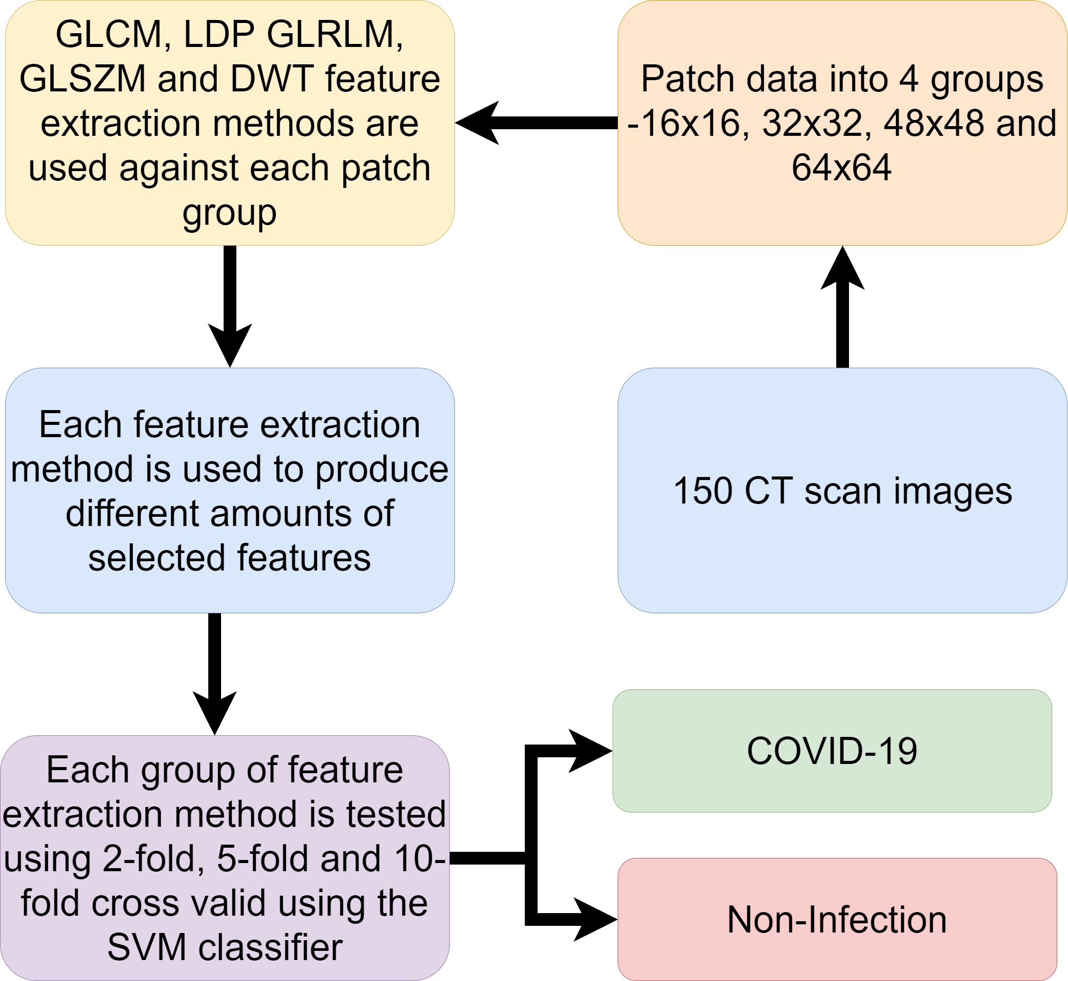
The research of Barstugan, Mucahid et al. [38] used machine learning on a dataset of 150 CT images. The dataset contains 53 infected cases. Patches of the images are taken. Different sized patches are used to create 4 different samples of different sized patches. The patch sizes are 16x16, 32x32, 48x48 and 64x64. The images were labelled as infected and non-infected in regards to COVID-19. The research used different methods of feature extraction on the images. These methods include Grey Level Co-occurrence Matrix (GLCM), Local Directional Patterns (LDP), Grey Level Run Length Matrix (GLRLM), Grey Level Size Zone Matrix (GLSZM) and Discrete Wavelet Transform (DWT). Support Vector Machine (SVM) algorithm was then used to classify the extracted features of each of the methods. SVM was experimented on the features using 2-fold, 5-fold and 10-fold cross validation. The highest accuracy result achieved was 99.64 %. This result was achieved using DWT with 10 fold cross validation using 48x48 patch dimension images. A flow chart of the methodology followed in this research is shown in figure 3.
The above papers show that CT scanning is capable of showing the signs of COVID-19 within a persons lungs. The research has also shown how AI can be used to assist in the determination of whether COVID-19 is present in the lungs. The studies have also shown how AI can also determine the level of infection and differentiate between Pneumonia and COVID-19 infections. Table III provides a break down of the above research papers.
| Title of Paper | Training data | Algorithms | Results |
|---|---|---|---|
| Lung infection quantification of covid-19 in ct images with deep learning | 249 CT images of COVID-19 showing different levels of infection. | Custom Convolutional neural network (CNN) called “VB-Net” | 91.6 % Accuracy |
| Artificial intelligence distinguishes COVID-19 from community acquired pneumonia on chest CT | 400 COVID-19 CT images, 1396 Pneumonia CT images and 1173 non infected CT images | Custom Convolutional neural network (CNN) called “COVNet” | 90 % sensitivity of COVID-19 samples. |
| Coronavirus (covid-19) classification using ct images by machine learning methods | 150 CT images including 53 COVID-19 cases. | Support Vector Machine | 99.64 % Accuracy |
2.2 X-ray Imaging
X-ray images are able to provide an analysis of the health of the lungs and is used frequently to diagnose pneumonia [39]. The same strategy is used with X-ray images of the lungs to display the visual indicators of COVID-19 [40, 41]. Similar to CT scans, X-ray equipment is also expensive and requires professionals to analyse the X-ray image.
The paper entitled ”Automatic detection of coronavirus disease (covid-19) using x-ray images and deep convolutional neural networks” used X-ray images of COVID-19 infected and non infected patients to create a dataset which was used to predict COVID-19 automatically in patients. The X-ray images are used in a ResNet-50 Convolutional Neural Network (CNN) which successfully obtained results of 98 % accuracy [42].
The paper of Zhang, Jianpeng, et al. [43] used Deep learning on a dataset of X-ray images of 70 patients confirmed to have COVID-19. Additional images of patients with Pneumonia are added from a public chest X-ray dataset. The model is used to identify between patients with COVID-19 and Pneumonia. The proposed deep learning model was able to achieve a sensitivity of 90 % detecting COVID-19 and a specificity of 87.84 % in detecting non COVID-19 cases.
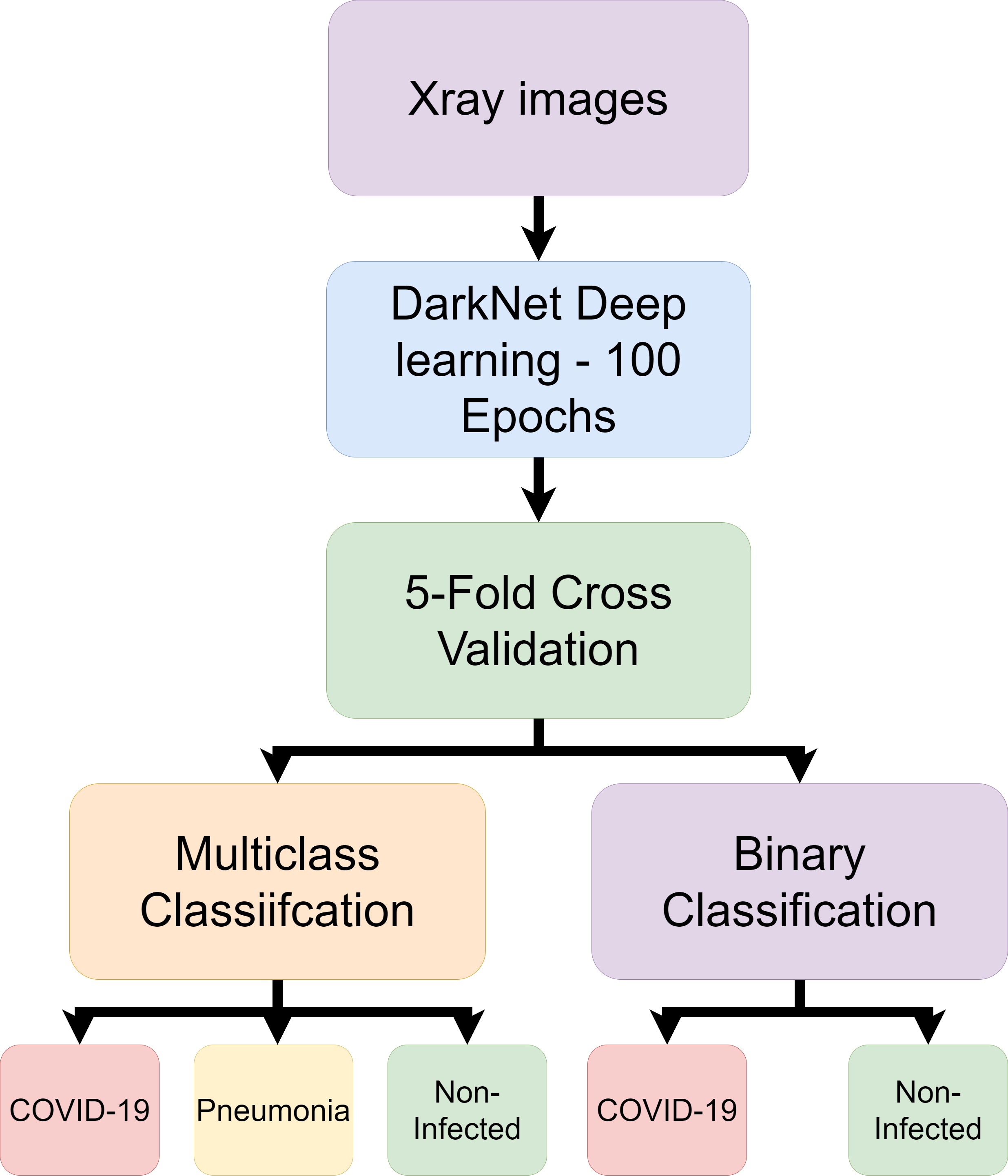
Ozturk, Tulin, et al. [44] also conducted experiments of using deep learning to classify X-ray images of patients. The experiments made use of a custom deep learning model named DarkNet to perform binary and multi-class classifications. The binary classification seeks to distinguish between COVID-19 and no findings of disease. The multi-class classification distinguishes between no findings of disease and the presence of Pneumonia. The experiments used a publicly available dataset of COVID-19 X-ray scans and another publicly available dataset for non-infected and pneumonia X-ray scans. The complete dataset included 127 COVID-19 samples and 500 each of non-infected and Pneumonia. The deep learning used the DarkNet neural network. The data was divided between 80 % training and 20 % testing. The deep learning was ran for 100 epochs using 5 fold cross validation. The results produced an accuracy score of 98 % for binary classification and an accuracy score of 87.02 % for multi class classification. It is expected that the result will fall as the number of classifications increase as the AI will need to recognise more features to distinguish between classes. The complete process followed in this work is detailed in figure 4.
2.3 Camera Technology
Camera technology can be used to provide non contact sensing by observing the chest movements of an individual [45]. This can provide assistance in the detection of COVID-19 as one of the symptoms include an increase in breathing rate.
The paper ”Combining Visible Light and Infrared Imaging for Efficient Detection of Respiratory Infections such as COVID-19 on Portable Device” used RGB-thermal camera footage for the detection of COVID-19. The footage was used with machine learning to detect normal and abnormal breathing from people wearing protective masks. The research collected real world data and applied deep learning to achieve a good result of 83.7 % accuracy which is the highest result in the literature in regards to breathing detection using RGB-thermal cameras using deep learning. This research can provide a scanning method which can be used to control the spread of the virus [46].
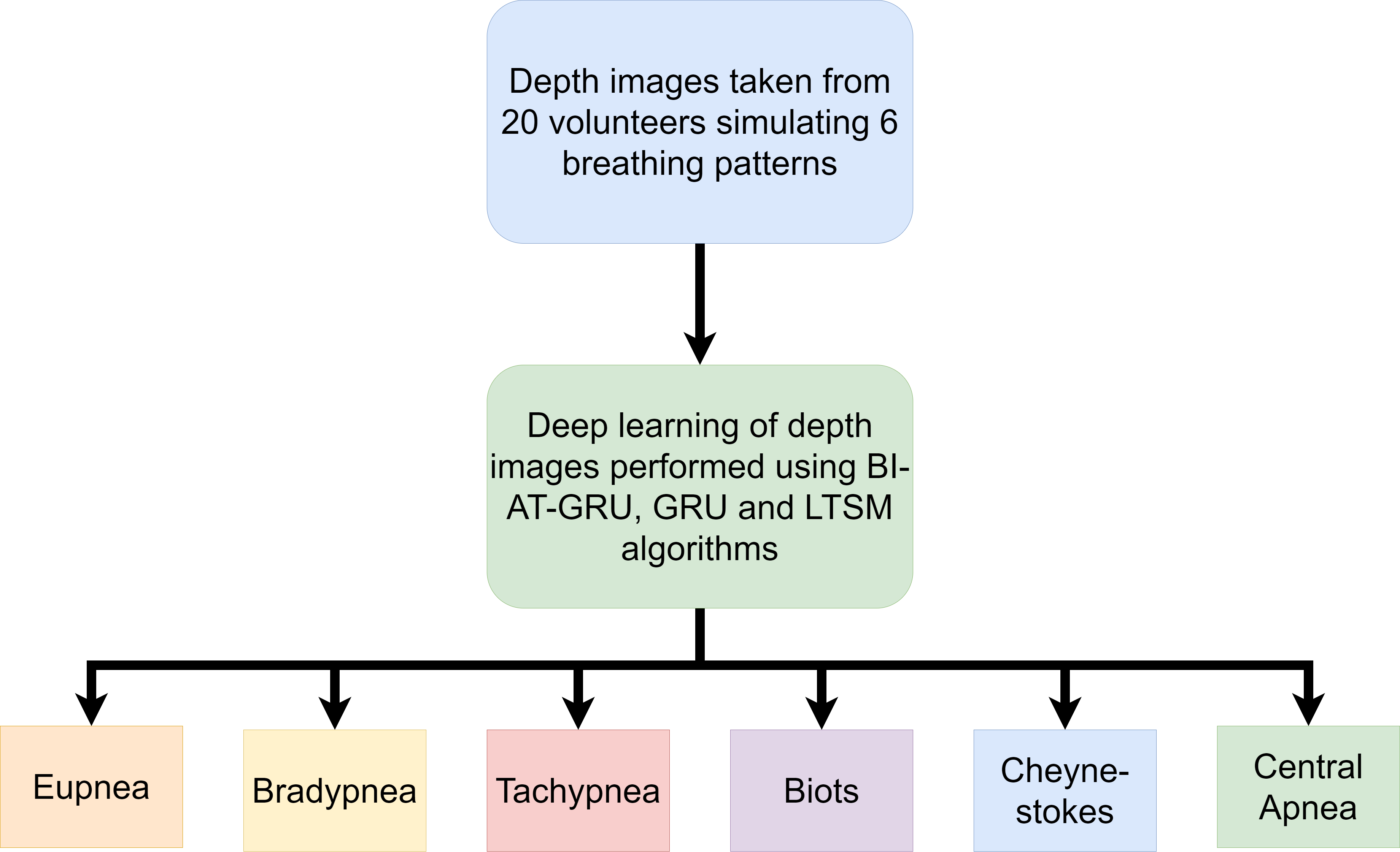
Wang, Yunlu, et al. [47] used Microsoft Kinect cameras to take depth images of volunteers breathing. A total of 20 volunteers were asked to sit on a chair and simulate 6 different breathing patterns. The breathing patterns were Eupnea, Bradypnea, Tachypnea, Biots, Cheyne-stokes and Central Apnea. Patients of COVID-19 display the rapid breathing pattern of Tachypnea. During data collection, a Spirometer was used to ensure the breathing pattern was being simulated correctly. The images taken using the camera are used in a deep learning neural network to classify abnormal breathing patterns associated with COVID-19. The deep learning model used was the BI-AT-GRU algortithm. Gated Recurrent Unit (GRU) is a simplified version of Long Term Short Memory(LTSM) algorithm. The BI-AT-GRU algorithm results achieved the best accuracy score of 94.5 %. This research shows how depth images can be used to identifiy the Tachypnea breathing pattern observed in COVID-19 patients. The process map for this research is shown in figure 5.
The primary disadvantage of using this method is the cost of thermal and depth cameras and the camera operators. Although the price of these cameras is falling gradually, it still remains substantially high [48]. The reseach done with cameras has shown that the devices can be used with AI in the detection of COVID-19 without contact with the body.
2.4 Ultrasound Technology
Ultrasound technology can be applied to detect respiratory failure of the lungs. An ultrasound machine is a device that uses high frequency sound waves to image body movements [49]. The sound waves bounce off different parts of the body which create echoes that are detected by the probe and used to create a moving image. Lung ultrasounds has seen great development in recent years [50]. The use of Ultrasound Technology can be used in the detection of COVID-19 in a non contact method where the risk of healthcare professionals becoming infected from patients can be decreased [51, 52]. Ultrasound technology can be performed using smart phones for the signal and processing of ultrasound images in a portable setting [53]. The disadvantage of ultrasound technology is that patients must prepare themselves before an ultrasound can effectively create an image of the body [54]. This preparation can include not eating for a few hours before.
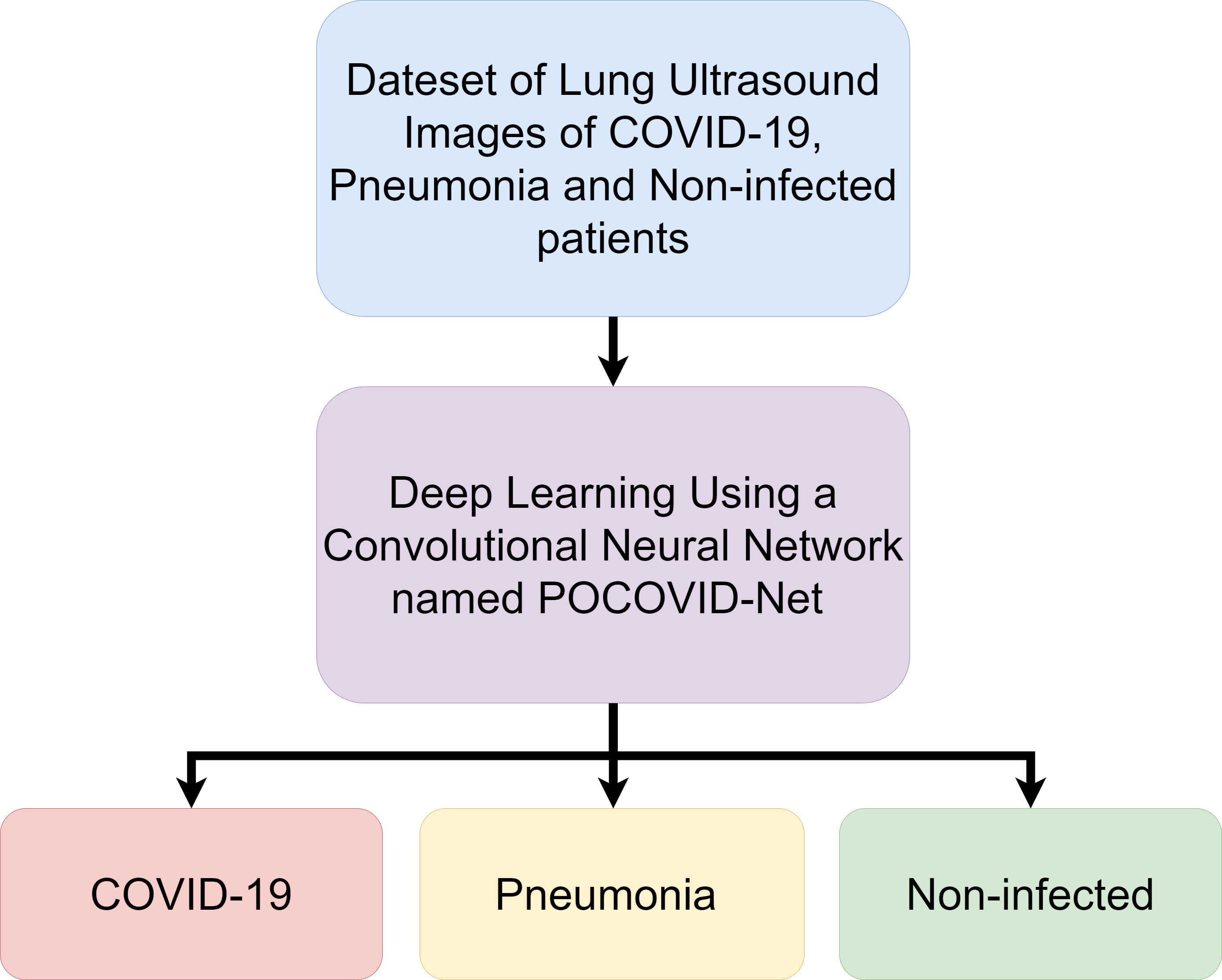
The work of Born, Jannis, et al. [55] shows that ultrasound technology can be used with deep learning to distinguish the differences in COVID-19, Pneumonia and no infection. The research collects a dataset of lung ultrasound images which contain video recordings of lung ultrasound scans. The dataset includes a total of 64 video recordings with 39 of the recordings of COVID-19 patients, 14 videos of Pneumonia patients and 11 videos of non-infected patients. The paper has developed a convolutional neural network named POCOVID-Net. The deep learning algorithm was able to achieve an accuracy score of 89 %. Figure 6 shows a simplified flow graph of the experiment undertaken in this paper.
2.5 Radar Technology
Radar Technology can be used to monitor the respiratory system within a home environment and provide a quick response if abnormalities are found which suggest COVID-19 being present. Radar systems use frequency-modulated continuous-wave (FMCW) to observe the Doppler effect when a person moves [56, 57, 58, 59]. This can be used to monitor the fine movements associated with breathing. Research done shows that radar technology can achieve 94 % accuracy for the detection of breathing rates and 80 % accuracy for heart rate detection [60, 61, 30]. The Israeli military force has made use of radar systems for monitoring the vital signs of COVID-19 patients. The goal of using this method is to prevent medical staff from becoming infected while caring for patients [62, 63]. Using radar technology to monitor vital signs can provide non-interference monitoring, however the disadvantages of radar systems is that it has high power requirements and the technology comes at a high cost [64].
2.6 Radio Frequency Signals
The use of radio frequency (RF) signal sensing can be used to detect the vital signs of individuals by sensing the minute movements of the chest made while breathing as the heart beats ( [65, 66, 67, 68, 69]). This technique can be used for monitoring the vital signs of patients independent of their activities [70]. The RF signals detect the movement by observing the Channel State Information (CSI) which can show amplitudes of the RF signals while movement occurs between a RF transmitter and receiver. [71, 72]. The Emerald system has been developed to monitor patients using RF signals. The system uses RF signals to detect the breathing rate of COVID-19 patients and then uses AI to infer the breathing rate of the patient. This allows for doctors treating the patients to be able to monitor the patient from a safe distance. This method prevents the risk of infection to staff and provides the patient comfort as they do not need to wear monitoring devices [73]. RF signals can be vulnerable to other movements within the room. The other movements create noise in the CSI which can then in turn cause false readings.
2.7 Future Directions
This section will detail some of the future directions which may be suitable for expanding on the research presented in this paper. The research has highlighted how the detection of COVID-19 is possible using various techniques. This section will now discuss how this research can be taken further to work within real life scenarios.
-
•
One of the biggest challenges with CT scanning as a means to diagnose COVID-19 is the lack of portability. This means that although the method is non contact, its use still requires individuals to travel to a location where the machine is available. As the CT images are able to provide high resolution, the AI can be used to for the detection of COVID-19. Therefore future directions of this method should look to creating highly accurate models that can eventually lead to the automation of COVID-19 detection. This can allow for faster diagnosis. Which can allow for more patients to be tested and increase availability of staff operating and analysing CT scans.
-
•
X-rays similarly to CT scans are not portable. Like CT scans professionals are required to operate these machines and analyse the X-ray images. The research presented in this paper has shown that AI can be used to make predictions if COVID-19 is present in the lungs. This can be useful similarly to CT scans where AI can be applied to make the predictions and speed up the process. The more data collected, the more advanced the model will become. Perhaps initially the predictions will need to be confirmed by humans but eventually the checks can become less frequent. Since the research above have displayed an ability of AI to distinguish between not just COVID-19 and non infected but also pneumonia at high accuracy, then the AI has proved to be capable of accurate classifications.
-
•
Thermal and Depth cameras are able to detect the irregular breathing patterns that are associated with COVID-19 symptoms. The issue here is that even though the camera can detect the irregular breathing pattern it is unable to categorically define COVID-19 as the cause for individuals displaying the irregular breathing patterns. In a real life situation, the camera method may be better suited to monitoring vulnerable people who are considered high risk from COVID-19. Then once the monitoring system has identified the irregular breathing patterns and alarm can be raised with a career or family member. Then appropriate action can be taken for greater accuracy such as diagnosis with CT scanning or X-ray scanning.
-
•
Ultrasound technology is able to take moving images of the lungs and detect COVID-19. This can also be made portable by using mobile devices. AI can be applied to recognise if COVID-19 or pneumonia is present in the lungs. This research can be further applied to develop applications on a mobile that device can capture an ultrasound of the lungs then compared to a AI model to predict if COVID-19 is present. Although not all phones may not have the necessary hardware to achieve this, the non contact method can allow for others to be able to use the devices for diagnosis at a safe distance.
-
•
Radar technology is able to identify the breathing patterns of individuals. Much like camera technology, the identification of breathing patterns are able to raise cause of concern but it cannot isolate COVID-19 as the sole cause. Radar technology can again be used to monitor individuals but due to the high costs its more likely to be used as a monitoring system within a hospital and not a home environment.
-
•
Any future directions should consider the use of RF signals as a means to detect the breathing patterns which give indication of COVID-19 symptoms. The RF systems can be implemented inexpensively using existing WiFi technology present within many homes. This allows for monitoring of individuals without the costs incurred in implementing radar or camera technologies highlighted in this paper.
3 Conclusion
The works listed in this paper have shown that the COVID-19 virus can be detected using contact-less techniques. Techniques such as CT scans and X-ray imaging provide high accuracy and high image resolution but the cost of the equipment is high and not portable. Thermal and depth camera technology has been used to detect breathing patterns which is associated with COVID-19 symptoms. However these cameras are expensive and needs to be operated by a professional. Radar technology also able to detect the breathing patterns but carries disadvantages of high operating expenses and capital expenditures. RF signals provide low cost and high accuracy as compared with other non-invasive technologies. The technologies are able to work on AI which can allow for skilled professionals to be available to assist in other areas of healthcare during the pandemic. The non-contact methods also protect healthcare workers from contracting the disease. The future direction of non-contact detection should look at the use of RF systems as the cost is cheap and it is easier to implement within a home environment in comparison to other methods. This gives the advantage of allowing the users to remain within isolation.
4 Acknowledgement
William Taylor’s studentship is funded by CENSIS UK through Scottish funding council in collaboration with British Telecom. This work is supported in parts by EPSRC DTG EP/N509668/1 Eng, EP/T021020/1 and EP/T021063/1
References
- [1] Tanu Singhal “A review of coronavirus disease-2019 (COVID-19)” In The Indian Journal of Pediatrics Springer, 2020, pp. 1–6
- [2] Lei Pan et al. “Clinical characteristics of COVID-19 patients with digestive symptoms in Hubei, China: a descriptive, cross-sectional, multicenter study” In The American journal of gastroenterology 115 Wolters Kluwer Health, 2020
- [3] Neo Poyiadji et al. “COVID-19–associated acute hemorrhagic necrotizing encephalopathy: CT and MRI features” In Radiology Radiological Society of North America, 2020, pp. 201187
- [4] Zhe Xu et al. “Pathological findings of COVID-19 associated with acute respiratory distress syndrome” In The Lancet respiratory medicine 8.4 Elsevier, 2020, pp. 420–422
- [5] Xiao Wu et al. “Exposure to air pollution and COVID-19 mortality in the United States” In medRxiv Cold Spring Harbor Laboratory Press, 2020
- [6] Joel Hellewell et al. “Feasibility of controlling COVID-19 outbreaks by isolation of cases and contacts” In The Lancet Global Health Elsevier, 2020
- [7] Shibo Jiang, Shuai Xia, Tianlei Ying and Lu Lu “A novel coronavirus (2019-nCoV) causing pneumonia-associated respiratory syndrome” In Cellular & molecular immunology 17.5 Nature Publishing Group, 2020, pp. 554–554
- [8] Muhammad Altaf Khan and Abdon Atangana “Modeling the dynamics of novel coronavirus (2019-nCov) with fractional derivative” In Alexandria Engineering Journal Elsevier, 2020
- [9] Hiroshi Nishiura, Natalie M Linton and Andrei R Akhmetzhanov “Initial cluster of novel coronavirus (2019-nCoV) infections in Wuhan, China is consistent with substantial human-to-human transmission” Multidisciplinary Digital Publishing Institute, 2020
- [10] Muhammad Adnan Shereen et al. “COVID-19 infection: origin, transmission, and characteristics of human coronaviruses” In Journal of Advanced Research Elsevier, 2020
- [11] Chih-Cheng Lai et al. “Severe acute respiratory syndrome coronavirus 2 (SARS-CoV-2) and corona virus disease-2019 (COVID-19): the epidemic and the challenges” In International journal of antimicrobial agents Elsevier, 2020, pp. 105924
- [12] Ping Liu et al. “Are pangolins the intermediate host of the 2019 novel coronavirus (SARS-CoV-2)?” In PLoS Pathogens 16.5 Public Library of Science San Francisco, CA USA, 2020, pp. e1008421
- [13] Kangpeng Xiao et al. “Isolation and characterization of 2019-nCoV-like coronavirus from Malayan pangolins” In BioRxiv Cold Spring Harbor Laboratory, 2020
- [14] Coronavirus Pneumonia Emergency Response Epidemiology Novel “The epidemiological characteristics of an outbreak of 2019 novel coronavirus diseases (COVID-19) in China” In Zhonghua liu xing bing xue za zhi= Zhonghua liuxingbingxue zazhi 41.2, 2020, pp. 145
- [15] A Spinelli and G Pellino “COVID-19 pandemic: perspectives on an unfolding crisis” In The British Journal of Surgery Wiley-Blackwell, 2020
- [16] Ying-Ying Zheng, Yi-Tong Ma, Jin-Ying Zhang and Xiang Xie “COVID-19 and the cardiovascular system” In Nature Reviews Cardiology 17.5 Nature Publishing Group, 2020, pp. 259–260
- [17] Di Dong et al. “The role of imaging in the detection and management of COVID-19: a review” In IEEE Reviews in Biomedical Engineering IEEE, 2020
- [18] Jing Cai et al. “Indirect virus transmission in cluster of COVID-19 cases, Wenzhou, China, 2020” In Emerg Infect Dis 26.6, 2020
- [19] World Health Organization “Modes of transmission of virus causing COVID-19: implications for IPC precaution recommendations: scientific brief, 27 March 2020”, 2020
- [20] Soheil Kooraki, Melina Hosseiny, Lee Myers and Ali Gholamrezanezhad “Coronavirus (COVID-19) outbreak: what the department of radiology should know” In Journal of the American college of radiology Elsevier, 2020
- [21] Jennifer Beam Dowd et al. “Demographic science aids in understanding the spread and fatality rates of COVID-19” In Proceedings of the National Academy of Sciences 117.18 National Acad Sciences, 2020, pp. 9696–9698
- [22] Marcel Salathé et al. “COVID-19 epidemic in Switzerland: on the importance of testing, contact tracing and isolation.” In Swiss medical weekly 150.11-12 EMH Swiss Medical Publishers, Ltd., 2020, pp. w20225
- [23] Joseph A Lewnard and Nathan C Lo “Scientific and ethical basis for social-distancing interventions against COVID-19” In The Lancet. Infectious diseases Elsevier, 2020
- [24] John Willan, Andrew John King, Katie Jeffery and Nicola Bienz “Challenges for NHS hospitals during covid-19 epidemic” British Medical Journal Publishing Group, 2020
- [25] Xinyue Yang et al. “Human Posture Recognition in Intelligent Healthcare” In Journal of Physics: Conference Series 1437.1, 2020, pp. 012014 IOP Publishing
- [26] Qammer H Abbasi, Masood Ur Rehman, Khalid Qaraqe and Akram Alomainy “Advances in body-centric wireless communication: Applications and state-of-the-art” Institution of EngineeringTechnology, 2016
- [27] William Taylor et al. “An Intelligent Non-Invasive Real-Time Human Activity Recognition System for Next-Generation Healthcare” In Sensors 20.9 Multidisciplinary Digital Publishing Institute, 2020, pp. 2653
- [28] Bo Tan et al. “Exploiting WiFi channel state information for residential healthcare informatics” In IEEE Communications Magazine 56.5 IEEE, 2018, pp. 130–137
- [29] John J Marini and Luciano Gattinoni “Management of COVID-19 respiratory distress” In Jama, 2020
- [30] Dou Fan et al. “Breathing rhythm analysis in body centric networks” In IEEE Access 6 IEEE, 2018, pp. 32507–32513
- [31] Wenjing Yang et al. “The role of imaging in 2019 novel coronavirus pneumonia (COVID-19)” In European Radiology Springer, 2020, pp. 1–9
- [32] Buddhisha Udugama et al. “Diagnosing COVID-19: the disease and tools for detection” In ACS nano 14.4 ACS Publications, 2020, pp. 3822–3835
- [33] Guilherme Duprat Ceniccola et al. “Current technologies in body composition assessment: advantages and disadvantages” In Nutrition 62 Elsevier, 2019, pp. 25–31
- [34] David J Brenner “Radiation risks potentially associated with low-dose CT screening of adult smokers for lung cancer” In Radiology 231.2 Radiological Society of North America, 2004, pp. 440–445
- [35] Feng Shi et al. “Review of artificial intelligence techniques in imaging data acquisition, segmentation and diagnosis for covid-19” In IEEE Reviews in Biomedical Engineering IEEE, 2020
- [36] Fei Shan et al. “Lung infection quantification of covid-19 in ct images with deep learning” In arXiv preprint arXiv:2003.04655, 2020
- [37] Lin Li et al. “Artificial intelligence distinguishes COVID-19 from community acquired pneumonia on chest CT” In Radiology Radiological Society of North America, 2020, pp. 200905
- [38] Mucahid Barstugan, Umut Ozkaya and Saban Ozturk “Coronavirus (covid-19) classification using ct images by machine learning methods” In arXiv preprint arXiv:2003.09424, 2020
- [39] Prabira Kumar Sethy and Santi Kumari Behera “Detection of coronavirus disease (covid-19) based on deep features” In Preprints 2020030300, 2020, pp. 2020
- [40] Linda Wang and Alexander Wong “COVID-Net: A Tailored Deep Convolutional Neural Network Design for Detection of COVID-19 Cases from Chest X-Ray Images” In arXiv preprint arXiv:2003.09871, 2020
- [41] Biraja Ghoshal and Allan Tucker “Estimating uncertainty and interpretability in deep learning for coronavirus (COVID-19) detection” In arXiv preprint arXiv:2003.10769, 2020
- [42] Ali Narin, Ceren Kaya and Ziynet Pamuk “Automatic detection of coronavirus disease (covid-19) using x-ray images and deep convolutional neural networks” In arXiv preprint arXiv:2003.10849, 2020
- [43] Jianpeng Zhang et al. “Covid-19 screening on chest x-ray images using deep learning based anomaly detection” In arXiv preprint arXiv:2003.12338, 2020
- [44] Tulin Ozturk et al. “Automated detection of COVID-19 cases using deep neural networks with X-ray images” In Computers in Biology and Medicine Elsevier, 2020, pp. 103792
- [45] Yunyoung Nam et al. “Monitoring of heart and breathing rates using dual cameras on a smartphone” In PloS one 11.3 Public Library of Science San Francisco, CA USA, 2016, pp. e0151013
- [46] Zheng Jiang et al. “Combining Visible Light and Infrared Imaging for Efficient Detection of Respiratory Infections such as COVID-19 on Portable Device” In arXiv preprint arXiv:2004.06912, 2020
- [47] Yunlu Wang et al. “Abnormal respiratory patterns classifier may contribute to large-scale screening of people infected with COVID-19 in an accurate and unobtrusive manner” In arXiv preprint arXiv:2002.05534, 2020
- [48] Heather E Elphick et al. “Exploratory study to evaluate respiratory rate using a thermal imaging camera” In Respiration 97.3 Karger Publishers, 2019, pp. 205–212
- [49] Alexander EJ Powles, David J Martin, Irving TP Wells and Christopher R Goodwin “Physics of ultrasound” In Anaesthesia & Intensive Care Medicine 19.4 Elsevier, 2018, pp. 202–205
- [50] Francesco Mojoli, Bélaid Bouhemad, Silvia Mongodi and Daniel Lichtenstein “Lung ultrasound for critically ill patients” In American journal of respiratory and critical care medicine 199.6 American Thoracic Society, 2019, pp. 701–714
- [51] Gino Soldati et al. “Is there a role for lung ultrasound during the COVID-19 pandemic?” In Journal of Ultrasound in Medicine Wiley-Blackwell, 2020
- [52] Danilo Buonsenso, Davide Pata and Antonio Chiaretti “COVID-19 outbreak: less stethoscope, more ultrasound” In The Lancet Respiratory Medicine 8.5 Elsevier, 2020, pp. e27
- [53] Kyu Cheol Kim et al. “Smartphone-based portable ultrasound imaging system: A primary result” In 2013 IEEE International Ultrasonics Symposium (IUS), 2013, pp. 2061–2063 IEEE
- [54] Alicja Genc et al. “Ultrasound imaging in the general practitioner’s office–a literature review” In Journal of ultrasonography 16.64 Polish Ultrasound Society, 2016, pp. 78
- [55] Jannis Born et al. “POCOVID-Net: automatic detection of COVID-19 from a new lung ultrasound imaging dataset (POCUS)” In arXiv preprint arXiv:2004.12084, 2020
- [56] Chuanwei Ding et al. “Fall detection with multi-domain features by a portable FMCW radar” In 2019 IEEE MTT-S International Wireless Symposium (IWS), 2019, pp. 1–3 IEEE
- [57] Syed Aziz Shah, Xiaodong Yang and Qammer H Abbasi “Cognitive health care system and its application in pill-rolling assessment” In International Journal of Numerical Modelling: Electronic Networks, Devices and Fields 32.6 Wiley Online Library, 2019, pp. e2632
- [58] Xiaodong Yang et al. “Diagnosis of the Hypopnea syndrome in the early stage” In Neural Computing and Applications 32.3 Springer, 2020, pp. 855–866
- [59] Syed Aziz Shah et al. “Sensor Fusion for Identification of Freezing of Gait Episodes using Wi-Fi and Radar Imaging” In IEEE Sensors Journal IEEE, 2020
- [60] Mostafa Alizadeh et al. “Remote monitoring of human vital signs using mm-Wave FMCW radar” In IEEE Access 7 IEEE, 2019, pp. 54958–54968
- [61] Francesco Fioranelli, Julien Le Kernec and Syed Aziz Shah “Radar for Health Care: Recognizing Human Activities and Monitoring Vital Signs” In IEEE Potentials 38.4 IEEE, 2019, pp. 16–23
- [62] Shekh MM Islam “Can Radar Remote Life Sensing Technology Help to Combat COVID-19?”, 2020
- [63] Syed Aziz Shah and Francesco Fioranelli “Human Activity Recognition: Preliminary Results for Dataset Portability using FMCW Radar”, 2019
- [64] Paul D Christenson, Cai Xia Yang and Naima Kaabouch “A Low Cost Through-Wall Radar for Vital Signs Monitoring” In 2019 IEEE International Conference on Electro Information Technology (EIT), 2019, pp. 567–571 IEEE
- [65] Jian Liu et al. “Monitoring vital signs and postures during sleep using WiFi signals” In IEEE Internet of Things Journal 5.3 IEEE, 2018, pp. 2071–2084
- [66] Binbin Dong et al. “Monitoring of atopic dermatitis using leaky coaxial cable” In Healthcare technology letters 4.6 IET, 2017, pp. 244–248
- [67] Daniyal Haider et al. “Utilizing a 5G spectrum for health care to detect the tremors and breathing activity for multiple sclerosis” In Transactions on Emerging Telecommunications Technologies 29.10 Wiley Online Library, 2018, pp. e3454
- [68] Xiaodong Yang et al. “Detection of essential tremor at the -band” In IEEE journal of translational engineering in health and medicine 6 IEEE, 2018, pp. 1–7
- [69] Xiaodong Yang et al. “Monitoring of patients suffering from REM sleep behavior disorder” In IEEE Journal of Electromagnetics, RF and Microwaves in Medicine and Biology 2.2 IEEE, 2018, pp. 138–143
- [70] Jian Liu et al. “Tracking vital signs during sleep leveraging off-the-shelf wifi” In Proceedings of the 16th ACM International Symposium on Mobile Ad Hoc Networking and Computing, 2015, pp. 267–276
- [71] Jijun Zhao et al. “R-DEHM: CSI-based robust duration estimation of human motion with WiFi” In Sensors 19.6 Multidisciplinary Digital Publishing Institute, 2019, pp. 1421
- [72] Nishtha Chopra et al. “THz time-domain spectroscopy of human skin tissue for in-body nanonetworks” In IEEE Transactions on Terahertz Science and Technology 6.6 IEEE, 2016, pp. 803–809
- [73] Adam Conner-Simons “CSAIL device lets doctors monitor COVID-19 patients from a distance” In CSAIL device lets doctors monitor COVID-19 patients from a distance — MIT CSAIL, 2020 URL: https://www.csail.mit.edu/news/csail-device-lets-doctors-monitor-covid-19-patients-distance